Academia.edu no longer supports Internet Explorer.
To browse Academia.edu and the wider internet faster and more securely, please take a few seconds to upgrade your browser.
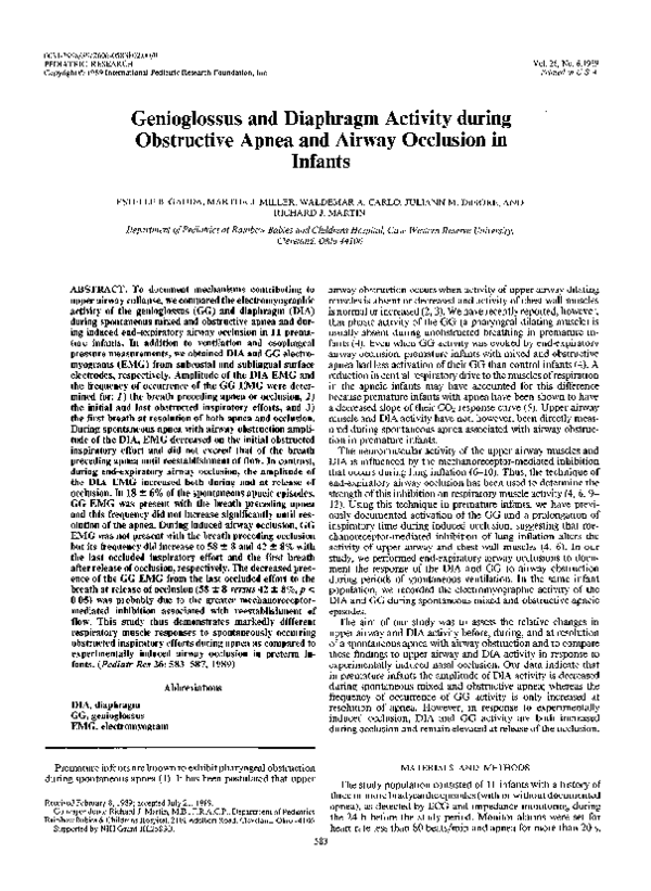
Genioglossus and Diaphragm Activity during Obstructive Apnea and Airway Occlusion in Infants
Genioglossus and Diaphragm Activity during Obstructive Apnea and Airway Occlusion in Infants
Genioglossus and Diaphragm Activity during Obstructive Apnea and Airway Occlusion in Infants
Genioglossus and Diaphragm Activity during Obstructive Apnea and Airway Occlusion in Infants
Genioglossus and Diaphragm Activity during Obstructive Apnea and Airway Occlusion in Infants
1989, Pediatric Research
Related Papers
Journal of applied physiology (Bethesda, Md. : 1985)
Site and mechanics of spontaneous, sleep-associated obstructive apnea in infants2000 •
To examine the mechanics of infantile obstructive sleep apnea (OSA), airway pressures were measured using a triple-lumen catheter in 19 infants (age 1-36 wk), with concurrent overnight polysomnography. Catheter placement was guided by correlations between measurements of magnetic resonance images and body weight of 70 infants. The level of spontaneous obstruction was palatal in 52% and retroglossal in 48% of all events. Palatal obstruction predominated in infants treated for OSA (80% of events), compared with 38.6% from infants with infrequent events (P = 0.02). During obstructive events, successive respiratory efforts increased in amplitude (mean intrathoracic pressures -11.4, -15.0, and -20.4 cmH(2)O; ANOVA, P < 0.05), with arousal after only 29% of the obstructive and mixed apneas. The soft palate is commonly involved in the upper airway obstruction of infants suffering OSA. Postterm, infant responses to upper airway obstruction are intermediate between those of preterm infant...
2004 •
Journal of Applied Physiology
Effect of upper airway negative pressure on inspiratory drive during sleep1998 •
Journal of applied physiology (Bethesda, Md. : 1985)
Hemodynamic effects of pressures applied to the upper airway during sleep2000 •
The increase in systemic blood pressure after an obstructive apnea is due, in part, to sympathetically mediated vasoconstriction. We questioned whether upper airway (UA) receptors could contribute reflexly to this vasoconstriction. Four unanesthetized dogs were studied during wakefulness and non-rapid-eye-movement (NREM) sleep. The dogs breathed via a fenestrated tracheostomy tube sealed around the tracheal stoma. The snout was sealed with an airtight mask, thereby isolating the UA when the fenestration was closed and exposing the UA to negative inspiratory intrathoracic pressure when it was open. The blood pressure response to three UA perturbations was studied: 1) square-wave negative pressures sufficient to cause UA collapse with the fenestration closed during a mechanical hyperventilation-induced central apnea; 2) tracheal occlusion with the fenestration open vs. closed; and 3) high-frequency pressure oscillations (HFPO) with the fenestration closed. During NREM sleep, 1) blood ...
Canadian Journal of Anaesthesia-journal Canadien D Anesthesie
Automated respiratory inductive plethysmography to evaluate breathing in infants at risk for postoperative apnea2008 •
Purpose: Although respiratory inductive plethysmography (RIP) is the method of choice for the assessment of sleep disordered breathing, it has not been applied to the study of infants at risk for postoperative apnea (POA). The purpose of this study was to apply RIP to evaluate breathing in these infants. An additional purpose was to implement, simultaneously, three novel algorithms to detect movement artifact, respiratory pauses, and thoracoabdominal asynchrony, since their combined output both detects respiratory pauses and classifies them as obstructive or central in origin. Methods: A prospective study design was employed to record the analogue output of RIP, saturation, and finger plethysmography in a convenience sample of infants. The data record underwent a dual analysis: 1) automated detection of respiratory events; and 2) visual coding of the cardiorespiratory data. A novel index, coined pause density, was calculated as the sum of all respiratory pauses. Results: Twenty infants, whose mean postconceptional ages and weights were 44.47±2.88 weeks and 4.21±0.99 kg, respectively, were recruited. Data recording ranged from four to 24 hr. Ten infants (term=5) experienced POA: central apnea=5, mixed obstructive apnea=6, and two former premature infants experienced both. Twenty-five central apneic events were detected, and the majority followed a sigh. Infants who experienced apnea also had high values of pause density. Conclusion: Respiratory inductive plethysmography may provide a useful method to evaluate breathing in infants at risk for POA. The study of short respiratory pauses may prove useful in predicting apnea risk. Objectif: Bien que la pléthysmographie inductive respiratoire (RIP) soit la méthode privilégiée pour évaluer les troubles respiratoires du sommeil, cette technique n’a pas été employée dans l’étude des nourrissons présentant un risque d’apnée postopératoire. L’objectif de cette étude était d’utiliser la RIP pour évaluer la respiration de ces nourrissons. Un objectif secondaire était de mettre en oeuvre trois nouveaux algorithmes pour détecter simultanément les artéfacts de mouvement, les pauses respiratoires et l’asynchronie thoraco-abdominale, étant donné que la combinaison de ces données détecte les pauses respiratoires et les classifie selon que leur origine soit obstructive ou centrale. Méthode: Un devis prospectif a été utilisé pour enregistrer la sortie analogique de la RIP, la saturation, et la pléthysmographie digitale dans un échantillon de commodité de nourrissons. Le fichier de données a subi une analyse double : 1) détection automatique des événements respiratoires ; et 2) codage visuel des données cardiorespiratoires. Un nouvel indicateur, appelé densité des pauses, qui est égal á la somme de toutes les pauses respiratoires, a été calculé. Résultats: Vingt nourrissons, dont l’âge post-conceptionnel et le poids moyen étaient respectivement de 44,47±2,88 semaines et 4,21±0,99 kg, ont été recrutés. L’enregistrement des données allait d’une durée de quatre à 24 h. Dix nourrissons (5 à terme) ont présenté une apnée postopératoire : apnée centrale=5, apnée mixte obstructive=6, et deux nourrissons nés prématurément ont présenté ces deux types d’apnée. Au total, 25 événements apnéiques centraux ont été détectés, et la majorité sont survenus à la suite d’un soupir. Chez les nourrissons ayant manifesté de l’apnée, nous avons également observé des valeurs élevées de densité des pauses. Conclusion: La pléthysmographie inductive respiratoire pourrait constituer une méthode utile pour évaluer la respiration des nourrissons présentant un risque d’apnée postopératoire. L’étude des courtes pauses respiratoires pourrait s’avérer utile pour prédire le risque d’apnée.
The American review of respiratory disease
Effect of laryngeal anesthesia on pulmonary function testing in normal subjects1988 •
Pulmonary function tests (PFT) were performed on 11 normal subjects before and after topical anesthesia of the larynx. The PFT consisted of flow volume loops and body box determinations of functional residual capacity and airway resistance, each performed in triplicate. After the first set of tests, cotton pledgets soaked in 4% lidocaine were held in the pyriform sinuses for 2 min to block the superior laryngeal nerves. In addition, 1.5 ml of 10% cocaine was dropped on the vocal cords via indirect laryngoscopy. PFT were repeated 5 min after anesthesia. Besides routine analysis of the flow volume loops, areas under the inspiratory (Area I) and expiratory (Area E) portions of the loops were calculated by planimetry. Area I, peak inspiratory flow (PIF), as well as forced inspiratory flow at 25, 50, and 75% forced vital capacity (FVC), decreased after anesthesia. Peak expiratory flow decreased after anesthesia, but Area E and forced expiratory flow at 25, 50, and 75% FVC were unchanged....
Wiley Encyclopedia of Biomedical Engineering
Obstructive Sleep Apnea: Electrical Stimulation Treatment2006 •
RELATED PAPERS
1994 •
Journal of applied physiology (Bethesda, Md. : 1985)
A volume-dependent apneic threshold during NREM sleep in the dog1994 •
Pediatric Pulmonology
Measurements of respiratory mechanics in the newborn: A simple approach1987 •
Clinics in Perinatology
Which Continuous Positive Airway Pressure System is Best for the Preterm Infant with Respiratory Distress Syndrome?2012 •
Conference proceedings : ... Annual International Conference of the IEEE Engineering in Medicine and Biology Society. IEEE Engineering in Medicine and Biology Society. Conference
Increased variability in respiratory parameters heralds obstructive events in children with sleep disordered breathing2013 •
Journal of Applied Physiology
Role of respiratory control mechanisms in the pathogenesis of obstructive sleep disorders2008 •
The Journal of Pediatrics
Effect of caffeine on control of breathing in infantile apnea1983 •
Critical Reviews in Oral Biology & Medicine
Oral and Pharyngeal Reflexes in the Mammalian Nervous System: Their Diverse Range in Complexity and the Pivotal Role of the Tongue2002 •
Bulletin of Mathematical Biology
Adjustment of the Human Respiratory System to Increased Upper Airway Resistance During Sleep2002 •
Journal of Applied Physiology
Effects of hypoxia and hypercapnia on nonnutritive swallowing in newborn lambs2007 •
Journal of Applied Physiology
Active glottal closure during central apneas limits oxygen desaturation in premature lambsSleep & breathing = Schlaf & Atmung
Increased thoracoabdominal asynchrony during breathing periods free of discretely scored obstructive events in children with upper airway obstruction2014 •
Journal of applied physiology (Bethesda, Md. : 1985)
Chronic recordings of hypoglossal nerve activity in a dog model of upper airway obstruction1999 •
The Journal of Pediatrics
The effect of a low continuous positive airway pressure on the reflex control of respiration in the preterm infant1977 •
Respiration Physiology
Prolonged active glottic closure after barbiturate-induced respiratory arrest in lambs1996 •
The Journal of Pediatrics
Physiologic effects of doxapram in idiopathic apnea of prematurity1986 •
Respiratory Physiology & Neurobiology
Apnea of prematurity – Perfect storm2013 •
The Journal of Pediatrics
Characteristics of hypoxemic episodes in very low birth weight infants on ventilatory support1997 •
Baillière's Clinical Anaesthesiology
10 Sudden infant death syndrome1995 •
2002 •
American Journal of Otolaryngology
Pediatric sleep-related breathing disorders2000 •
C23. COPD COMORBIDITIES: EXPANDING THE HORIZONS
Compensatory Responses To Upper Airway Obstruction In Chronic Obstructive Pulmonary Disease2012 •
2005 •
Update in Intensive Care Medicine
Long-term Outcomes of Mechanical Ventilation2005 •
Pediatric Pulmonology
Effects of non-invasive pressure support ventilation (NI-PSV) on ventilation and respiratory effort in very low birth weight infants2007 •
Journal of applied physiology (Bethesda, Md. : 1985)
Compensatory responses to upper airway obstruction in obese apneic men and women2012 •
Critical Care Medicine
Pressure support ventilation in patients with acute lung injury2000 •
Current Opinion in Pediatrics
Infant pulmonary function testing2003 •
The Journal of Physiology
Inhibition of inspiratory motor output by high-frequency low-pressure oscillations in the upper airway of sleeping dogs1999 •
1996 •
Respiratory Physiology & Neurobiology
Spontaneous central apneas occur in the C57BL/6J mouse strain2008 •

 Juliann Di Fiore