Indian Journal of Medical Microbiology, (2002) 20 (4):194-199
Original Article
EVALUATION OF A NEW PHAGE AMPLIFICATION TECHNOLOGY
FOR RAPID DIAGNOSIS OF TUBERCULOSIS
S Shenai, *C Rodrigues, AP Mehta
Abstract
Purpose: Rapid diagnosis of tuberculosis is essential to initiate timely and appropriate treatment to curb
the spread of this potentially life threatening disease. The purpose of this study was to evaluate a phage
amplification technology viz., FASTPlaque TB,™ for the diagnosis of tuberculosis. Methods: We
evaluated the clinical utility of this new assay by analyzing 50 respiratory and 40 non-respiratory
specimens, using FASTPlaque TB™ kit (Biotec Laboratories, UK) and the performance was compared
with TB Bactec 460 semi-automated liquid culture system and conventional LJ culture method. Results:
In case of respiratory specimens phage assay gave good specificity (100%) compared with TB Bactec
whereas with respect to LJ method the sensitivity and specificity were 93.1% and 88.2% respectively. In
case of non-respiratory specimens comparison of results obtained by phage assay showed sensitivity of
90.9% and specificity of 88.8% with respect to TB Bactec and 87.5% and 93.8% with respect to LJ method.
Conclusions: We believe that this new low cost assay may have widespread applicability, especially in
developing countries, due to its manual format and rapid reporting of results.
Tuberculosis (TB) continues to be one of the leading
infectious causes of death in the world today. It affects
one-third of the world’s population, of which 95% is in
the developing countries where resources are limited.1
In India, 13 million are infected and diseased, 3.5
million are positive for acid fast bacilli (AFB) with 2.2
million new TB cases being added every year.2 The
definitive diagnosis of TB continues to depend on
microscopy and culture. Smear microscopy remains the
mainstay of TB diagnosis in developing countries, but
suffers from low specificity, and variable sensitivity.3
Laboratory cultivation of Mycobacterium tuberculosis
is much more sensitive, but it is time consuming and
susceptible to contamination problems.4 Rise in TB
statistics and recent outbreaks of multidrug resistant
(MDR) TB have heightened the importance of rapid
diagnosis of this disease. Molecular methods for
detection of TB are proving rapid and sensitive, but the
high cost of these methods and requirement for
sophisticated equipment currently renders them
inappropriate for routine use in many countries with a
high burden of disease. 5 Hence, any test broadly
acceptable to the global TB diagnostic community needs
to be cost effective, accurate, simple and easy to
implement within the current infrastructure. This
challenge has prompted scientists to reconsider the use
*Corresponding author
PD Hinduja National Hospital and Medical Research
Center, Veer Savarkar Marg, Mahim, Mumbai - 400 016,
India.
Received: 16-01-2002
Accepted: 31-05-2002
of mycobacteriophages as tools in diagnosis and drug
susceptibility testing.
Gardner and Weiser isolated the first
mycobacteriophage in 1947 and since that time over 250
phages have been identified.6,7 The recent upsurge of
drug-resistant bacterial infection has prompted fresh
interest in the field of phage therapy but, unfortunately,
attempts to use lytic phages therapeutically during
tuberculosis infection have so far failed to elicit cure in
experimentally infected animals.7 Instead, the use of
these mycobacteriophages in investigative studies of
mycobacteria has become widespread and recently, their
potential as tools for drug susceptibility testing was
reported.1,8,9 FASTPlaque TB,TM a new rapid test for
diagnosis of TB, was launched in year 2000 by Biotec
Laboratories Ltd.
FASTPlaque TB™ is a novel, patent protected,
phage amplification technology that has been developed
for rapid detection and enumeration of M.tuberculosis
complex from respiratory specimens. This method uses
specific mycobacteriophages (viruses that infect
M.tuberculosis complex) to detect the presence of viable
TB bacilli in the clinical specimen.3,10 Mycobacteria are
mixed with phages, which are allowed to adsorb and
infect the cells. All unadsorbed extracellular phages are
then inactivated using a phagicidal chemical (virucide);
while the phages which have infected the viable TB
bacilli remain protected and continue to replicate. After
replication the progeny bacteriophages are released and
detected by mixing with fast growing non-pathogenic
helper cells (M.smegmatis) on an agar plate. The
www.ijmm.org
�October, 2002
Shenai et al - FASTPlaque TB™ Assay for the Diagnosis of Tuberculosis
mycobacteriophages in turn infect, replicate and lyse
these helper cells and lysis is detected as plaques (clear
zones). The number of plaques visualized from a given
sample is related to the number of viable tubercle bacilli
in the original sample.8,11,12 In M.smegmatis the lytic
cycle is completed within 90 minutes whereas lysis takes
approximately 13 hours in M.tuberculosis complex
thereby making results available rapidly in terms of
plaques.7 In this study, we have evaluated the clinical
utility of phage assay, using FASTPlaque TB kit, by
comparing the results with clinical data, smear
microscopy, and culture methods.
195
current details of medication (if taken) of all these
subjects were collected.
Decontamination and concentration of specimens
Materials and Methods
All specimens, which were likely to contain normal
or transient bacterial flora, were decontaminated by
standard N-acetyl-L-cysteine-NaOH method,13 while
specimens collected from sterile sites were centrifuged
and sediment was used to perform the FASTPlaque TB
test. Decontaminated and concentrated sediment was
resuspended in 2 mL of sterile 0.67 M phosphate buffer
(pH 6.8) and used for phage assay and cultures by TB
Bactec and Lowenstein Jensen (LJ) methods.
Patients
Microscopy
The study comprised of 50 respiratory (sputum,
bronchoalveolar lavage and endotracheal secretion) and
40 non-respiratory (pleural fluid, CSF, cold abscess,
lymph node, pus, urine etc.) specimens collected at PD
Hinduja National Hospital and Medical Research Center.
The clinical history, symptoms, radiological,
histopathological, other laboratory findings, and past and
Smears were made for all the clinical specimens
studied and stained with Ziehl-Neelsen carbol fuchsin
(ZN) staining method13 for AFB.
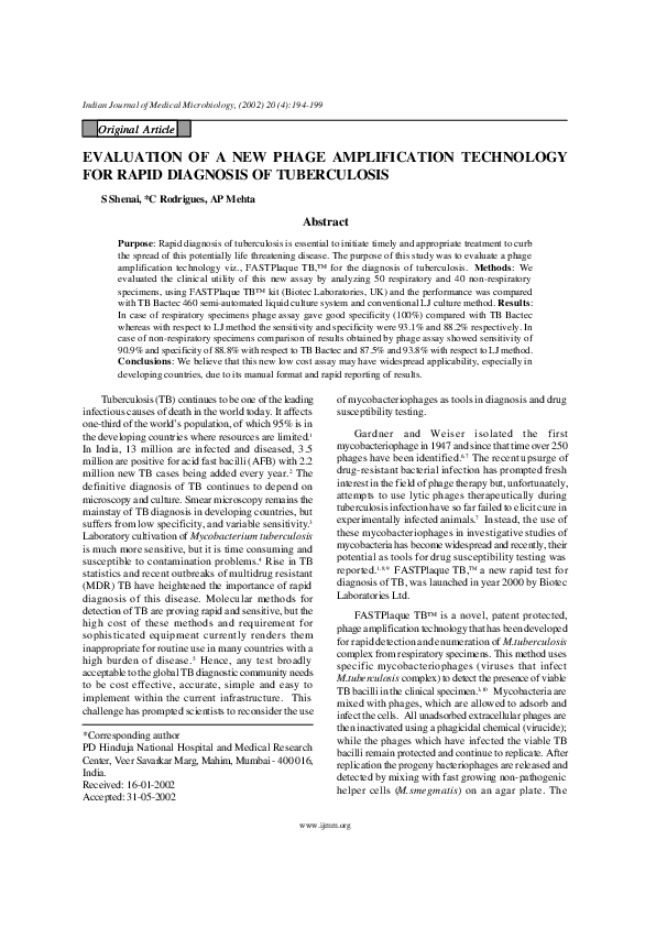
FASTPlaque TB Assay
The principle of the FASTPlaque TB assay is shown
in figure 1.
Infection
Actiphage
TB Bacilli
Neutralization of
Virusol and Addition
of Sensor Cells
Phage start
to replicate
in cells
Treatment
with Virusol
Plating of Mixture in a
Petri dish & overnight
incubation
Figure 1 : Principle of FASTPlaque TB assay
The assay was carried out by using FASTPlaque
TB™ kit (Biotec laboratories, UK). All respiratory
specimens were processed according to the instructions
given by the manufacturer whereas minor modifications
were made in the procedure while processing non-
respiratory specimens as this kit was not recommended
for non-respiratory specimens. Both Positive and
negative controls were also included in the assay and
tested as per the manufacturer’s instructions. Negative
control contained 1 mL of plain FASTPlaque TB broth,
www.ijmm.org
�196
Indian Journal of Medical Microbiology
whereas three positive controls were prepared by serial
dilution (10-2 ,10-4 ,10-6 ) of M.smegmatis respectively.
These controls were included to assess the integrity of
the phage and effectiveness of the phagicidal agent. For
the assay 1 mL of decontaminated and concentrated
sediment was mixed with 1mL of FASTPlaque TB broth
and incubated at 370 C overnight to enrich viable TB
bacilli present in the sample. In case of non-respiratory
specimens enrichment period was increased to 48 hours.
After enrichment 100 mL of mycobacteriophage solution
was added and incubated for further 1 hour to allow
infection to take place. Then 100 mL of virucide
solution was added for destruction of all bacteriophages,
which have not infected host cells and incubated at room
temperature for 15 min. Then 5 mL fast plaque TB
medium was added to neutralize excess of virucide,
followed by 1 mL of helper cells. After mixing
thoroughly it was added to the petridish and overlayered
with 5 mL of molten agar. On pouring, plates were
rotated several times, both clockwise and
counterclockwise. Plates were allowed to set and they
were incubated at 370 C and number of plaques was
counted after overnight incubation. A cutoff of 20
plaques was used to interpret the results as
recommended by the manufacturer (Fig.2).
Vol.20, No.4
incubated at 37 0 C. The slants were inspected every day
for first week and then weekly for 10 weeks. All culture
positives were confirmed by ZN microscopy and further
identification was done by standard biochemical tests.13
Statistical analysis
The sensitivity, specificity, positive predictive value
(PPV) and negative predictive value (NPV) for phage
assay were calculated by comparing with the AFB
smear, TB Bactec, LJ method and also by comparing
with the clinical evidence of disease. The following
formulae were used for calculations. Sensitivity was true
positives/(true positives + false negatives) x 100;
specificity was true negatives/(true negatives + false
positives) x 100; PPV was true positives/(true positives
+ false positives) x 100; and NPV was true negatives/
(true negatives + false negatives) x 100.
Results
The comparison of phage assay with AFB smear of
both respiratory and nonrespiratory specimens is shown
in table 1. Of the 50 respiratory specimens results
concurred in 41 cases whereas discrepancy was noticed
in nine cases. Of these nine AFB smear positive and
phage assay negative samples, six were identified, as
Mycobacteria Other Than Tuberculosis (MOTT)
whereas remaining three were late culture positives. A
sensitivity of 90.6% and excellent specificity of 100%
was observed in case of respiratory specimens.
Table 1 : Comparison of Phage assay with AFB
smears
Figure 2 : Positive (>20 plaques)
Negative (No plaques)
AFB
Smear
Respiratory
Samples (n = 50)
Phage Phage
positive negative
Non-respiratory
Samples (n = 40)
Phage Phage
positive negative
TB Bactec 460
Positive
29
06*+03
21
1*+13
0.5 mL of processed specimen was inoculated into
Bactec 12 B vial supplemented with PANTA (a mixture
of 5 different antibiotics polymyxin B, amphotericin B,
nalidixic acid, trimethoprim and azlocillin) and
incubated at 370 C. Reading was taken daily for first 3
weeks and thereafter once a week for culture positivity
till the end of 6 weeks. AFB smear was made from vials
with GI>30 and further identification of mycobacteria
grown in Bactec cultures was done by NAP (para-nitroalpha-acetylamino-beta-hydroxy-propiophenone)
test.14,15
Negative
00
12
01
04
LJ method
0.5 mL of processed specimen were inoculated on
2 slants of Lowenstein-Jensen (LJ) egg medium and
Respiratory samples
Sensitivity- 90.6%, Specificity- 100%, PPV - 100%, NPV 80%
Non-respiratory samples
Sensitivity- 61.8%, Specificity- 80%, PPV - 95.5%, NPV
23.5%
*MOTT
In case of non-respiratory specimens results
obtained by both methods were comparable in 25 cases
whereas disparity was observed in 15 cases, of which
only one was identified as MOTT. In the 13 AFB smear
positive and phage assay negative cases, all patients
were found to be on treatment. One AFB smear
negative phage assay positive case was clinically
www.ijmm.org
�October, 2002
Shenai et al - FASTPlaque TB™ Assay for the Diagnosis of Tuberculosis
diagnosed as a case of TB meningitis. In case of
respiratory samples, sensitivity, specificity, PPV and
NPV were 61.8%, 81%, 68.1% and 23.5% respectively.
Table 2 shows comparison of phage assay with
culture techniques (TB Bactec and LJ). Of the 50
respiratory specimens, 39 comparable results were
obtained by Bactec and phage assay showing sensitivity
of 90.6% and specificity of 100%. Of the 11 discrepant
results eight were identified as MOTTs and three were
late culture positives. Further comparison of phage assay
with culture on LJ showed 42 comparable results. Of
six LJ positive and phage assay negative specimens four
were MOTTs and two were late culture positives. Two
phage assay positive and LJ negative specimens were
identified as M.tuberculosis complex by TB Bactec. The
overall sensitivity and specificity of phage assay with
respect to LJ was 93.1% and 88.2%.
Table 2 : Comparison of Phage assay with TB
Bactec and LJ method
Respiratory
Samples (n = 50)
Phage Phage
positive negative
Bactec positive
Bactec negative
LJ positive
LJ negative
29
00
27
02
08*+03
10
04*+02
15
Non-respiratory
Samples (n = 40)
Phage Phage
positive negative
20
02
21
01
02
16
03
15
Respiratory samples
With respect to Bactec: sensitivity-90.6%, specificity-100%,
PPV-100%, NPV-76.9%
With respect to LJ: sensitivity-93.1%, specificity-88.2%, PPV93.1%, NPV-88.2%
Non-respiratory samples
With respect to Bactec: sensitivity-90.9%, specificity-88.8%,
PPV 90.9%, NPV-88.8%
With respect to LJ: sensitivity-87.5%, specificity-93.8%, PPV95.5%, NPV-83.3%
*MOTT
In case of non-respiratory specimens comparison of
Bactec with phage assay indicates discrepancy in four
cases. Two Bactec positive phage assay negative
specimens were late culture positives. Among two phage
assay positive (CSF and urine) Bactec negative one was
clinically diagnosed case of TB meningitis and in the
other case there was a mixed infection with
M.tuberculosis complex and Mycobacterium spp.
showing two different types of colonies on LJ.
However, only Mycobacterium spp. was grown in
Bactec where as M.tuberculosis complex was picked up
by phage assay. Further comparison of phage assay with
197
LJ shows discrepancy in four cases of which three late
culture positives were negative by phage assay. One
phage assay positive specimen was negative by LJ. The
overall sensitivity, specificity were 90.9% and 88% with
respect to Bactec and 87.5% and 88.2% respectively
with respect to LJ.
We further compared phage assay results with
clinical evidence of disease (TB) as indicated in table
3. All 90 patients were divided into two groups viz.,
disease present and disease absent. The presence of
disease was determined by positive AFB smear, positive
culture results, or those with history of TB, clinical,
radiological and other laboratory findings suggestive of
TB. Of the 50 respiratory samples studied 45 cases were
correctly diagnosed by phage assay whereas discrepancy
was observed in five cases. All these five were on
treatment of which three were late culture positives. The
overall sensitivity and specificity were 85.3% and 100%
respectively. In case of non-respiratory samples 15
clinically diagnosed cases of tuberculosis were negative
by phage assay decreasing the sensitivity to 59.5% and
NPV to 16.6%. Of these 15, 14 patients were on
treatment, and three of these were late culture positive.
Table 3 : Comparison of Phage assay with clinical
evidence of disease
Respiratory
Samples (n = 50)
Phage Phage
positive negative
Disease present
Disease absent
29
00
05
16
Non-respiratory
Samples (n = 40)
Phage Phage
positive negative
22
00
15
03
Respiratory samples: Sensitivity-85.3%, Specificity-100%,
PPV-100%, NPV-76.2%
Non-respiratory samples: Sensitivity-59.5%, Specificity100%, PPV-100%, NPV-16.7%
Discussion
Rapid and accurate diagnosis allows proper
management of a disease. Current methods of diagnosis
of tuberculosis are either time consuming or costly,
therefore, a rapid, reliable, simple and cost effective
method would be highly desirable, especially in
developing countries where prevalence of tuberculosis
is high. The phage assay is a simple technique, which
does not require any expensive instrumentation and can
be used in most of the routine mycobacteriology
laboratories. An additional advantage is the safety during
the assay procedure as large percentage of the bacilli are
rendered noninfective by mycobacteriophages. This is
in contrast to culture techniques where a substantial
increase in the number of infective particles is observed.9
www.ijmm.org
�198
Indian Journal of Medical Microbiology
Phage assay has a short detection time of 24 - 48
hours compared to LJ and TB Bactec. Results are
available in terms of plaques and are easy to interpret.
In our study, plaques varied in number from 35-300. In
majority of highly positive cases (3+ and 4+) by smear
more than 300 plaques were observed. Variations in
plaque number could be attributed to, the number of
viable TB bacilli present in the clinical specimen, and
presence of small clumps which appear to protect the
bacilli from phage infection thereby affecting the
kinetics of phage infection.
In this study, the clinical utility of phage assay has
been evaluated using FASTPlaque TB kit. Of the 50
respiratory specimens, 38 were positive for AFB by
smear, 32 were isolated as M.tuberculosis complex by
TB Bactec, 29 by LJ medium, phage assay respectively
showing concordance with culture methods. Of the 40
non-respiratory specimens, 35 were positive by smear
of which 22 were isolated as M.tuberculosis complex by
TB Bactec and by phage assay whereas 24 were isolated
as M.tuberculosis complex by LJ medium. In case of
non-respiratory specimens, though a good sensitivity and
specificity was observed with respect to culture methods,
discrepancy was seen when compared with AFB smear.
As indicated in table 1, there were 23 culture
positive and phage assay negative specimens (9
respiratory and 14 non-respiratory). Of these, six (3
respiratory and 3 non-respiratory) patients were on antiTB treatment and late culture positive. In Bactec the
growth was seen almost after 4-6 weeks and on LJ
medium after 6-8 weeks, demonstrating low number of
viable organisms in the sample. In case of nonrespiratory specimens of the remaining 11 discrepant
smear positive, one was MOTT and 10 were negative
by culture (LJ and Bactec). All these 10 patients were
also on anti TB treatment. Possible explanation for all
these “smear positive phage assay negative” cases could
be the low cell numbers (well below the analytical
sensitivity of the assay) due to effective anti-TB therapy.
This suggests that the assay could be used as an
important tool to monitor the treatment success as it
detects only viable bacilli. Phage assay showed
decreased sensitivity as compared to AFB smear (Table
1) which also picked up non-viable TB bacilli.
In case of respiratory specimens, of the eight
MOTTs isolated by TB Bactec (Table 2), four grew on
LJ medium, however, in case of non-respiratory
specimens only two MOTTs were isolated by both
Bactec and LJ. Of these 10 MOTTs only one was found
positive by the phage assay. Further investigation of this
case on LJ revealed the presence of two different types
of colonies indicating the presence of a mixed infection
Vol.20, No.4
of M.tuberculosis complex with MOTT. This highlights
the potential of phage assay to pick up only
M.tuberculosis complex even in the presence of other
contaminants (Mycobacterium spp.) thus underlining its
specificity for M.tuberculosis complex.
On further comparing the phage assay results with
clinical evidence of disease (Table 3) a specificity of
100% was observed for both respiratory as well as nonrespiratory specimens showing a very low incidence of
false positive results.16 But as the number of non -TB
cases studied here are less (16 respiratory and only 3
non-respiratory) more number of non-TB cases should
be tested to confirm this finding. Though the sensitivity
in case of respiratory samples was 85.3%, a very low
sensitivity of 59.5% was observed for non-respiratory
specimens. The possible explanation for this is that 40%
(16/40) of the non-respiratory specimens included in this
study were negative by both culture methods indicating
less than 10 viable mycobacteria/mL (as culture can
detect 10-100 organisms/mL). As the analytical
sensitivity of the phage assay is 100-300 bacilli/mL, all
these specimens were negative by phage assay.16 The
assay therefore is not useful in direct detection of
M.tuberculosis using paucibacillary specimens
containing less number of organisms and the clinical
information should be taken into consideration while
interpreting the results. In our study, better results were
observed in case of non-respiratory specimens by
increasing the enrichment period from 24 to 48 hours.
Perhaps increasing initial number of organisms present
in the sample by culturing it for few days in liquid media
may help in increasing the sensitivity of non-respiratory
specimens.
The sensitivity, specificity of FASTPlaque TB test
with respect to culture and clinical evidence of disease
in case of respiratory specimens were excellent and also
in agreement with the results obtained from studies
performed by Mole et al.3 Due to the non availability
of data on non-respiratory specimens, comparison of our
results with other studies could not be done.
We conclude that the phage assay is simple to
perform and inexpensive as it does not require any
sophisticated or dedicated equipment. Results are
available within 48-72 hours allowing earlier reporting
and aiding appropriate therapeutic decision making. It
is highly specific for MTB complex and can be used as
a rapid screen for TB in case of respiratory specimens.
As it detects only viable TB bacilli it might be used as
a sensitive tool for monitoring the treatment success.
Further additional research is required in order to apply
it for direct detection of M.tuberculosis complex from
paucibacillary specimens.
www.ijmm.org
�October, 2002
Shenai et al - FASTPlaque TB™ Assay for the Diagnosis of Tuberculosis
Acknowledgement
We are grateful to BIOTEC Laboratories Ltd. and
Medispan Ltd. for providing us consumables to perform
199
this study. We are thankful to National Health &
Education Society, PD Hinduja National hospital and
Medical Research Centre, for their encouragement and
support.
References
1. Eltringham LJ, Wilson SM, Drobniewski FA.
Evaluation of bacteriophage-based assay (phage
amplified biological assay) as a rapid screen for
resistance to isoniazid, ethambutol, streptomycin,
pyrazinamide, and ciprofloxacin among clinical
isolates of Mycobacterium tuberculosis. J Clinic
Microbiol 1999;37(11):3528-3532.
2. Udwadia ZF. India. In : Clinical tuberculosis, 2nd
edition. PDO Davis, Ed. (Chapman & Hall, London)
1998:591-605.
3. Mole R, Maskell TW. Review Phage as a diagnostic
- the use of phage in TB diagnosis. J Chem Tech
Biotech 2001;76:683-688.
4. Thornton CG, MacLellan KM, Brink TL, Passen S.
In vitro comparison of NALC-NaOH, Tween 80 and
C18 -Carboxypropylbetaine for processing of
specimens for recovery of mycobacteria. J Clinc
Microbiol 1998;36:3558-3566.
5. McNerney R, Kiepiela P, Bishop KS, Nye PM,
Stoker NG. Rapid screening of Mycobacterium
tuberculosis for susceptibility to rifampicin and
streptomycin. Int J Tuberc Lung Dis 2000;4(1):17.
6. McNerney R, Wilson SM, Sidhu AM, et al .
Inactivation of mycobacteriophage D29 using
ferrous ammonium sulphate as tool for the detection
of viable Mycobacterium smegmatis a n d
M.tuberculosis. Res Microbiol 1998;149:487-495.
7. McNerney R. TB: the return of the phage. A review
of fifty years of mycobacteriophage research. Int J
tuberc lung dis 1999;3(3):179-184.
8. Jacob WR, Barletta R, Udani R, et al. Rapid
assessment of drug susceptibilities of
Mycobacterium tuberculosis by means of luciferase
reporter phages. Science1993;260:819-822.
9. Wilson SM, Suwaidi ZA, McNerney R, Porter J,
Drobniewski F. evaluation of a new rapid
bacteriophage-based method for the drug
susceptibility testing of Mycobacterium
tuberculosis. Nature medicine 1997;3:465-468.
10. Albert H, Heydenrych A, Mole R, Trollip A,
Blumberg L. Evaluation of FASTPlaque TB-RIF
TM, a rapid, manual test for the determination of
rifampicin resistance from Mycobacterium
tuberculosis cultures. Int J Tuberc Lung Dis
2001;5(10):906-911.
11. Eltringham IJ, Drobniewski FA, Mangan JA,
Butcher PD, Wilson SM. Evaluation of reverse
transcription-PCR and a bacteriophage-based assay
for rapid phenotypic detection of rifampicin
resistance in clinical isolates of Mycobacterium
tuberculosis. J Clinic Microbiol 1999;37:35243527.
12. Watterson SA, Wilson SM, Yates MD, Drobniewski
FA. Comparison of three molecular assays for rapid
detection of rifampicin resistance in Mycobacterium
tuberculosis. J Clinic Microbiol 1998;36(7):1-7.
13. Kent PT, Kubica GP. Public Health
Mycobacteriology. A Guide for the Level III
Laboratory. Centres for Disease Control, Atlanta,
GA, USA. 1985.
14. Siddiqi SH. BACTEC NAP test. In : Clinical
Microbiology Procedures Hand book, Vol. 1. HD
Isenberg, (Ed). (American Society for
Microbiology, Washington DC) 1992:3.13.1-3.13.4.
15. Watterson SA, Wilson SM, Yates MD, Drobniewski
FA. Comparison of three molecular assays for rapid
detection of rifampicin resistance in Mycobacterium
tuberculosis. J Clinic Microbiol 1998;36(7):1-7.
16. BIOTEC FASTPlaque TB TM . An innovative
breakthrough for the detection of tuberculosis.
Technical data report. BIOTEC Laboratories Ltd.
UK. 2000.
www.ijmm.org
�
 Shubhada Shenai