Human Molecular Genetics, 2010, Vol. 19, No. 5
doi:10.1093/hmg/ddp537
Advance Access published on December 4, 2009
943–952
Allele-specific CDH1 downregulation
and hereditary diffuse gastric cancer
Hugo Pinheiro1, Renata Bordeira-Carriço1, Susana Seixas1, Joana Carvalho1, Janine Senz2,
Patrı́cia Oliveira1, Patrı́cia Inácio1, Leonor Gusmão1, Jorge Rocha1,3, David Huntsman2,
Raquel Seruca1,4 and Carla Oliveira1,4,�
1
Institute of Molecular Pathology and Immunology, University of Porto (IPATIMUP), Porto 4200-465, Portugal,
Hereditary Cancer Program, British Columbia Cancer Agency, Vancouver, Canada V5Z 4E6, 3Department of Zoology
and Anthropology, Faculty of Sciences, University of Porto, Porto 4169-007, Portugal and 4Faculty of Medicine,
University of Porto, Porto 4200-319, Portugal
2
Received August 24, 2009; Revised November 10, 2009; Accepted December 1, 2009
Hereditary diffuse gastric cancer (HDGC) is an autosomal dominant cancer susceptibility syndrome characterized by early-onset diffuse gastric cancer (DGC) and lobular breast cancer. E-cadherin (CDH1) heterozygous germline mutations and deletions are found in 40% of families. Independent of CDH1 alterations,
most HDGC tumours display mislocalized or absent E-cadherin immunoexpression, therefore undetected
defects at the CDH1 locus may still be involved. We aimed at determining whether CDH1 mutation-negative
probands display germline CDH1 allele-specific expression (ASE) imbalance, using a single-nucleotide
primer extension-based procedure and tried to uncover the underlying molecular defect. CDH1 ASE analysis
was performed using three intragenic SNPs in RNA extracted from the blood of 21 cancer-free individuals and
22 HDGC probands (5 CDH1 mutation carriers and 17 CDH1 negative). Germline promoter methylation, deletions and haplotype-related susceptibility at the CDH1 locus were analysed. Both CDH1 alleles from
cancer-free individuals displayed equivalent expression levels, whereas monoallelic CDH1 expression
or high allelic expression imbalance (AI) was present in 80% of CDH1 mutant and 70.6% (n 5 12) of CDH1negative HDGC probands. Germline deletions and promoter hypermethylation were found in 25% of probands displaying high CDH1 AI. No particular haplotype was found to be associated with CDH1 high AI.
Germline CDH1 AI is highly frequent among CDH1 mutation-negative probands but was not seen in
cancer-free individuals. This implicates the CDH1 locus in the majority of mutation-negative HDGC families.
INTRODUCTION
Hereditary diffuse gastric cancer (HDGC) (OMIM No. 137215)
is an autosomal dominant cancer-associated syndrome characterized by clustering of early-onset diffuse gastric cancer
(DGC) (1) and lobular breast cancer (LBC) (2). Approximately
40% of HDGC families harbour heterozygous germline inactivating alterations of E-cadherin (CDH1) segregating with the
disease (3,4). We have recently reported that HDGC is not
only caused by CDH1 mutations but also by large deletions
affecting the CDH1 locus (4). Although many additional highand low-penetrance genes have been studied in HDGC, we
and others failed to identify other germline genetic causes for
cases that remain without molecular diagnosis.
DGC occurring in CDH1 germline mutation carriers displays abnormal or absent E-cadherin protein expression, due
to the inactivation of the remaining wild-type allele through
somatic promoter methylation, loss of heterozygozity or a
second mutation (5 – 7). In our experience, tumours from
families with clustering of DGC display similar morphological
features and abnormal E-cadherin expression pattern, independent of harbouring germline CDH1 alterations (unpublished
data). Therefore, we believe that other CDH1 germline
genetic and epigenetic defects may be the cause of DGC
clustering in families that remain genetically unexplained.
Recently, autosomal genes have been demonstrated to be
the subject of random monoallelic inactivation (8). Yet,
CDH1 was not one of those genes and was shown to be
�
To whom correspondence should be addressed at: IPATIMUP, Rua Roberto Frias s/n, 4200-465 Porto, Portugal. Tel: þ351 225570700;
Fax: þ351 225570799; Email: carlaol@ipatimup.pt
# The Author 2009. Published by Oxford University Press. All rights reserved.
For Permissions, please email: journals.permissions@oxfordjournals.org
�944
Human Molecular Genetics, 2010, Vol. 19, No. 5
biallelically expressed in normal conditions (8). Approximately 10% of 4000 human autosomes analysed display
random monoallelic expression a feature shared with
imprinted genes or those encoded by the X-chromosome (8 –
10). Allelic expression imbalance (AI) for breast susceptibility
genes BRCA2 and BRCA1 and for the colon cancer susceptibility gene APC, has also been associated as cancer-associated
risk factors (11,12). More recently, germline AI of TGFBR1
was shown to confer increased risk of colorectal cancer, and
two major haplotypes were predominantly found among
cases displaying AI. Nevertheless, none of the previous
reports identified the AI-causing mechanism (13,14). The
measurement of CDH1 allele-specific expression (ASE) in
the germline of CDH1-negative HDGC probands could be
used to identify individuals with potential heterozygous
germline genetic and epigenetic CDH1 abnormalities. Tan
et al. (15) used common single-nucleotide polymorphisms
within the CDH1 transcripts (cSNPs) to demonstrate the AI
of CDH1 and other autosomal genes in a familial pancreatic
cancer patient.
We studied whether patients with familial clustering of
gastric cancer (GC) mainly of the diffuse type (HDGC) that
tested negative for CDH1 germline alterations display germline CDH1 AI and attempted to identify the genetic abnormality underlying this phenomenon.
RESULTS
In this study, we aimed at investigating whether families with
GC aggregation, namely HDGC families, that proved negative
for CDH1 germline alterations display CDH1 AI in RNA
derived from peripheral blood lymphocytes (PBLs). Highly
polymorphic SNPs at the CDH1 mRNA (coding SNPs:
rs1801552 and rs33964119, and 30 -UTR SNP rs1801026)
were selected and used as allele discriminators for CDH1 AI
determination.
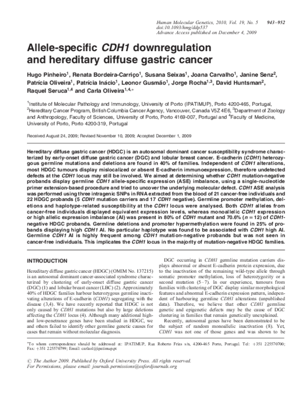
Cancer-free individuals display equivalent germline RNA
expression of CDH1 maternal and paternal alleles
Three SNPs were genotyped from PBLs’ RNA from 50 control
cancer-free individuals to select a series of heterozygous individuals for ASE analysis. Twenty-one of the control cancerfree individuals were heterozygous at SNP rs1801552 (n ¼
14), rs33964119 (n ¼ 1) and/or rs1801026 (n ¼ 10), and
their cDNAs used to determine the relative expression of
CDH1 maternal and paternal alleles. In all cases, T and C
alleles were identically represented (Fig. 1A), and a range of
normalcy values was defined with an upper boundary for
normal allelic expression ratio. The mean expression ratio in
the cancer-free individuals’ germline RNA was 1.32 + 0.14,
and the ratio between alleles did not change significantly
when a different SNP was used in the same sample
(Fig. 1B). Moreover, these RNA results were similar to
those obtained when using matched genomic DNA (gDNA)
(Fig. 1C). The fact that the ratio remains equivalent independent of nucleic acid and SNPs used demonstrates that this
assay is suitable for ASE quantitative measurement.
HDGC CDH1 germline mutation carriers display germline
CDH1 AI
We applied the ASE quantification method, established for
cancer-free individuals, in PBLs’ RNA from five HDGC probands shown elsewhere to be germline CDH1 mutation carriers (Table 1). Our aim was to understand whether CDH1
AI would reflect the presence of a CDH1 germline mutation.
After confirming that all five probands were constitutively
heterozygous for SNP rs1801552 using polymerase chain reaction (PCR) sequencing, ASE analysis was conducted in germline RNA: 80% (4/5) of CDH1 mutation carriers showed high
AI, which was not observed in gDNA using the same primer
extension assay (Fig. 2A). One of the five mutation carriers
lacked AI and displayed similar ASE mean ratios in RNA
and DNA (data not shown). This specific patient carried a missense mutation, whereas the other four were carriers of truncating CDH1 mutations.
CDH1-negative probands display germline CDH1 AI
Of 27 CDH1 mutation-negative probands, 17 showed to be
heterozygous for at least one of the SNPs (n ¼ 16 for
rs1801552, n ¼ 1 for rs33964119 and n ¼ 1 for rs1801026)
and represented families with: (i) two or more GC cases
with at least one DGC in first-degree relatives diagnosed
before age 50 (n ¼ 9); (ii) three or more GC in first-degree
relatives with at least one DGC diagnosed at any age (n ¼
1); (iii) DGC before 45 years of age (n ¼ 1); (iv) LBC
before 45 years of age (n ¼ 1); (v) clustering of GC cases of
unknown histology (n ¼ 5) (Table 1).
Fifteen out of 17 (88.2%) heterozygous CDH1-negative
probands presented AI with mean ratios above the proposed
normal values (Fig. 2B), whereas gDNA data showed equivalent representation of both alleles (Fig. 2C). In 70.6% (12/17)
of patients with AI, the mean ratio was higher than 6,
whereas the remaining demonstrated a more modest overrepresentation of one allele (Table 1). Complete reduction to
monoallelic expression was observed in 41.2% (7/17) of
cases (Table 1). The analysis of gDNA in these cases was
repeated using a different DNA sample and the heterozygous
state at the SNP position confirmed (data not shown). In
four mutation-negative probands, we used a different singlebase extension (SBE) primer to the same SNP to show that
results were primer independent (Fig. 2C).
CDH1 germline promoter hypermethylation and germline
large deletions occur in probands with germline CDH1 AI
To disclose putative epigenetic and structural mechanisms that
could underlie the presence of high AI in the germline of
CDH1-negative probands, we studied the CDH1 promoter
methylation status as well as the presence of genomic
rearrangements at the gene locus in PBLs’ DNA.
We studied germline CDH1 promoter hypermethylation in 12
probands with high CDH1 AI. A single proband from Family 11
displayed promoter hypermethylation at least in 11/19 (57.9%)
CpG sites (Fig. 3A). Although not completing the HDGC criteria, this family has a history of at least two GC of unknown histology, and several cases of breast and colorectal cancer, which
�Human Molecular Genetics, 2010, Vol. 19, No. 5
945
Figure 1. CDH1 ASE analysis in cancer-free individuals. (A) CDH1 allelic expression ratio in cancer-free individuals. (B) ASE in RNA samples from four
heterozygous individuals. (C) Allele-specific quantification in gDNA samples from four heterozygous individuals.
Table 1. Features of probands from GC families selected for ASE analysis
Family ID
Geographic
origin
Inclusion
criteriaa
CDH1 germline alteration
(type)
ASE ratio
rs1801552
ASE ratio
rs33964119
ASE ratio
rs1801026
21 cancer-free
individuals
Family 1 (16)
Family 2 (17)
Family 3 (18)
Family 4 (19)
Family 5 (20)
Family 6
Family 7
Family 8
Family 9
Family 10
Family 11
Portugal
NA
NA
1.27 + 0.06
1.42 + 0.13
1.38 + 0.02
Brazil
Canada
Canada
Canada
Portugal
Portugal
Portugal
Portugal
China
Canada
Canada
i
i
i
ii
i
i
v
i
i
i
v
10.00 + 0.00
10.00 + 0.00
9.07 + 1.24
8.53 + 1.97
2.11 + 0.89
8.87 + 1.51
10.00 + 0.00
10.00 + 0.00
6.67 + 2.02
10.00 + 0.00
8.63 + 1.82
NA
NA
NA
NA
NA
NA
NA
NA
NA
NA
NA
NA
NA
NA
NA
NA
10 + 0.00
NA
NA
NA
NA
NA
Family 12
Family 13
Family 14
Family 15
Family 16
Family 17
Family 18
Family 19
Family 20
Family 21 (4)c
Family 22 (4)d
Canada
Ireland
Italy
Ireland
Portugal
Canada
Canada
Spain
Portugal
Canada
Canada
v
i
iv
ii
v
v
iii
i
i
i
i
Mutation (1137G>A)
Mutation (2276delG)
Mutation (1792C>T)
Mutation (187C>T)
Mutation (2269G>A)
Negative
Negative
Negativeb
Negative
Negative
Promoter
hypermethylation
Negative
Negative
Negative
Negative
Negativeb
Negative
Negative
Negative
Negative
Deletion
Deletion
10.00 + 0.00
10.00 + 0.00
9.93 + 0.09
8.44 + 2.08
3.24 + 0.64
2.87 + 1.36
2.48 + 0.54
1.96 + 0.08
1.69 + 0.30
10 + 0.00
NA
NA
NA
NA
NA
NA
NA
NA
NA
NA
NA
10 + 0.00
NA
NA
NA
NA
NA
NA
NA
NA
NA
NA
NA
NA, not applicable; high A1 values and CDH1 alterations are highlighted in bold.
See Results section.
b
Mutation negative and not tested for large deletions.
c
Family 1.
d
Family 4 in reference (4).
a
�946
Human Molecular Genetics, 2010, Vol. 19, No. 5
Figure 2. CDH1 allele-specific quantification in CDH1-mutated and CDH1-negative probands. (A) ASE in CDH1 mutation carriers’ RNA and matched DNA.
(B) Conventional boxplot (with whiskers) of allele expression ratio in cancer-free individuals (n ¼ 21; IQR ¼ 0.17; Q1 ¼ 1.24; Q3 ¼ 1.41), CDH1 mutation
carriers (n ¼ 5; IQR ¼ 3.23; Q1 ¼ 6.77; Q3 ¼ 10) and CDH1 mutation-negative probands (n ¼ 17; IQR ¼ 7.02; Q1 ¼ 2.96; Q3 ¼ 9.98). P-values refer to
Wilkoxon test (BC correction: �3).
may also occur in the tumour spectrum of HDGC. Even so, all
affected members in this family have deceased, and therefore
a transgenerational study could not be performed. We had
access to PBLs’ DNA from a probands’ brother and granddaughter (both non-affected by cancer so far) and observed
that neither had CDH1 promoter hypermethylation.
To understand whether CDH1 hypermethylation could be
allele-specific as well as to determine the extent of methylation in CDH1 alleles of this case, 24 cloned alleles were
sequenced (Fig. 3B). A significant proportion of alleles displayed variable allele-to-allele hypermethylation, supporting
that germline allele-specific methylation could be occurring
in this patient. To address this issue, we took advantage of
patients’ heterozygozity at the common CDH1 promoter
SNP rs16260 (2161C/A) to perform amplification refractory
mutation system (ARMS)-based PCR (ARMS-PCR) and
amplify each allele separately from bisulphite-converted
DNA. With this approach, we showed that the methylated
clones observed in Figure 3B are likely all derived from the
2161A allele, since only this allele was hypermethylated
(Fig. 3C). In parallel with the current study, a large series of
CDH1 mutation-negative HDGC probands, which included
15/17 probands from the present CDH1-negative series, was
screened for large genomic rearrangements at the CDH1
locus on chromosome 16 by MLPA and array CGH (4). By
crossing our CDH1 AI data with the data arising from the
rearrangement mapping, we verified that 2/12 (16.7%) probands with high CDH1 AI displayed large deletions at the
CDH1 locus. One deletion (193.593 bp) was found in
Family 21 (referred as Family 1 in our previous report) and
encompassed the full sequence of CDH3, extending to position
IVS2þ57.595 in CDH1 (4). The other deletion found in
Family 22 (referred as Family 4 in our previous report) was
a 150 bp removal, encompassing the CDH1 TSS, with breakpoints located 125 bp upstream and 25 bp downstream of the
TSS (4).
Our results show that 25% (3/12) of probands previously
described as mutation negative display high CDH1 AI,
�Human Molecular Genetics, 2010, Vol. 19, No. 5
947
Figure 3. Germline CDH1 promoter hypermethylation analysis. (A) Partial electropherogram from Family 11 showing methylation at 9/19 CpG sites. (B) Schematic representation of CpG dinucleotides encompassed by the flanking PCR. Each line of circles represents an isolated allele; an open circle, a non-methylated
CpG; and a black circle, a methylated CpG. (C) ARMs PCR scheme covering 13 CpG. The rs16260_A allele is methylated in at least eight CpG sites and
partially methylated in three (circles with a black dot).
which is likely generated by germline CDH1 methylation and
large genomic rearrangements at the gene locus.
Germline CDH1 AI is not associated with a specific
extended haplotype
A finding derived from the present ASE analysis was the
exclusive underexpression of the rs1801552_C allele in all
samples, independent of the level of AI detected as well as
of the CDH1 germline alteration status, when PBLs’ RNA
was compared with gDNA (Fig. 2A and C). This result was
consistent when both flanking and SBE primers were
changed (Fig. 2C, last panel), supporting the hypothesis of a
rs1801552_C allele-associated downregulation, possibly reminiscent of an ancestral disease-associated haplotype and potentially associated with the occurrence of germline CDH1
alterations always in the C-allele. Supporting this idea, the
haplotype reconstruction in Family 11 (displays high CDH1
AI and allele-specific CDH1 germline promoter methylation)
showed that the hypermethylated rs16260_A CDH1 promoter
allele was in phase with the underexpressed rs1801552_C
allele.
To test this hypothesis, and assuming that, whatever the
mechanism, downregulation of CDH1 was occurring always
in the ancestral rs1801552_C-allele, we genotyped 12
SNPs, encompassing distinct haplotype blocks along CDH3
and CDH1 regions from chromosome 16 (Fig. 4A, Supplementary Material, Fig. S1) in 14 probands with high
CDH1 AI (four with germline CDH1 mutation, one with
germline CDH1 promoter methylation and nine without
CDH1 alteration). Haplotypes were reconstructed using
PHASE 2.02 software (16,17). HapMap haplotypes from a
control population (n ¼ 120) with a known phase were
used to improve inference of probands’ haplotypes. The
GOLD (graphical overview of linkage disequilibrium) application (18,19) shows, for our SNP data set, a common region
of strong linkage disequilibrium (jD0 j � 0.84) between
rs1801552, rs7203904 and rs2276330 (Supplementary
Material, Fig. S2). However, the block structure observed
in the HapMap control data (Supplementary Material, Figs
S1 and S2A) for the region encompassing rs1801552,
rs7203904 and rs2276330 was not maintained for the group
of 14 probands with high CDH1 AI alone (Supplementary
Material, Fig. S2B). Even when the nine probands with
high CDH1 AI were analysed separately, similar results
were obtained, suggesting that the proband series with high
CDH1 AI is somewhat more diverse than the HapMap one
(Supplementary Material, Fig. S2B1, 2C and 2A). Nevertheless, when we assessed the breakdown of rs1801552_T and
rs1801552_C haplotypes (Fig. 4B—left and right panels,
respectively) from probands and controls is made clear that
the higher diversity of CDH1 high AI may be explained by
rs1801552_C haplotypes alone. Moreover, the lack of a
homogeneous region in the vicinity of rs1801552_C,
common to all probands, refutes the hypothesis of high AI
being associated with a specific extended haplotype.
�948
Human Molecular Genetics, 2010, Vol. 19, No. 5
Figure 4. Haplotype analysis. (A) Adapted NCBI representation of 193 kb encompassing 12 coding and noncoding SNPs chosen along CDH3 and CDH1 loci.
(B) Decay over distance of common haplotypes from rs1801552_T and rs1801552_C alleles. The most frequent haplotype is represented above the lines for each
considered subgroup (HapMap, high CDH1 AI, germline mutant and methylated and CDH1 negative).
DISCUSSION
To date, �40% of families with well-defined clustering of
DGC, described as HDGC, display CDH1 germline inactivation (4). A lower frequency of CDH1 alterations has also
been described in isolated early-onset DGC patients, in
families that do not fulfil the widely accepted clinical criteria
and in families with clustering of early-onset LBC, without
DGC involvement (3,20). Most reported germline CDH1
alterations lead to the production of truncated inactive peptides that lack normal function.
Given that a fair proportion of eligible families for germline
CDH1 mutation screening lack such alterations despite displaying E-cadherin protein expression impairment in their
tumours (data not shown), we propose that, in these cases,
CDH1 is still compromised due to alterations that escape
detection by conventional techniques.
Mutations leading to protein truncation constitute the
majority of inactivating mutations in cancer susceptibility
genes (3,21– 24). Transcripts containing premature termination codons (PTCs), mainly as a result of frameshift and nonsense mutations, are eliminated by the nonsense-mediated
mRNA decay (NMD) (25). Owing to the instability of
PTC-containing transcripts, many mutations remained undetected by RNA-based techniques and only became discovered
upon usage of DNA-based methods. Conversely, as the latter
rely mostly on the amplification of individual exons with intronic flanking primers, large genomic deletions and changes in
more distant noncoding regions were also missed, until
recent technical improvements have been achieved (MLPA
and arrayCGH) allowing their detection. Undiscovered alterations may therefore be revealed by techniques that permit individual assessment of paternal and maternal allelic expression.
Without disregarding this interesting possibility, one has to
consider that monoallelic expression with random choice
between maternal and paternal alleles is no longer exclusive
of imprinted and X-inactivated genes, as a group of autosomal
gene families has been recently identified as a target of monoallelic downregulation (8).
In the last decade, at least three studies have been published
re-enforcing the role of germline AI as a major risk factor for
cancer development in sporadic and hereditary cohorts. These
studies identified AI in BRCA1, BRCA2, APC and lately in
TGFBR1, using well-selected cohorts of cancer patients and
cancer-free control individuals (11 – 13). All these studies
made use of different methodological approaches to analyse
ASE in patients’ germline RNA; nevertheless, none described
the molecular mechanism underlying the observed effect on
RNA monoallelic downregulation.
In the present study, we established a reliable and straightforward assay to evaluate CDH1 ASE using SNPs within
CDH1 mRNA. We demonstrated that CDH1 expression is
biallelic in PBLs from heterozygous cancer-free individuals
and that both CDH1 alleles express equivalent RNA levels,
as mentioned previously (8). Moreover, the contribution of
each allele was found to be independent from the SNP
tested or SBE primers used (Fig. 1B and 2C). This result
enabled us to set an upper artificial boundary for CDH1
�Human Molecular Genetics, 2010, Vol. 19, No. 5
normal expression ratio in cancer-free individuals. A valuable
test of the approach reliability came from results obtained in
four HDGC CDH1 germline-truncating mutation carriers,
which showed that CDH1 AI in mutant probands was statistically higher than that of cancer-free individuals (P ¼
2.11e203), as expected after PTC degradation by NMD
(25,26). Similar results have been previously obtained for
BRCA1, BRCA2 and APC (11,12).
Having established the assay, we had the proper tool to disclose whether this finely tuned biallelic expression was getting
disrupted in our series of CDH1-negative probands. Our main
concern was to select a series of patients that, although negative for CDH1 germline alterations, was, in all features,
similar to any series previously used for CDH1 mutation
screening. This selection was expected to increase the probability of finding a mechanism of disease, although it was
highly dependent on RNA availability from probands’ PBLs.
After the selection of 17 CDH1-negative probands, from
different geographic backgrounds and fulfilling well-defined
clinical criteria (see Results), we investigated the possibility
of these probands presenting germline CDH1 AI. An impressive and unexpected proportion of these patients (70.6%)
revealed AI ratios .6, with recurrent downregulation of the
rs1801552_C allele. Coincidently, a recent study reporting
ASE in a familial pancreatic cancer patient showed that the
same rs1801552_C allele was underexpressed (15). Therefore,
this allele was assumed as a preferential target for a germline
downregulation mechanism so far unidentified.
Recent reports demonstrated that hMLH1 or hMSH2 monoallelic germline hypermethylation caused HNPCC and could
be transmitted through generations (27,28). These studies
established epimutations as the cause of a novel cancer susceptibility pattern of inheritance (27,28). Assuming that CDH1
could be the target of a similar epigenetic phenomenon, we
have analysed CDH1 germline promoter hypermethylation in
probands with high AI. The PBLs’ DNA extracted from the
Family 11 proband displayed CDH1 hypermethylation with
a striking pattern of clonal heterogeneity similar to that somatically found in sporadic carcinomas, cancer cell lines, and
tumour foci from HDGC CDH1 germline mutation carriers,
leading to CDH1 expression downregulation (7,29). We
believe however that this proband harbours germline hypermethylation as the cloning experiment revealed that a large
proportion of the sequences cloned were methylated. This
result would not be expected if a small amount of cancer
cells would be circulating in the blood, and is pointing to a
germline defect rather than to somatic cancer-associated
methylation reminiscent from cancer-circulating cells. The
most interesting aspect in this case was that CpG methylation
was occurring only at the rs16260_A allele, raising the
hypothesis that allele-specific methylation is generating
germline-specific downregulation of the rs1801552_Ccontaining allele. Further supporting this hypothesis is the
fact that haplotype reconstruction by PHASE 2.02 software
revealed that both alleles are indeed in phase. To assess
CDH1 germline hypermethylation in a larger series, we
extended the methylation analysis but failed to identify this
phenomenon in 56 additional GC family probands (data not
shown); therefore, we believe this is a rare mechanism in
this setting, according to previously reported findings (30).
949
Nevertheless, the overall frequency of CDH1 germline
methylation (1.47%—1/68) herein identified does not differ
from that reported for hMLH1 and hMSH2 in HNPCC-like
probands (0.6 and 3.2%, respectively) (27,28).
We also found that 2/12 (16.7%) high CDH1 AI probands
display large genomic deletions encompassing CDH1, so, in
summary, we identified the potential mechanism underlying
high AI in 25% (3/12) of the cases in this series, suggesting
that AI most likely represents a marker for both germline
genetic and epigenetic defects in CDH1.
Recently, germline heterozygous deletions of TACSTD1
30 -exons were found in 0.9% of HNPCC-suspected families,
causing transcriptional read-through that resulted in hMSH2
downregulation (31,32). In the light of our CDH1 AI results,
it is expected that the application of ASE analysis to
hMSH2, in the aforementioned HNPCC families, would have
spotted monoallelic expression of the gene. Conversely, it is
also possible that a similar structural rearrangement, in the surroundings of CDH1 gene, could generate AI in some probands
of our series.
Recently it was shown that TGFBR1 AI results in globally
reduced expression of the gene, alters SMAD-mediated
TGFb signalling, is dominantly inherited, segregates in
families and occurs in sporadic colorectal cancer (13). Two
major TGFBR1 haplotypes were found predominant among
TGFBR1 AI cases suggesting ancestral mutations; nevertheless, no causative germline change was identified (13).
As we failed to find a mechanism underlying the nine
remaining cases displaying high CDH1 AI, and given that
the rs1801552_C-containing allele was recurrently downregulated, we reasoned that this allele could be in linkage disequilibrium with a putative aetiological variant, either in CDH1 or
in its proximity. The rs1125557_A allele (163þ37235G.A)
localized in CDH1 intron 2 was recently claimed as a susceptibility variant for sporadic DGC, mainly in homozygous AA
individuals (33). This SNP was studied in our series and contrarily to what is reported, the percentage of AA individuals
was similar between probands (28%) and controls (33%).
Moreover, our quest for high CDH1 AI-related haplotypes
generated no significant difference between haplotypic distribution in probands and in the HapMap reference/control
group. As no haplotype is overrepresented within
CDH1-negative probands, high CDH1 AI is most probably
being caused by haplotype independent mechanisms.
Another interesting hypothesis arises from a study describing that two cis-regulatory FGFR2 SNPs are able to alter
binding affinity for transcription factors Oct-1/Runx2 and C/
EBPb (34), and that they synergize to augment FGFR2
expression, thereby increasing the propensity for tumour formation (34). Whether a similar phenomenon is occurring in
our patients, and another SNP exists that synergizes with
CDH1 rs1801552_C allele, still needs to be addressed.
A last hypothesis to cause CDH1 AI could be the existence
of a trans-acting factor binding differentially to rs1801552_C
and rs1801552_T alleles, as it was recently described for
DEAF-1 (35). This protein was reported to allele-specific
trans-regulate GDF5, through the differential binding to a
GDF5 sequence encompassing a common SNP (rs143383).
In the context of CDH1-negative probands, individuals carrying the common rs1801552_C allele would only be at risk of
�950
Human Molecular Genetics, 2010, Vol. 19, No. 5
developing cancer in a background with abnormal expression
of a trans-acting factor, with differential binding affinity for
the rs1801552_C allele.
In summary, our results show that: (i) high CDH1 AI may
be used as a marker for increased susceptibility to familial
aggregation of GC, namely HDGC, as it occurs specifically
in CDH1-truncating mutation carriers (100%) and
CDH1-negative probands (70.6%), but not in cancer-free individuals; (ii) germline allele-specific CDH1 promoter methylation and deletion encompassing the CDH1 locus underlie
high CDH1 AI in 25% of CDH1-negative probands; (iii)
high CDH1 AI determination provides a simple, cost-effective
and efficient tool to perceive indirectly changes of CDH1
expression that escape detection in gDNA-based screenings;
and (iv) the remainder 75% of cases with high AI are not
related to an extended susceptibility haplotype.
As AI detection does not vary upon the SNP used, any
coding or even pre-mRNA SNP can be used to test ASE,
and virtually every single individual can be analysed with
this strategy. ASE quantification of gene transcripts opens
the way to the detection of both trans- and cis-acting regulatory variations (35,36) and overcomes the difficulties resulting
from non-genetic factors affecting mRNA levels and from
mRNA instability, which can differ between samples.
In conclusion, we believe that most CDH1-negative probands, especially those presenting tumours with E-cadherin
expression impairment, similar to that observed in CDH1
germline alteration carriers, do have a germline CDH1
expression defect caused by either direct or indirect mechanisms targeting the CDH1 genomic sequence.
MATERIALS AND METHODS
Patients and samples
The collection of clinical data from patients and families and
PBL samples from patients and controls was approved by the
appropriate Ethics Committee from each of the centres participating in this work: University of British Columbia, Vancouver,
Canada; Institute of Molecular Pathology and Immunology of
the University of Porto (IPATIMUP), Porto, Portugal; and Hospital Geral de Santo António, Porto, Portugal. Family history of
cancer was obtained with informed consent.
Thirty-two probands from families displaying aggregation
of GC mainly of diffuse histology (HDGC) and early onset
isolated cases (aged ,45 years) with either DGC or LBC
were selected for the study. Twenty-seven of the 32 probands
tested negative for CDH1 germline point mutations, whereas 5
have been described elsewhere to carry germline point
mutations in the CDH1 gene (26,37 – 40). A series of 50
cancer-free individuals was used as controls.
For germline CDH1 hypermethylation analysis, we gathered
68 probands, including the 27 CDH1-negative probands from
the aforementioned series and 41 other probands collected at
IPATIMUP.
Nucleic acid preparation
gDNA was extracted from PBLs from patients and controls
using a standard protocol. Total RNA was isolated from
PBLs using the TriPure Isolation Agent (Roche Diagnostics,
Basel, Switzerland) or the whole blood and bone marrow protocols—300 ml blood sample protocol from PURESCRIPT
RNA Isolation Kit (GENTRA, MN, USA).
Approximately 300 ng of total RNA was used to synthesize
first-strand cDNA with Superscript II Reverse Transcriptase
and random hexamer primers (Invitrogen, CA, USA).
CDH1 SNP genotyping of cancer-free individuals and
patients
Three CDH1 SNPs were genotyped in all patients and controls: two CDH1 coding SNPs rs1801552-2076C/T [minor
allele frequency (MAF) T: 0.408 in HapMap phase II CEU
panel from individuals with European ancestry] and
rs33964119-2253C/T (MAF T: 0.045 in HapMap phase II
European EGP CEPH panel); and a noncoding SNP
rs1801026-g.� þ54C.T located at the 30 -UTR before the
polyadenylation signal (MAF T: 0.205 in HapMap phase II
European EGP CEPH panel) by PCR using standard conditions. MAFs were obtained from the dbSNP home page at
http://www.ncbi.nlm.nih.gov/projects/SNP/. PCR products
were sequenced using BigDye Terminator v3.1 Cycle Sequencing Kits (Applied Biosystems, CA, USA) and run in an ABI
Prism 3130 DNA automated sequencer (Applied Biosystems).
Primer sequences and amplicon sizes are listed in Supplementary Material, Table S1.
ASE analysis
Primer extension analysis relies on the incorporation of a
single dideoxynucleotide triphosphate (ddNTP) (complementary to the base of interest) that is selected to allow the differential extension of a primer annealed next to the polymorphic
site (SBE primer). ASE products can be distinguished, and
quantitatively measured, based on the presence of a specific
fluorophore coupled to the incorporated ddNTP. PCR products
encompassing the polymorphic site were used as templates for
primer extension with fluorescently labelled dNTPs which are
incorporated, causing chain termination in alleles having a C,
G, A or T in this position. Purified PCR products were submitted to SNaPshot reaction using SNaPshot Multiplex Kit
(Applied Biosystems) and an SBE-specific primer following
manufacturer’s instructions. Final products were analysed in
a 310 Genetic Analyser (Applied Biosystems) using the size
standard GeneScan 120 LIZ (Applied Biosystems). The peak
area corresponding to each allele was quantified with the GeneScan results analyser software. The ratio of ddNTP incorporation by each allele was calculated by taking the ratio of the
areas corresponding to T and C alleles. For samples presenting
complete downregulation of one allele in germline RNA, a
ratio value of 10 was attributed to characterize AI.
As a validation step, a PCR amplicon encompassing the
same SNPs was generated from gDNA from patients and controls and submitted to the same analysis to demonstrate equivalent representation of each allele through equivalent ddNTP
incorporation. Moreover, to exclude ASE bias mediated by a
specific primer sequence, ASE results obtained from patients
and control samples were confirmed using either different
primer sequences for the primer extension assay or, whenever
�Human Molecular Genetics, 2010, Vol. 19, No. 5
possible, another SNP. Primer sequences and amplicon sizes
are listed in Supplementary Material, Table S2.
Promoter methylation analysis
DNA from probands’ PBLs belonging to 68 CDH1-negative
families mainly displaying GC aggregation, originating from
Canada (10), Ireland (2), Spain (1), Brazil (1), China (1),
Italy (1) and Portugal (52), was screened for hypermethylation
at the CpG island 3 of the CDH1 promoter. Approximately
50– 100 ng of sodium bisulphite-modified DNA [EpiTect
Bisulfite Kit (Qiagen, Hilden, Germany)—sodium bisulphite
conversion of unmethylated cytosines in DNA from solutions
with low concentrations of DNA] was used for PCR and
sequencing to screen to screen for methylation at 19 CpG
sites (position –56 bp to position þ115 bp from the CDH1
TSS), with flanking primers not containing CpG sites.
Samples displaying methylated CpG sites were resubmitted to
an independent PCR that was cloned into pCRw 2.1-TOPOw
vector (Invitrogen). Colony-PCR with Universal M13 primers
was performed to determine the level and extent of CDH1 promoter methylation. Primer sequences and amplicon sizes are
listed in the Supplementary Material, Table S3.
951
Table S5. The phased haplotypes from the CEU panel haplotypes from HapMap phase II database for European populations were used with our multilocus genotype data to
statistically infer the probands’ haplotypes, using the software
PHASE 2.02 (16,17). Linkage disequilibrium statistic D0 was
calculated from the inferred haplotypes using the DNAsp
program, version 5.0 (18).
Statistical analysis
R statistical program was used for the construction of a standard boxplot, plotted with whiskers and outliers. Owing to
the non-normal distribution of the data, a non-parametric
test, in particular the Wilcoxon rank sum test was used to
calculate significance. The P-values obtained were further
corrected using the Bonferroni correction due to the multiple
testing performed.
SUPPLEMENTARY MATERIAL
Supplementary Material is available at HMG online.
ACKNOWLEDGEMENTS
Amplification refractory mutation system-PCR
To analyse allele-specific CDH1 hypermethylation, heterozygozity at the common CDH1 promoter SNP rs16260
(2161C/A) was used to perform ARMS-PCR. Primers were
designed to discriminate between templates which differed
in a specific single nucleotide (41), thereby amplifying each
allele separately from bisulphite-converted DNA. Briefly,
primers flanking rs16260 SNP were used in a first-round
PCR with 50 ng of bisulphite-converted DNA. Products
were re-amplified with the same reverse primer and specific
forward primers to the polymorphic A or C allele. Sequencing
of each PCR product with the same reverse primer allowed
analysis of 13 methylated CpG sites in independent alleles
[the last three CpG sites had been previously analysed by
cloning and sequencing (Fig. 3B and C)]. Primer sequences
and amplicon sizes are listed in Supplementary Material,
Table S4.
We gratefully acknowledge patients, families and their caregivers for their willing participation in this project and who
provided consent regarding the use of the information obtained
from the study. We thank Luis Maia, Marta Novais and Teresa
Coelho for providing biological material from cancer-free
individuals.
Conflict of Interest statement. None declared.
FUNDING
This work was supported by The Portuguese Foundation for
Science and Technology (FCT) (grant number PTDC/
SAU-GMG/72168/2006; PhD grant numbers: SFRH/BD/
41223/2007-HP, SFRH/BD/46462/2008-RC, SFRH/BD/
44074/2008-JC, SFRH/BD/32984/2006-PO; and salary
support from the programme Ciência 2007 to C.O.); and
The Canadian Cancer Society/National Cancer Institute of
Canada operating grant (grant number 018381).
Haplotype analysis
Twelve coding and noncoding SNPs with heterozygous frequencies ranging from 0.217 to 0.648 (HapMap phase II
CEU panel from individuals with European ancestry, http://
www.ncbi.nlm.nih.gov/projects/SNP/) scattered along CDH1
and CDH3 loci and covering a region of approximately
193 kb (67.227.572– 67.420.442) were genotyped in a multiplex PCR, according to manufacturer’s instructions
(QIAGEN Multiplex PCR Kit, Quiagen). SNP-specific
primers for primer extension assay were designed to be used
in a SNaPshot multiplex reaction by adding different size
tags to each primer (SBE primers). Purified PCR products
and SBE primers were used for primer extension assay according to the SNaPshot Multiplex Kit (Applied Biosystems).
Selected SNPs, primers (PCR primers and SBEs) and respective amplicon sizes are listed in Supplementary Material,
REFERENCES
1. Guilford, P., Hopkins, J., Harraway, J., McLeod, M., McLeod, N.,
Harawira, P., Taite, H., Scoular, R., Miller, A. and Reeve, A.E. (1998)
E-cadherin germline mutations in familial gastric cancer. Nature, 392,
402– 405.
2. Pharoah, P.D., Guilford, P. and Caldas, C., International Gastric Cancer
Linkage Consortium (2001) Incidence of gastric cancer and breast cancer
in CDH1 (E-cadherin) mutation carriers from hereditary diffuse gastric
cancer families. Gastroenterology, 121, 1348–1353.
3. Oliveira, C., Seruca, R. and Carneiro, F. (2006) Genetics, pathology, and
clinics of familial gastric cancer. Int. J. Surg. Pathol., 14, 21– 33.
4. Oliveira, C., Senz, J., Kaurah, P., Pinheiro, H., Sanges, R., Haegert, A.,
Corso, G., Schouten, J., Fitzgerald, R., Vogelsang, H. et al. (2009)
Germline CDH1 deletions in hereditary diffuse gastric cancer families.
Hum. Mol. Genet., 18, 1545– 1555.
5. Grady, W.M., Willis, J., Guilford, P.J., Dunbier, A.K., Toro, T.T., Lynch,
H., Wiesner, G., Ferguson, K., Eng, C., Park, J.G. et al. (2000)
�952
6.
7.
8.
9.
10.
11.
12.
13.
14.
15.
16.
17.
18.
19.
20.
21.
22.
23.
24.
Human Molecular Genetics, 2010, Vol. 19, No. 5
Methylation of the CDH1 promoter as the second genetic hit in hereditary
diffuse gastric cancer. Nat. Genet., 26, 16– 17.
Barber, M., Murrell, A., Ito, Y., Maia, A.T., Hyland, S., Oliveira, C., Save,
V., Carneiro, F., Paterson, A.L., Grehan, N. et al. (2008) Mechanisms and
sequelae of E-cadherin silencing in hereditary diffuse gastric cancer.
J. Pathol., 216, 295– 306.
Oliveira, C., Sousa, S., Pinheiro, H., Karam, R., Bordeira-Carriço, R.,
Senz, J., Kaurah, P., Carvalho, J., Pereira, R., Gusmão, L. et al. (2009)
Quantification of epigenetic and genetic 2nd hits in CDH1 during
hereditary diffuse gastric cancer syndrome progression. Gastroenterology,
136, 2137–2148.
Gimelbrant, A., Hutchinson, J.N., Thompson, B.R. and Chess, A. (2007)
Widespread monoallelic expression on human autosomes. Science, 318,
1136– 1140.
Reik, W. and Walter, J. (2001) Genomic imprinting: parental influence on
the genome. Nat. Rev. Genet., 2, 21– 32.
Lyon, M.F. (1986) X chromosomes and dosage compensation. Nature,
320, 313.
Chen, X., Weaver, J., Bove, B.A., Vanderveer, L.A., Weil, S.C., Miron,
A., Daly, M.B. and Godwin, A.K. (2008) Allelic imbalance in BRCA1
and BRCA2 gene expression is associated with an increased breast cancer
risk. Hum. Mol. Genet., 17, 1336–1348.
Yan, H., Dobbie, Z., Gruber, S.B., Markowitz, S., Romans, K., Giardiello,
F.M., Kinzler, K.W. and Vogelstein, B. (2002) Small changes in
expression affect predisposition to tumourigenesis. Nat. Genet., 30,
25–26.
Valle, L., Serena-Acedo, T., Liyanarachchi, S., Hampel, H., Comeras, I.,
Li, Z., Zeng, Q., Zhang, H.T., Pennison, M.J. and Sadim, M. (2008)
Germline allele-specific expression of TGFBR1 confers an increased risk
of colorectal cancer. Science, 321, 1361– 1365.
Castellvı́-Bel, S. and Castells, A. (2009) Allele-specific expression as a
new genetic susceptibility mechanism for colorectal cancer.
Gastroenterology, 136, 2397– 2399.
Tan, A.C., Fan, J.B., Karikari, C., Bibikova, M., Garcia, E.W., Zhou, L.,
Barker, D., Serre, D., Feldmann, G., Hruban, R.H. et al. (2008)
Allele-specific expression in the germline of patients with familial
pancreatic cancer: an unbiased approach to cancer gene discovery. Cancer
Biol. Ther., 7, 135– 144.
Stephens, M., Smith, N.J. and Donnelly, P. (2001) A new statistical
method for haplotype reconstruction from population data. Am. J. Hum.
Genet., 68, 978– 989.
Stephens, M. and Donnelly, P. (2003) A comparison of bayesian methods
for haplotype reconstruction from population genotype data. Am. J. Hum.
Genet., 73, 1162–1169.
Librado, P. and Rozas, J. (2009) DnaSP v5: a software for comprehensive
analysis of DNA polymorphism data. Bioinformatics, 25, 1451–1452.
Abecasis, G.R. and Cookson, W.O. (2000) GOLD—graphical overview of
linkage disequilibrium. Bioinformatics, 16, 182– 183.
Masciari, S., Larsson, N., Senz, J., Boyd, N., Kaurah, P., Kandel, M.J.,
Harris, L.N., Pinheiro, H.C., Troussard, A., Miron, P. et al. (2007)
Germline E-cadherin mutations in familial lobular breast cancer. J. Med.
Genet., 44, 726– 731.
Gayther, S.A., Warren, W., Mazoyer, S., Russell, P.A., Harrington, P.A.,
Chiano, M., Seal, S., Hamoudi, R., van Rensburg, E.J., Dunning, A.M.
et al. (1995) Germline mutations of the BRCA1 gene in breast and ovarian
cancer families provide evidence for a genotype–phenotype correlation.
Nat. Genet., 11, 428– 433.
Gayther, S.A., Mangion, J., Russell, P., Seal, S., Barfoot, R., Ponder, B.A.,
Stratton, M.R. and Easton, D. (1997) Variation of risks of breast and
ovarian cancer associated with different germline mutations of the
BRCA2 gene. Nat. Genet., 15, 103–105.
Powell, S.M., Petersen, G.M., Krush, A.J., Booker, S., Jen, J., Giardiello,
F.M., Hamilton, S.R., Vogelstein, B. and Kinzler, K.W. (1993) Molecular
diagnosis of familial adenomatous polyposis. N. Engl. J. Med., 329,
1982– 1987.
Peltomäki, P. and Vasen, H.F. (1997) Mutations predisposing to
hereditary nonpolyposis colorectal cancer: database and results of a
collaborative study. The International Collaborative Group on Hereditary
Nonpolyposis Colorectal Cancer. Gastroenterology, 113, 1146–1158.
25. Frischmeyer, P.A. and Dietz, H.C. (1999) Nonsense-mediated mRNA
decay in health and disease. Hum. Mol. Genet., 8, 1893–1900.
26. Karam, R., Carvalho, J., Bruno, I., Graziadio, C., Senz, J., Huntsman, D.,
Carneiro, F., Seruca, R., Wilkinson, M.F. and Oliveira, C. (2008) The
NMD mRNA surveillance pathway downregulates aberrant E-cadherin
transcripts in gastric cancer cells and in CDH1 mutation carriers.
Oncogene, 27, 4255–4260.
27. Hitchins, M., Williams, R., Cheong, K., Halani, N., Lin, V.A., Packham,
D., Ku, S., Buckle, A., Hawkins, N., Burn, J. et al. (2005) MLH1 germline
epimutations as a factor in hereditary nonpolyposis colorectal cancer.
Gastroenterology, 129, 1392–1399.
28. Chan, T.L., Yuen, S.T., Kong, C.K., Chan, Y.W., Chan, A.S., Ng, W.F.,
Tsui, W.Y., Lo, M.W., Tam, W.Y., Li, V.S. and Leung, S.Y. (2006)
Heritable germline epimutation of MSH2 in a family with hereditary
nonpolyposis colorectal cancer. Nat. Genet., 38, 1178– 1183.
29. Graff, J.R., Gabrielson, E., Fujii, H., Baylin, S.B. and Herman, J.G. (2000)
Methylation patterns of the E-cadherin 50 CpG island are unstable and
reflect the dynamic, heterogeneous loss of E-cadherin expression during
metastatic progression. J. Biol. Chem., 275, 2727– 2732.
30. Yamada, H., Shinmura, K., Goto, M., Iwaizumi, M., Konno, H., Kataoka,
H., Yamada, M., Ozawa, T., Tsuneyoshi, T. and Tanioka, F. (2009)
Absence of germline mono-allelic promoter hypermethylation of the
CDH1 gene in gastric cancer patients. Mol. Cancer, 8, 63.
31. Ligtenberg, M.J., Kuiper, R.P., Chan, T.L., Goossens, M., Hebeda, K.M.,
Voorendt, M., Lee, T.Y., Bodmer, D., Hoenselaar, E.,
Hendriks-Cornelissen, S.J. et al. (2009) Heritable somatic methylation and
inactivation of MSH2 in families with Lynch syndrome due to deletion of
the 30 exons of TACSTD1. Nat. Genet., 41, 112– 117.
32. Niessen, R.C., Hofstra, R.M., Westers, H., Ligtenberg, M.J., Kooi, K.,
Jager, P.O., de Groote, M.L., Dijkhuizen, T., Olderode-Berends, M.J.,
Hollema, H. et al. (2009) Germline hypermethylation of MLH1 and
EPCAM deletions are a frequent cause of Lynch syndrome. Genes Chrom.
Cancer, 48, 737 –744.
33. Nasri, S., More, H., Graziano, F., Ruzzo, A., Wilson, E., Dunbier, A.,
McKinney, C., Merriman, T., Guilford, P., Magnani, M. and Humar, B.
(2008) A novel diffuse gastric cancer susceptibility variant in E-cadherin
(CDH1) intron 2: a case– control study in an Italian population. BMC
Cancer, 8, 138.
34. Meyer, K.B., Maia, A.T., O’Reilly, M., Teschendorff, A.E., Chin, S.F.,
Caldas, C. and Ponder, B.A. (2008) Allele-specific up-regulation of
FGFR2 increases susceptibility to breast cancer. PLoS Biol., 6, e108.
35. Egli, R.J., Southam, L., Wilkins, J.M., Lorenzen, I., Pombo-Suarez, M.,
Gonzalez, A., Carr, A., Chapman, K. and Loughlin, J. (2009) Functional
analysis of the osteoarthritis susceptibility-associated GDF5 regulatory
polymorphism. Arthritis Rheum., 60, 2055– 2064.
36. Milani, L., Gupta, M., Andersen, M., Dhar, S., Fryknäs, M., Isaksson, A.,
Larsson, R. and Syvänen, A.C. (2007) Allelic imbalance in gene
expression as a guide to cis-acting regulatory single nucleotide
polymorphisms in cancer cells. Nucleic Acids Res., 35, e34.
37. Suriano, G., Yew, S., Ferreira, P., Senz, J., Kaurah, P., Ford, J.M.,
Longacre, T.A., Norton, J.A., Chun, N., Young, S. et al. (2005)
Characterization of a recurrent germ line mutation of the E-cadherin gene:
implications for genetic testing and clinical management. Clin. Cancer
Res., 11, 5401–5409.
38. Humar, B., Graziano, F., Cascinu, S., Catalano, V., Ruzzo, A.M.,
Magnani, M., Toro, T., Burchill, T., Futschik, M.E., Merriman, T. and
Guilford, P. (2002) Association of CDH1 haplotypes with susceptibility to
sporadic diffuse gastric cancer. Oncogene, 21, 8192–8195.
39. Gayther, S.A., Gorringe, K.L., Ramus, S.J., Huntsman, D., Roviello, F.,
Grehan, N., Machado, J.C., Pinto, E., Seruca, R., Halling, K. et al. (1998)
Identification of germ-line E-cadherin mutations in gastric cancer families
of European origin. Cancer Res., 58, 4086–4089.
40. Simões-Correia, J., Figueiredo, J., Oliveira, C., van Hengel, J., Seruca, R.,
van Roy, F. and Suriano, G. (2008) Endoplasmic reticulum quality
control: a new mechanism of E-cadherin regulation and its implication in
cancer. Hum. Mol. Genet., 17, 3566–3576.
41. Newton, C.R., Heptinstall, L.E., Summers, C., Super, M., Schwarz, M.,
Anwar, R., Graham, A., Smith, J.C. and Markham, A.F. (1989)
Amplification refractory mutation system for prenatal diagnosis and
carrier assessment in cystic fibrosis. Lancet, 2, 1481–1483.
�
 Joana Carvalho