The Scientific World Journal
Volume 2012, Article ID 976513, 8 pages
doi:10.1100/2012/976513
The cientificWorldJOURNAL
Research Article
The Effect of the Weight of Equipment on Muscle Activity of
the Lower Extremity in Soldiers
Tobias Lindner,1 Christoph Schulze,1, 2 Sandra Woitge,1, 3 Susanne Finze,1
Wolfram Mittelmeier,1 and Rainer Bader1
1 Department
of Orthopaedics, University Medicine Rostock, Doberaner Straße 142, 18057 Rostock, Germany
Institute of Sports Medicine, Dr.-Rau-Allee 32, 48231 Warendorf, Germany
3 Rostock Military Medical Centre, Hohe Düne 30, 18119 Rostock, Germany
2 Bundeswehr
Correspondence should be addressed to Tobias Lindner, tobias.lindner@med.uni-rostock.de
Received 14 June 2012; Accepted 15 July 2012
Academic Editors: C. Y. Guezennec and D. Rafferty
Copyright © 2012 Tobias Lindner et al. This is an open access article distributed under the Creative Commons Attribution License,
which permits unrestricted use, distribution, and reproduction in any medium, provided the original work is properly cited.
Due to their profession and the tasks it entails, soldiers are exposed to high levels of physical activity and strain. This can result in
overexertion and pain in the locomotor system, partly caused by carrying items of equipment. The aim of this study was to analyse
the extent of muscle activity in the lower extremities caused by carrying specific items of equipment. For this purpose, the activity
of selected groups of muscles caused by different items of equipment (helmet, carrying strap, backpack, and rifle) in the upper and
lower leg was measured by recording dynamic surface electromyograms. Electrogoniometers were also used to measure the angle of
the knee over the entire gait cycle. In addition to measuring muscle activity, the study also aimed to determine out what influence
increasing weight load has on the range of motion (ROM) of the knee joint during walking. The activity of recorded muscles of
the lower extremity, that is, the tibialis anterior, peroneus longus, gastrocnemius lateralis, gastrocnemius medialis, rectus femoris,
and biceps femoris, was found to depend on the weight of the items of equipment. There was no evidence, however, that items of
equipment weighing a maximum of 34% of their carrier’s body weight had an effect on the ROM of the knee joint.
1. Introduction
Due to the high level of physical strain to which they are
exposed and the specific physical tasks they are required
to perform, soldiers run an increased risk of sustaining
injuries, including overexertion injuries, to the locomotor
system [1–3]. In this context, various predisposing factors
such as personal fitness level, age, sex, smoking behaviour,
or biomechanical characteristics such as the shape of the
foot or spinal curvature play an important role [1, 2, 4–7].
One of the main causes of symptoms and injuries is strain
resulting from carrying various items of equipment over long
distances [3, 4]. Electromyographic tests on how backpack
weight affects various muscles [8–11] have been carried
out, as have tests on the kinematic and kinetic effects of
equipment items [9, 12–15]. These tests reveal, for example,
that the weight of equipment is significant for step length,
step frequency, range of joint movement, and the orientation
of the body axes in space [14].
In previous studies, it has also been established that the
weight of equipment items also influences the activity of the
trunk muscles [8, 16]. Knapik et al. [17] found that loadbearing systems that are supported on the hips influence
the activity of the trapezius and erector spinae muscles.
Schulze et al. [18] showed that soldiers’ footwear can cause
specific changes in the muscle activity of the lower extremity.
Increased strain on lower extremity muscles is closely
linked to the development of exertion-related symptoms,
for example, shin splints or patellofemoral pain syndrome,
which have a higher than average occurrence in soldiers [1].
In this context, there is a direct link between modified activity
of the tibialis anterior muscle and the development of shin
splints, whereas the activity of the rectus femoris muscle
is of significance in connection with the development of
�2
functional knee pain [19, 20]. Modified muscular activity
of the gastrocnemius lateralis muscle, in combination with
impaired movement in the knee joint, negatively promotes
the development of Achilles tendinopathies [21].
The aim of this study was to demonstrate, by means
of electromyography, the effect of a successive increase in
strain produced by specific items of equipment (helmet,
carrying strap, backpack, and rifle) on the activity of
selected muscle groups in the lower extremity, that is, the
tibialis anterior, peroneus longus, gastrocnemius lateralis,
gastrocnemius medialis, rectus femoris, and biceps femoris
muscles, during walking. In addition, a goniometer was used
to determine whether strain produced by equipment items
changes the range of motion (ROM) of the knee joint during
the gait cycle.
2. Materials and Methods
The Scientific World Journal
soldier were shaved, slightly roughened, cleaned with an
alcohol pad, and air-dried. The electrodes were then placed
both longitudinally and axially over the muscle belly of
interest at a distance of approximately 40 mm (centre-tocentre) [23]. The EMG data were sampled at a frequency of
1500 Hz. The signals were amplified, filtered (10–400 Hz)
and transmitted to a personal computer via a wireless
transmitter.
In addition, the participants were equipped with an
uniaxial electrogonimeter (Noraxon, Scotsdale, Arizona,
USA) placed across the lateral side of the left knee to record
flexion and extension angles for each gait cycle. All walking
exercises took place on a standard motor-driven treadmill
(Tempest, Kettler, Ense-Parsit, Germany) equipped with an
inbuilt velocity/speed control. Surface EMG recordings were
taken at each loading setup while the subjects were walking
on the treadmill at a constant speed of 0.89 m/s (3.2 km/h).
2.1. Participants. Thirty-seven German Air Force soldiers
participated in this study on a voluntary basis. Five soldiers
did not complete the analysis; the data obtained prior to
them leaving the cohort was included in the evaluation.
The participants were aged between 20 and 53 years (mean
age: 29 years; median: 26 years). Their weight was between
62.5 and 112.0 kg (mean weight: 81.5 kg; median weight:
81.0 kg), their height between 163 and 193 cm (mean
height: 177.8 cm; median: 179.0 cm), and their body mass
index (BMI) between 21 and 34 kg/m2 (mean: 25.9 kg/m2 ;
median: 26.0 kg/m2 ). All participants had completed their
initial training and had been declared fit for duty when
they participated in the study. Prior to the actual study
recruitment, the soldiers underwent a physical examination
to detect orthopaedic diseases and ROM of the ankle joint,
knee, hip, and shoulder. We also examined the curvature of
the spinal columns. The study protocol was approved by the
Ethical Committee of the University of Rostock (file number:
A 2009 36). All participants were fully informed about the
content of the study and gave their written consent.
2.4. Procedures. Following a two-minute warm-up walk on
the treadmill at full test pace, EMG recordings were started
with the first equipment setup. Muscle activity and knee
angle were recorded using the EMG system. Each recording
consisted of at least five double steps. After each loading
setup, the participants added a piece of equipment in the
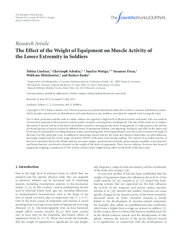
order described in Figure 1. The warm-up period and the
EMG, together with the knee angle recordings, were then
restarted.
2.2. Equipment. In the first test conditions (reference) the
participants wore shorts, standard combat boots, and socks.
In the order shown in Figure 1, all the participants wore
the standard equipment of a soldier, consisting of a helmet,
carrying strap, backpack, and rifle, consecutively. The weight
of each item of additional equipment is specified in Table 1.
2.6. Statistical Analysis. Descriptive statistics (median, standard deviation, minimum, and maximum) were calculated
for each dataset. After the Friedman test rejected the hypothesis of equality of the means for EMG mean amplitude,
peak, and integral for different load conditions, the Wilcoxon
test for pairwise comparison against control setup was
performed. For this reason, it is important to mention that
the measurements were dependent on the particular test
subject. All P values are the result of two-tailed statistical
tests, with values of P < 0.05 regarded as significant. All
data were stored and analysed using the statistics program
Statistical Package for Social Sciences for Windows (SPSS)
Version 15.0 (SPSS Inc. Chicago, Illinois, USA).
2.3. Instruments. Dynamic surface electromyograms
(EMGs) of the peroneus longus, gastrocnemius lateralis,
gastrocnemius medialis, tibialis anterior, rectus femoris,
and biceps femoris muscles of the right leg were taken in
accordance with the “standards for reporting EMG data”
[22] of the International Society of Electrophysiology
and Kinesiology using a wireless EMG system (Noraxon
Telemyo 2400T, Noraxon, Scotsdale, Arizona, USA). Bipolar
recordings were made, using disposable, self-adhesive
Ag/AgCl electrodes (Blue Sensor P, Ambu, Germany) with
an active electrode of diameter 7 mm. Before positioning
the electrodes, their specified locations on the skin of each
2.5. Data Processing. The software MyoResearch XP
(Noraxon, Scotsdale, Arizona, USA) was used for subsequent
processing of the EMG data. To determine the magnitude
of muscle activity, the EMG data for each muscle was
fully wave-rectified and smoothed by applying the root
mean square calculation (RMS-EMG). The amount of
muscle activity was determined by calculating mean EMG
amplitude, peak, and area under the EMG curve (AUC
or iEMG). All EMG data were normalised to the EMG
measurement of the reference condition (a).
3. Results
3.1. Electromyography. Analysis of the EMG data clearly
revealed that the equipment items used influenced the
activity of the muscles under examination. Mean amplitude,
�The Scientific World Journal
3
(1)
(a)
(b)
(c)
(d)
(2)
(e)
Figure 1: Equipment in order of investigation: (a) reference, (b) helmet, (c) load-carrying strap, (d) backpack, (e-1) rifle (in front of the
body), and (e-2) rifle (slung over the right shoulder).
Table 1: Weight of different equipment items used in the measurements.
Equipment setup
Without equipment in shorts, combat boots and socks
+ Helmet
+ Carrying strap
+ Backpack
+ Weapon G36
Individual weight of the
piece of equipment, kg
—
1.5
1
15
Weight of the equipment
in total, kg
—
1.5
2.5
17.5
3.6
3.6
21.1
21.1
(a) Carried in front of the body
(b) Slung over the shoulder
peak, and area under the curve (AUC) changed to varying
degrees as a result of carrying different equipment items.
Generally, relatively light items (helmet, carrying strap, and
rifle)—in contrast to heavy items such as a backpack—
caused little change in activity in the relevant muscles under
observation.
Figures 2, 3, and 4 show the changes in EMG amplitude,
EMG maximum and integrated EMG signal of the various
muscles, depending on the various equipment items.
3.2. The Tibialis Anterior Muscle. When wearing a helmet,
the mean (4%) and AUC EMG value (4.2%) of the tibialis
anterior muscle (TA) fell slightly. In comparison to the
reference measurement, the peak value remained the same.
After adding the load-carrying strap, the activity of the
TA changed very little (mean, peak, and integral). When
the backpack was added, activity increased significantly for
all three evaluated EMG parameters by approximately 16%
in comparison to the control. Additionally, carrying the
weapon, be it in front of the body or over the shoulder, had
no influence on the activity of the TA muscle.
Setup number
a (Reference)
b
c
d
e-1
e-2
3.3. The Peroneus Longus Muscle. The treadmill analysis
showed, in comparison to the reference value (P < 0.001),
a significant increase in the activity of the peroneus longus
(PL) muscle under an increasingly heavy equipment load
when carrying the backpack. The helmet and load-carrying
strap, on the other hand, did not cause any significant
increase in activity in comparison to the reference value
(Table 2). However, there was a significant increase in activity
between the helmet and carrying strap load levels. Carrying
the weapon, both in front of the body and slung over the
shoulder, caused no significant differences in comparison to
the backpack, except for a significantly different AUC value
between the rucksack and carrying the weapon in front of
the body (weapon e-1) and between the peak EMG values of
the weapon when carried in front of the body or slung over
the shoulder.
3.4. The Gastrocnemius Lateralis and Gastrocnemius Medialis Muscles. The two muscles, gastrocnemius laterialis
(GL) and medialis (GM), did not show any significant
increase in activity in comparison to the activity in the
reference measurement when the subjects were wearing
�4
The Scientific World Journal
Table 2: Summary of significant effects on the EMG of different muscles.
Setup
EMG
Reference (a)
Helmet (b)
Carrying strap (c)
Backpack (d)
Weapon (e-1)
Weapon (e-2)
Mean peak AUC Mean peak AUC Mean peak AUC Mean peak AUC Mean peak AUC Mean peak AUC
Muscle
TA
PL
Reference (a)
GL
GM
RF
BF
Helmet (b)
Carrying strap
(c)
TA
∗
◦
∗
PL
◦
◦
◦
GL
◦
◦
◦
GM
◦
◦
◦
RF
◦
◦
◦
BF
◦
◦
◦
TA
◦
◦
◦
◦
◦
◦
PL
◦
◦
◦
∗∗∗
∗
∗∗
GL
◦
◦
◦
∗
◦
◦
GM
◦
∗
◦
∗
◦
∗∗∗
RF
∗
∗
BF
∗∗
◦
∗
∗∗∗ ∗∗ ∗∗∗
∗∗ ∗∗∗
◦
∗∗∗
TA ∗∗∗ ∗∗∗ ∗∗∗ ∗∗∗ ∗∗∗ ∗∗∗ ∗∗∗ ∗∗∗ ∗∗∗
PL ∗∗∗ ∗∗∗ ∗∗∗ ∗∗∗ ∗∗∗ ∗∗∗ ∗∗
Backpack (d)
∗∗
∗∗
GL ∗∗∗ ∗∗∗ ∗∗∗ ∗∗∗ ∗∗∗ ∗∗∗ ∗∗∗ ∗∗∗ ∗∗∗
GM ∗∗∗ ∗∗∗ ∗∗∗ ∗∗∗ ∗∗∗ ∗∗∗ ∗∗∗ ∗∗∗ ∗∗∗
RF ∗∗∗ ∗∗∗ ∗∗∗ ∗∗∗ ∗∗∗ ∗∗∗ ∗∗∗ ∗∗∗ ∗∗∗
BF
Weapon (e-1)
∗∗
∗∗
∗∗
∗∗
∗
∗∗
∗P
∗
◦
◦
◦
PL ∗∗∗ ∗∗∗ ∗∗∗ ∗∗∗ ∗∗∗ ∗∗∗ ∗∗
◦
◦
∗
∗∗
∗∗
GL ∗∗∗ ∗∗∗ ∗∗∗ ∗∗∗ ∗∗∗ ∗∗∗ ∗∗∗ ∗∗∗ ∗∗∗
◦
◦
◦
GM ∗∗∗ ∗∗∗ ∗∗∗ ∗∗∗ ∗∗∗ ∗∗∗ ∗∗∗ ∗∗∗ ∗∗∗
◦
◦
◦
RF ∗∗∗ ∗∗∗ ∗∗∗ ∗∗∗ ∗∗∗ ∗∗∗ ∗∗∗ ∗∗∗ ∗∗∗
◦
◦
◦
BF ∗∗∗ ∗∗∗ ∗∗
◦
◦
◦
∗∗
∗∗
∗∗
∗
∗
∗∗∗
◦
◦
◦
◦
◦
◦
◦
∗∗
◦
◦
◦
◦
∗
◦
GL ∗∗∗ ∗∗∗ ∗∗∗ ∗∗∗ ∗∗∗ ∗∗∗ ∗∗∗ ∗∗∗ ∗∗∗
◦
◦
◦
◦
◦
◦
GM ∗∗∗ ∗∗∗ ∗∗∗ ∗∗∗ ∗∗∗
◦
◦
◦
◦
◦
◦
RF ∗∗∗ ∗∗∗ ∗∗∗ ∗∗∗ ∗∗∗ ∗∗∗ ∗∗∗ ∗∗∗ ∗∗∗
◦
◦
◦
◦
◦
◦
BF ∗∗∗ ∗∗ ∗∗∗ ∗∗∗ ∗∗ ∗∗∗
∗
◦
◦
◦
◦
◦
∗
∗∗
∗∗ P
∗∗
∗∗∗ ∗∗∗
PL ∗∗∗ ∗∗∗ ∗∗∗ ∗∗∗ ∗∗∗ ∗∗∗
◦P
∗
TA ∗∗∗ ∗∗∗ ∗∗∗ ∗∗∗ ∗∗∗ ∗∗∗ ∗∗∗ ∗∗∗ ∗∗∗
TA ∗∗∗ ∗∗∗ ∗∗∗ ∗∗∗
Weapon (e-2)
∗
∗
∗
∗∗∗ ∗∗∗ ∗∗∗
∗
∗
∗∗
∗∗∗ P
> 0.05,
< 0.05,
< 0.01, and
< 0.001.
TA: tibialis anterior; BF: biceps femoris; PL: peroneus longus; RF: rectus femoris; GL: gastrocnemius lateralis; GM: gastrocnemius medialis.
the helmet and carrying strap. Only when carrying the
backpack was there a significant difference in comparison
to the reference value for the mean, peak, and integral
values. The activity of the GL and GM increased by 32%
or 24% (both mean) in comparison to the reference value
(P < 0.001).
As with the TA and PL muscles, there was no
significant change in activity in the GL and GM
�The Scientific World Journal
5
Muscle activity (mean RMS-EMG amplitude)
250
Reference (%)
200
150
100
50
0
Reference
M. tibialis ant.
M. peroneus longus
M. gastrocnemius lat.
Helmet
Backpack
Load carrying
strap
Equipment setup
M. gastrocnemius med.
M. rectus femoris
M. biceps femoris
Rifle in front of
the body
Rifle slung over
the shoulder
Figure 2: Mean EMG amplitude in percent compared to the mean EMG of the reference measurements.
muscles in comparison to the initial state when the
weapon was added.
3.5. The Rectus Femoris Muscle. The slight decrease in
activity in the rectus femoris muscle (RF) when the subjects
were wearing the helmet was insignificant in comparison
to the reference value. The slight increase in activity after
adding the carrying strap, however, was significant both in
comparison to the reference value (mean EMG P = 0.023)
and in comparison to the helmet load level (P < 0.001). After
the backpack was added, activity in the RF muscle increased
by 75% (mean EMG) or 76% (AUC) in comparison to the
value of the initial state (P < 0.001). Again, the change in
muscle activity caused by adding the weapon, regardless of
how it was carried, was not significant in comparison to the
load level values of the backpack load level.
3.6. The Biceps Femoris Muscle. Similarly to the RF, there was
a significant increase (mean and AUC) in the activity of the
biceps femoris muscle (BF) after the carrying strap was added
(P = 0.001, P = 0.002). The changes in the peak value for the
BF were not significant in comparison to the initial state. In
addition, significant muscle activity differences were revealed
between carrying strap and backpack load levels (mean EMG
P = 0.045 and AUC P = 0.021). As was the case for all
muscles examined, there was no further increase in activity
in the BF when the weapon was added.
3.7. Knee Angle Measurements. When the subjects were
carrying the various items of equipment, mean values of the
range of motion of the knee joint were between min. 55.1◦ ±
8.2◦ (weapon carried in front of the body) and 56.8◦ ± 6.6◦
(load-carrying strap), that is no significant differences were
detected between the examined load levels.
4. Discussion
Military service places high demands on the physical fitness
of soldiers. They frequently have to bear weighted loads
and items of equipment during their everyday professional
life. This is a potential risk factor for the occurrence of
overexertion syndromes in the locomotor system [24, 25],
particularly if the weight is carried over long distances. The
aim of this study was to analyse the activity of selected
muscles in the lower extremity on the basis of increasing
weight caused by typical equipment items worn or carried
by soldiers, for example helmet, carrying strap, backpack,
and rifle. Al-Khabbaz et al. [26] showed that backpack
weights of up to 20% of the carrier’s bodyweight do not
cause an increase in muscle activity in EMG measurements
while standing. Simpson et al. [27], on the other hand,
used measurements taken while subjects were walking to
determine that there is an increase in activity (integrated
EMG) of the vastus lateralis and gastrocnemius medialis
muscles in female recreational walkers as a result of carrying
a backpack weighing between 20 and 40% of their body
weight. In our study, the muscles under examination (TA,
PL, GL, GM, BF, and RF) showed the greatest increase in
activity after adding the backpack. The greatest increase in
muscle activity at this level of equipment load was detected
in the rectus femoris muscle. This muscle plays a major
�6
The Scientific World Journal
Muscle activity (peak)
250
Reference (%)
200
150
100
50
0
Reference
M. tibialis ant.
M. peroneus longus
M. gastrocnemius lat.
Helmet
Load carrying
strap
Backpack
Rifle in front of
the body
Rifle slung over
the shoulder
Equipment setup
M. gastrocnemius med.
M. rectus femoris
M. biceps femoris
Figure 3: Peak EMG in percent compared to the peak EMG values of the reference measurements.
role in the stretching of the knee [28]. Due to the relatively
high additional weight of the backpack, which is between 15
and 30% of the personal body weight of the soldiers under
examination, muscle activity also increased considerably as
a result of the heavy load. A possible consequence of this
increase in activity in soldiers can be frequent occurrence
of functional knee pain [20]. The EMG changes in the
gastrocnemius lateralis, gastrocnemius medialis, and tibialis
anterior muscles revealed in this study could be indicative of
the development of overexertion syndromes in the Achilles
tendon and around the edge of the shinbone, because there
is a link between a change in activity in the aforementioned
muscles and the development of these symptoms [19, 21].
Due to its relatively low weight, the helmet showed no
measurable influence on the muscle activity of the lower
extremity, with the exception of the tibialis anterior muscle.
A major proportion of the weight of the helmet is carried
by the local muscles of the neck and upper back and leads
to measurable differences in muscle activity in that area.
Thuresson et al. [29] verified these increases in activity
by means of EMG measurements of the neck muscles in
helicopter pilots.
The effect of carrying a rifle on lower extremity activity
has not been examined in previous EMG studies. Birrell and
Haslam [24] examined the effect of carrying a rifle on ground
reaction forces while walking, and discovered that effect to
be significant. Our results showed that carrying a weapon
exerts no additional influence on the activity of the examined
muscles of the lower extremity. While the weight of the
weapon is carried by the upper extremity, thus influencing
the kinematics of the subjects’ gait [24], the weapon, which in
itself weighs 3.6 kg, has only a relatively low additional weight
load—as is the case with a helmet and carrying strap—on
muscular activity in the lower extremity.
In studies dealing with exertion-dependent changes in
the movement range of the knee joint caused by the weight
of equipment items [9, 12, 14, 15, 30], results have been
inconsistent. While Attwells et al. [14] und Kinoshita et al.
[12] observed greater ROM in the knee joint with increasing
load, Ghori and Luckwill [9] found a decrease in ROM under
load. Other authors, for example Majumdar et al. [15] or
Tilbury-Davis and Hooper [30] did not observe any loaddependent change in the ROM of the knee joint. Our results
also did not show any increase or decrease in the ROM in the
various load situations. Majumdar et al. [15] claimed that the
reason for the lack of differences is that the additional weight
of between 6.5 and 27.2% of the subjects’ body weight was
too low. Tilbury-Davis and Hooper [30] presumed that the
differences in Kinoshita’s findings [24] can be attributed to
the subjects being in better training condition and therefore
having greater strength. With greater loads of between 47%
and 64% of their body weight, these subjects were also able
to maintain a normal gait pattern.
4.1. Limitations of the Study. In the examinations conducted,
it must be borne in mind that this was a dynamic study
and that gait phase-specific differences were also taken into
account [31]. In dynamic studies, the centre of gravity is
deflected in a sinusoidal movement in the transversal and
sagittal planes [31]. The maximum is always reached in
the middle standing phase on the side of the standing leg
[31]. In the event of an unevenly distributed increasing
�The Scientific World Journal
7
Muscle activity (integrated EMG)
250
Reference (%)
200
150
100
50
0
Reference
M. tibialis ant.
M. peroneus longus
M. gastrocnemius lat.
Helmet
Load carrying
strap
Backpack
Rifle in front of
the body
Rifle slung over
the shoulder
Equipment setup
M. gastrocnemius med.
M. rectus femoris
M. biceps femoris
Figure 4: Integrated EMG in percent compared to the mean integrated EMG of the reference measurements.
load, appropriate stabilisation work becomes necessary. Thus
stabilisation work, which increases in conjunction with the
load, is also a component of the measured muscle activity
and cannot be distinguished from the muscle activity that is
brought about by changing the loads.
5. Conclusions
The equipment items used in our study are essential for
soldiers to carry during military operations. For this reason,
their influence on the activity of different muscles in the
lower extremity was examined. By adding equipment items
consecutively, we determined that relatively light items
(helmet, carrying strap, and rifle) caused only minor changes
in muscle activity. In contrast, heavy items such a backpack
cause a considerable change in activity in the relevant
muscles under observation. In our studies, the backpack,
which weighed 15 kg, caused a mean 75% increase in muscle
activity in comparison with the reference measurement. The
loads that soldiers have to carry during marches should
therefore be kept as low as possible given the possible
reduction in the risk of musculoskeletal disorders.
Conflict of Interests
All authors disclose any financial or personal relationships
they may have with other people or organisations that could
inappropriately influence this work.
Acknowledgments
The authors would like to thank Professor Dr. Guenther
Kundt for supporting them during the statistical evaluation
of the EMG data. They would also like to thank the subjects
who volunteered for this study.
References
[1] K. R. Kaufman, S. Brodine, and R. Shaffer, “Military trainingrelated injuries: surveillance, research, and prevention,” American Journal of Preventive Medicine, vol. 18, no. 3, pp. 54–63,
2000.
[2] K. R. Kaufman, S. K. Brodine, R. A. Shaffer, C. W. Johnson,
and T. R. Cullison, “The effect of foot structure and range
of motion on musculoskeletal overuse injuries,” American
Journal of Sports Medicine, vol. 27, no. 5, pp. 585–593, 1999.
[3] J. J. Knapik, K. L. Reynolds, and E. Harman, “Soldier load
carriage: historical, physiological, biomechanical, and medical
aspects,” Military Medicine, vol. 169, no. 1, pp. 45–56, 2004.
[4] H. Taanila, J. Suni, H. Pihlajamäki et al., “Musculoskeletal
disorders in physically active conscripts: a one-year followup study in the Finnish Defence Forces,” BMC Musculoskeletal
Disorders, vol. 10, no. 1, article 89 89, 2009.
[5] D. N. Cowan, B. H. Jones, and J. R. Robinson, “Foot morphologic characteristics and risk of exercise-related injury,”
Archives of Family Medicine, vol. 2, no. 7, pp. 773–777, 1993.
[6] D. N. Cowan, B. H. Jones, P. N. Frykman et al., “Lower limb
morphology and risk of overuse injury among male infantry
trainees,” Medicine and Science in Sports and Exercise, vol. 28,
no. 8, pp. 945–952, 1996.
[7] B. H. Jones, D. N. Cowan, J. P. Tomlinson, J. R. Robinson,
D. W. Polly, and P. N. Frykman, “Epidemiology of injuries
�8
[8]
[9]
[10]
[11]
[12]
[13]
[14]
[15]
[16]
[17]
[18]
[19]
[20]
[21]
[22]
[23]
The Scientific World Journal
associated with physical training among young men in the
army,” Medicine and Science in Sports and Exercise, vol. 25, no.
2, pp. 197–203, 1993.
J. Bobet and R. W. Norman, “Effects of load placement on back
muscle activity in load carriage,” European Journal of Applied
Physiology and Occupational Physiology, vol. 53, no. 1, pp. 71–
75, 1984.
G. M. U. Ghori and R. G. Luckwill, “Responses of the lower
limb to load carrying in walking man,” European Journal of
Applied Physiology and Occupational Physiology, vol. 54, no. 2,
pp. 145–150, 1985.
M. Holewijn, “Physiological strain due to load carrying,”
European Journal of Applied Physiology and Occupational
Physiology, vol. 61, no. 3-4, pp. 237–245, 1990.
E. Harman, K. Han, P. Frykman, M. Johnson, F. Russel, and M.
Rosenstein, “The effects on gait timing, kinetics, and muscle
activity of various loads carried on the back,” Medicine &
Science in Sports & Exercise, vol. 24, no. 5, p. 129, 1992.
H. Kinoshita, “Effects of different loads and carrying systems
on selected biomechanical parameters describing walking
gait,” Ergonomics, vol. 28, no. 9, pp. 1347–1362, 1985.
P. E. Martin and R. C. Nelson, “The effect of carried loads on
the walking patterns of men and women,” Ergonomics, vol. 29,
no. 10, pp. 1191–1202, 1986.
R. L. Attwells, S. A. Birrell, R. H. Hooper, and N. J. Mansfield,
“Influence of carrying heavy loads on soldiers’ posture,
movements and gait,” Ergonomics, vol. 49, no. 14, pp. 1527–
1537, 2006.
D. Majumdar, M. S. Pal, and D. Majumdar, “Effects of military
load carriage on kinematics of gait,” Ergonomics, vol. 53, no. 6,
pp. 782–791, 2010.
C. Schulze, T. Lindner, K. Schulz, S. Woitge, W. Mittelmeier,
and R. Bader, “Influence of increased load wearing on human
posture and muscle activation of trunk and lower limb,” Swiss
Medical Weekly, vol. 142, supplement 193, pp. 4–5, 2012.
J. Knapik, E. Harman, and K. Reynolds, “Load carriage using
packs: a review of physiological, biomechanical and medical
aspects,” Applied Ergonomics, vol. 27, no. 3, pp. 207–216, 1996.
C. Schulze, T. Lindner, K. Schulz et al., “The influence in
airforce soldiers through wearing certain types of army-issue
footwear on muscle activity in the lower extremities,” The
Open Orthopaedics Journal, vol. 5, pp. 302–306, 2011.
J. T. Andrish, J. A. Bergfeld, and J. Walheim, “A prospective
study on the management of shin splints,” Journal of Bone and
Joint Surgery Series A, vol. 56, no. 8, pp. 1697–1700, 1974.
A. C. S. Ribeiro, D. B. Grossi, B. Foerster, C. Candolo, and V.
Monteiro-Pedro, “Electromyographic and magnetic resonance
imaging evaluations of individuals with patellofemoral pain
syndrome,” Revista Brasileira de Fisioterapia, vol. 14, no. 3, pp.
221–228, 2010.
L. B. Azevedo, M. I. Lambert, C. L. Vaughan, C. M. O’Connor,
and M. P. Schwellnus, “Biomechanical variables associated
with Achilles tendinopathy in runners,” British Journal of
Sports Medicine, vol. 43, no. 4, pp. 288–292, 2009.
R. Merletti, “Standards for reporting EMG data,” Journal of
Electromyography and Kinesiology, vol. 9, no. 1, pp. 3–5, 1999.
H. J. Hermens, B. Freriks, C. Disselhorst-Klug, and G. Rau,
“Development of recommendations for SEMG sensors and
sensor placement procedures,” Journal of Electromyography
and Kinesiology, vol. 10, no. 5, pp. 361–374, 2000.
[24] S. A. Birrell and R. A. Haslam, “The influence of rifle carriage
on the kinetics of human gait,” Ergonomics, vol. 51, no. 6, pp.
816–826, 2008.
[25] A. Polcyn, C. Bensel, E. Harman, J. Obusek, C. Pandorf, and
P. Frykman, “Effects of weight carried by soldiers: combined
analysis of four studies on maximal performance, physiology
and biomechanics,” Tech. Rep. TR-02/010, US Army Research
Institute of Environmental Medicine, Natick, Mass, USA,
2002.
[26] Y. S. S. M. Al-Khabbaz, T. Shimada, and M. Hasegawa, “The
effect of backpack heaviness on trunk-lower extremity muscle
activities and trunk posture,” Gait and Posture, vol. 28, no. 2,
pp. 297–302, 2008.
[27] K. M. Simpson, B. J. Munro, and J. R. Steele, “Backpack load
affects lower limb muscle activity patterns of female hikers
during prolonged load carriage,” Journal of Electromyography
and Kinesiology, vol. 21, no. 5, pp. 782–788, 2011.
[28] T. Schiebler, Anatomie, Springer, New York, NY, USA, 2005.
[29] M. Thuresson, B. Äng, J. Linder, and K. Harms-Ringdahl,
“Neck muscle activity in helicopter pilots: effect of position
and helmet-mounted equipment,” Aviation Space and Environmental Medicine, vol. 74, no. 5, pp. 527–532, 2003.
[30] D. C. Tilbury-Davis and R. H. Hooper, “The kinetic and
kinematic effects of increasing load carriage upon the lower
limb,” Human Movement Science, vol. 18, no. 5, pp. 693–700,
1999.
[31] J. Perry, Ganganalyse—Norm und Pathologie des Gehens,
Urban und Fischer, Jena, Germany, 2003.
�
 Tobias Lindner