Colloids and Surfaces A: Physicochem. Eng. Aspects 212 (2003) 219 �/226
www.elsevier.com/locate/colsurfa
Preparation and characterization of magnetite nanoparticles
coated by amino silane
Ming Ma �, Yu Zhang, Wei Yu, Hao-ying Shen, Hai-qian Zhang, Ning Gu
National Laboratory of Molecular and Biomolecular Electronics, Southeast University, Nanjing 210096, PR China
Received 13 January 2002; received in revised form 21 May 2002; accepted 18 June 2002
Abstract
Magnetite nanoparticles were prepared by coprecipitation of Fe2� and Fe3� with NH4OH, and then, amino silane
was coated onto the surface of the magnetite nanoparticles. Transmission electronic microscopy shows the average size
of 7.5 nm in diameter. Powder X-ray diffraction and electronic diffraction measurements show the spinel structure for
the magnetite nanoparticles. FT �/IR spectra indicate that amino silane molecules have been bound onto the surface of
the magnetite nanoparticles by Fe�/O�/Si chemical bonds. Energy dispersive X-ray spectroscopy (SEM �/EDS) indicates
atomic ratio of 96.75:3.25 for Fe:Si, implying a nearly monolayer coating of amino silane on the magnetite particle
surface according to a rough calculation. By an enzyme-linked assay, it was proved that the amino silane-coated
magnetite nanoparticles could significantly improve the protein immobilization.
# 2002 Elsevier Science B.V. All rights reserved.
Keywords: Magnetite nanoparticles; Core-shell structure; Surface coating; Amino silane; Protein immobilization
1. Introduction
Magnetic particles (microspheres, nanospheres
and ferrofluids) are widely studied for their
applications in various fields in biology and
medicine such as enzyme and protein immobilization, genes, radiopharmaceuticals, magnetic resonance imaging MRI, diagnostics, immunoassays,
RNA and DNA purification, magnetic cell separation and purification, magnetically controlled
transport of anti-cancer drugs as well as hyperthermia generation [1 �/3]. These magnetic beads are
� Corresponding author
E-mail address: maming@seu.edu.cn (M. Ma).
generally of core �/shell type: biological species
cells, nucleic acids, proteins are connected to the
magnetic core through an organic or polymeric
shell. The shells are either biocompatible in general
(such as dextran, PEG, etc.), or possessing active
groups which can be conjugated to biomolecules
such as proteins and enzymes. Therefore, the
investigation of magnetic nanoparticles with organic coating is of significance for applications.
In this work we prepared magnetite nanoparticles coated with a near monolayer of amino silane,
which has active group of �/NH2 that can connect
biomolecules, drugs and so on. And the morphology, structure and composition of the coated
magnetite nanoparticles were characterized by
TEM, ED, XRD, FT-IR and SEM �/EDS.
0927-7757/02/$ - see front matter # 2002 Elsevier Science B.V. All rights reserved.
PII: S 0 9 2 7 - 7 7 5 7 ( 0 2 ) 0 0 3 0 5 - 9
�220
M. Ma et al. / Colloids and Surfaces A: Physicochem. Eng. Aspects 212 (2003) 219 �/226
Furthermore, to prove that amina silane-coated
magnetite nanoparticles can be conjugated to
some biomolecule, an enzyme-linked colorimetric
assay was carried out after the enzyme horseradish
peroxidase (HRP) of different concentrations was
used to interact with the coated and the uncoated
magnetite nanoparticles.
magnetite particles were measured by energy
dispersive X-ray spectroscopy (SEM �/EDS,
EDAX, PV9100). Fourier transform infrared
spectroscopy (FT-IR, Nicolet, 750) of the samples
were used to study the chemical bonds between
Fe3O4 and APTS.
2.4. HRP-immobilized and its activity assays
2. Experimental
2.1. Synthesis of magnetite nanoparticles
Magnetite was made according to the method of
Molday [4]. Typically, a solution of mixture of
FeCl3 (0.01 M) and FeSO4 (0.006 M) at pH 1.7
was prepared under N2 protecting. Then, ammonia aqueous solution (1.5 M) was dropped into it
with violently stirring until the pH of the solution
raised to 9. The obtained magnetite was washed
immediately with water for 5 times and ethanol for
2 times by magnetic separation. Finally, part of
magnetite nanoparticles were dispersed in ethanol
with concentration of 0.0128 M, and the others
were dried into powder at room temperature under
vacuum.
2.2. APTS-coated magnetite nanoparticles
25 ml magnetite colloid ethanol solution prepared above was diluted to 150 ml by ethanol and
1 ml H2O. The solution was then treated by
ultrasonic wave for 30 min. 35 ml 3-aminopropyltriethoxysilane ( NH2(CH2)3Si(OC2H5)3, APTS)
was added into it with rapid stirring for 7 h. The
result solution was washed with ethanol for 5
times, and then dried into powder at room
temperature under vacuum.
2.3. Characterization
The particle size and morphology of the samples
were determined by transmission electronic microscopy (TEM, JEOL, JEM-200CX, 200 kV). Powder X-ray diffraction (XRD, Rigaku, D/Max-RA,
Cu Ka) and electronic diffraction ( ED) were used
to determine the crystal structure of the samples.
The elemental analysis and APTS loading on
The APTS-coated magnetite nanoparticles and
uncoated magnetite nanoparticles as control were
all dispersed in phosphate-buffered saline (PBS,
0.01 M, pH 7.4) with identical concentrations of 2
g l �1. Enzyme horseradish peroxidase HRP of
different concentrations were added into 200 ml
magnetite-PBS solutions. The mixtures were incubated in 37 8C for 1 h, and then retracted in
4 8C refrigeratory for 4 h. Then the mixtures were
washed carefully by PBS for 4 times and shifted to
other vessels to remove the dissociated enzymes.
Developed by addition of substrates, namely
3,3?,5,5?-tetramethylbenzidine and hydrogen peroxide (TMB-H2O2), for 10 min, the reaction was
stopped by 2 mol l �1 H2SO4. The optical density
at 450 nm was read immediately in an automatic
plate reader (Stat fax-2100, Beiken Company). All
samples were tested in duplicate, arithmetic means
and standard deviations of absorbance values were
calculated (x9/s).
3. Results and discussion
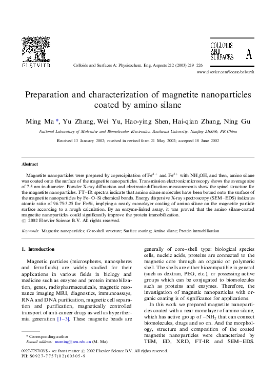
Fig. 1 is the TEM and ED photography of the
magnetite nanoparticles coated with APTS, which
shows that most of the particles are quasi-spherical
and with an average diameter of 7.5 nm. The
distribution of particle diameters is shown in Fig.
2.
According to the ED pattern, the d -spacing can
be calculated in the following eqution [5],
Ll�dR
(1)
where L is the distance between the test sample
and the film ( L �/137 cm), l is the wavelength of
electron beam (l �/0.0251 Å), R is the radius of
the diffraction ring. The calculation results are
shown in Table 1, which accord with the XRD
�M. Ma et al. / Colloids and Surfaces A: Physicochem. Eng. Aspects 212 (2003) 219 �/226
Fig. 1. TEM and ED photography of the magnetite nanoparticles coated with APTS.
pattern of the sample shown in Fig. 3 and indicate
the inverse cubic spinel structure of Fe3O4. Compared with the theoretical values, the reduction of
d -spacing obtained experimentally is due to lattice
constrictions for nanosized particles [7].
Since there are large surface-to-volume atomic
ratio, high surface activity, and amount of dangling bonds on nanoparticle surface, the atoms on
the surface are apt to adsorb ions or molecules in
solution. For Fe3O4 nanoparticles dispersed in a
neutral aqueous solution, the bare atoms of Fe and
O on the particle surface would adsorb OH � and
H � respectively, so that there is OH-rich surface.
The �/OH on the surface can react with APTS as
the process shown in Fig. 4. Therefore, the
magnetite nanoparticles can be coated with
APTS molecules by chemical bond. The fact was
proven by comparison of FT-IR spectra of the
coated and uncoated Fe3O4 nanoparticles shown
in Fig. 5. It can be seen that, compared with the
uncoated sample, the coated Fe3O4 nanoparticles
221
Fig. 2. Diameter distribution of magnetite nanoparticles coated
with APTS.
possess absorption bands in 2971.8 and 2925.5
cm �1 due to stretching vibration of C �/H bond,
band in 1091.5 cm �1 due to the stretching vibration of C �/N bond, band in 1051.0 cm �1 due to the
stretching vibration of Si �/O bond, band in 885.2
cm �1 due to the bending vibration of �/NH2
group. All of these reveal the existence of APTS.
In addition, in Fig. 5(a) and (b) the absorption
bands near 3400 and 1630 cm �1 refer to the
vibration of remainder H2O in the samples. And
there also exists the contribution of �/NH2 for the
band near 3400 cm �1 in Fig. 5(a).
Previously, it was reported that the characteristic absorption bands of the Fe �/O bond of bulk
Fe3O4 were in 570 and 375 cm �1 [8]. However, in
Fig. 5(b) these two bands shift to high wavenumbers of about 600 and 440 cm �1 respectively, and
the band near 600 cm �1 is split into two peaks of
631.4 and 582.9 cm �1. A principal effect of finite
size of nanoparticles is the breaking of a large
number of bonds for surface atoms, resulting in
the rearrangement of inlocalized electrons on the
�222
M. Ma et al. / Colloids and Surfaces A: Physicochem. Eng. Aspects 212 (2003) 219 �/226
Table 1
ED and XRD data for the magnetite nanoparticles coated with APTS
R (cm)
ED results-d (Å)
XRD results-d (Å)
Theory values-d (Å)
Crystalline plane (hlk) [6]
1
0.73
4.74
4.78
4.85
(111)
2
1.19
2.89
2.83
2.97
(220)
3
1.39
2.47
2.50
2.53
(311)
4
1.67
2.06
2.08
2.10
(400)
5
2.04
1.69
1.70
1.71
(422)
Fig. 3. XRD pattern of the magnetite nanoparticles coated with APTS.
Fig. 4. The procedure of the coating reaction of APTS with magnetite nanoparticles.
6
2.19
1.57
1.60
1.62
(511)
7
2.37
1.47
1.48
1.48
(440)
�M. Ma et al. / Colloids and Surfaces A: Physicochem. Eng. Aspects 212 (2003) 219 �/226
Fig. 5. FT-IR spectra of the coated (a) and uncoated (b) magnetite nanoparticles.
223
�224
M. Ma et al. / Colloids and Surfaces A: Physicochem. Eng. Aspects 212 (2003) 219 �/226
Fig. 6. SEM �/EDS elemental analysis of APTS-coated Fe3O4 nanoparticles on Au substrate.
particle surface [9]. And the lattice constrictions
have been observed as indicated in Table 1. As a
result, the surface bond force constant increases as
Fe3O4 is reduced to nanoscale dimension, so that
the absorption bands of IR spectra shift to higher
wavenumbers. So the blue-shift of absorption
bands of the Fe �/O bond of the Fe3O4 nanopar-
ticles can be observed. In addition, the split of the
bands is attributed to the split of the energy levels
of the quantized Fe3O4 nanoparticles[10].
It is also found that the characteristic absorption bands of the Fe �/O bond of APTS-coated
Fe3O4 shift to high wavenumbers of 636.4 and
590.1 cm �1 compared with that of uncoated
Fig. 7. HRP-linked colorimetric assays of APTS-coated and uncoated Fe3O4 nanoparticles.
�M. Ma et al. / Colloids and Surfaces A: Physicochem. Eng. Aspects 212 (2003) 219 �/226
Fe3O4(in 631.4 and 582.9 cm �1). The phenomenon can be explained according to the formation
of Fe �/O �/Si bonds where Fe �/O �/H groups on the
surface of the Fe3O4 particles are replaced by Fe �/
O �/Si(O �/)2 �/R as shown in Fig. 4. More electronegativity of �/Si(O �/)2 �/ than H leads to the
enhancement of bond force constant for Fe �/O
bonds[11], so that the absorption bands shift to
high wavenumbers.
Since we know the mean diameter of the
magnetite nanoparticles is 7.5 nm, the number of
Fe atoms in every Fe3O4 particle can be calculated
by means of following formula,
4
NFe �
3
pR3 Na
V Fe3 O4
�3� 8945
(2)
where V̄ Fe3 O4/refers to the molar volume of bulk
Fe3O4, R is the mean radius of Fe3O4 nanoparticles, Na is Avogadro’s number. If there is a
monolayer of APTS molecules coated on the
Fe3O4 particle, the number of APTS molecules
on the surface of every Fe3O4 nanoparticle can be
calculated by following formula,
NAPTS �
SFe3 O4
SAPTS
�
4pR2
SAPTS
�442
(3)
where SFe3 O4 is the surface area of Fe3O4 particle,
SAPTS is the area of surface coverage of about 40
Å2 per APTS molecule reported in the literature
[12]. So the atomic ratio of Fe/Si is/NFe =NAPTS �
20:2: Fig. 6 shows a typical SEM �/EDS elemental
analysis of APTS-coated magnetite nanoparticles.
From the peak area of Fe and Si, the atomic ratio
of Fe/Si is obtained to be 96.75/3.25 �/30. This
indicates that the surface APTS coverage ratio of
Fe3O4 nanoparticles is about 67.3%. Probably, the
incompleteness (a near monolayer) of the surface
coating is owing to the incompleteness of surface
hydroxylation and the existence of the spatial
resistance for the surface coating reaction.
Fig. 7 shows the result of the HRP-linked
magnetite nanoparticles colorimetric assays. Obviously, the absorbances of HRP-linked APTScoated magnetite nanoparticles are higher than
HRP-linked uncoated magnetite nanoparticles. It
is revealed that the amount of adsorbed HRP on
225
the APTS-coated magnetite nanoparticles is 1.4 �/
2.0 times higher than that of the uncoated
magnetite nanoparticles according to the measured
absorbance value. The increase of absorption is
attributed to the contribution of APTS whose
active group of �/NH2 can be conjugated to HRP
by chemical band, and the uncoated magnetite
nanoparticles connect the HRP by static adsorption only.
4. Conclusions
APTS-coated magnetite nanoparticles with 7.5
nm average diameter were prepared and characterized by TEM, ED, XRD, FT-IR, and SEM �/
EDS. Especially, FT-IR spectra were utilized to
prove the formation of Fe �/O �/Si chemical bonds.
A near monolayer APTS-coating on the particle
surface was also indicated according to the comparison of the experimental analysis by SEM �/
EDS with a simple calculation.
By an enzyme-linked assay, it has been proved
that these APTS-coated magnetite nanoparticles
could significantly improve the protein immobilization.
Acknowledgements
This work was supported by the National
Natural
Science
Foundation
of
China
(No.69890220, No.60171005) and the High Technology Research Subject of Jiangsu Province in
China (BG2001006). I am also very grateful to
Prof. Hong Jian-min of Center of Analysis and
Test, Nanjing University for his helping in TEM
experiments.
References
[1] U. Häfeli, W. Schütt, J. Teller, M. Zborowski, Scientific
and Clinical Applications of Magnetic, Plenum, New
York, 1997.
[2] R. Weissleder, A. Bogdanov, E.A. Neuwelt, M. Papisov,
Adv. Drug Del. Rev. 16 (1995) 321 �/334.
[3] A. Jordan, R. Scholz, P. Wust, H. Schirra, et al., J. Mag.
Mag. Mater. 194 (1999) 185 �/196.
�226
M. Ma et al. / Colloids and Surfaces A: Physicochem. Eng. Aspects 212 (2003) 219 �/226
[4] R.S. Molday, US Patent 4452773, 1984.
[5] Q. Dai, N. He, K.P. Weng, B.P. Lin, et al., J. of Inclusion
Phenomena and Macrocyclic Chemistry 35 (1999) 11 �/21.
[6] R. Fan, X.H. Chen, Z. Gui, et al., Mater. Res. Bul. 36
(2001) 497 �/502.
[7] X.Y. Qin, Nanostu. Mater. 2 (1993) 99.
[8] R.D. Waldron, Phys. Rev. 99 (1955) 1727.
[9] G. Xiong, S.J. Yu, X.J. Yang, et al., J. Fouc. Maters 29
(1998) 92 �/95(Chinese).
[10] Z.M. Gao, T.H. Wu, S.Y. Peng, Acta Phys. Chim. Sin. 11
(1995) 395 �/399.
[11] W.S. Wei, J. Yang, T.J. Wang, et al., Acta Phys. Chim.
Sin. 17 (2001) 507 �/510.
[12] F.J. Boerio, L. Armogan, S.Y. Cheng, J. Colloid interface
Sci. 73 (1980) 416.
�
 zhang hai