A. Silvest rini Biavat i* , A. Signori* * ,
A. Cast aldo* * * , G. M at arese* * * * , M . M igliorat i*
* Department of Orthodontics, University of Genoa, Italy
* * Department of Health Sciences, Section of Biostatistics, University
of Genoa, Italy
* * * Department of Orthodontics, University of Trieste, Italy
* * * * Department of Orthodontics, University of M essina, Italy
e-mail: armando.silvestrini@tin.it
Incidence
and distribution
of deciduous molar
ankylosis, a longitudinal
study
A BSTRACT
Aim To study incidence and distribution of deciduous molar
ankylosis.
M a t e r i a l s a n d m e t h o d s Study design: longitudinal
retrospective study. A total of 512 consecutive subjects (aged
5 to 15 years) w ere examined at the Orthodontics and
Paediatric Dentistry Department of the Genoa University
School of Dentistry; for each subject an ortopantomography
x-ray w as taken.
Result s Thirty-four children w ere affected by deciduous
molars ankylosis (6.6% ). A statistically significant difference
was revealed between the distributions: the lower deciduous
molars were ankylosed more frequently than the upper ones
(P<0.001); the second deciduous molars were ankylosed more
frequently than the first molars (P<0.001). No statistical
significance w as found betw een sex and number of
infraoccluded teeth (P=0.74).
Conclusion This study found an incidence of deciduous molar
ankylosis of about 6.6% ; the lower deciduous molars and
second deciduous molars were ankylosed more frequently
(P<0.001).
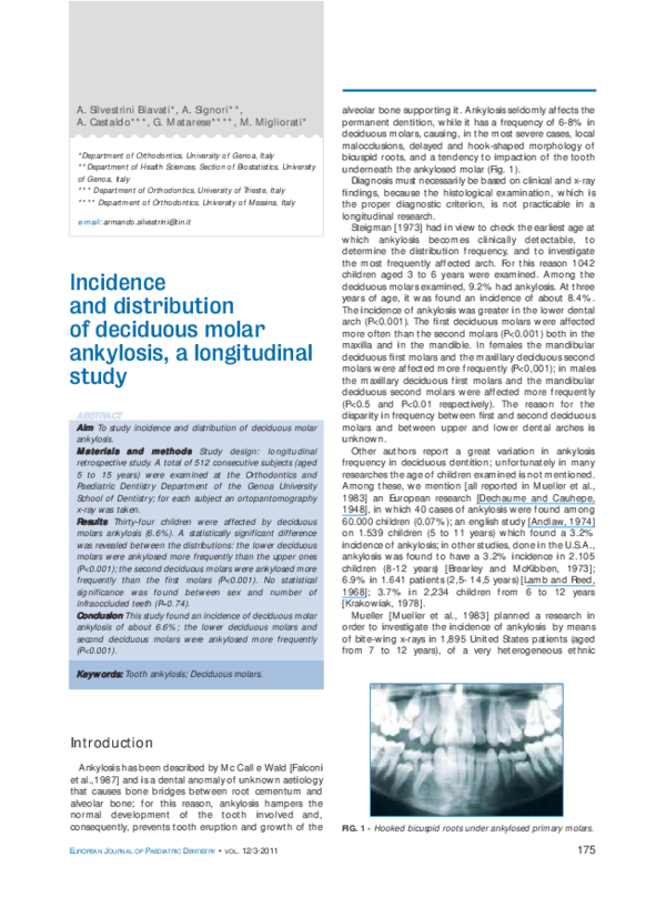
alveolar bone supporting it. Ankylosis seldomly affects the
permanent dentition, w hile it has a frequency of 6-8% in
deciduous molars, causing, in the most severe cases, local
malocclusions, delayed and hook-shaped morphology of
bicuspid roots, and a tendency to impaction of the tooth
underneath the ankylosed molar (Fig. 1).
Diagnosis must necessarily be based on clinical and x-ray
findings, because the histological examination, w hich is
the proper diagnostic criterion, is not practicable in a
longitudinal research.
Steigman [1973] had in view to check the earliest age at
w hich ankylosis becomes clinically det ect able, t o
determine the distribution frequency, and to investigate
the most frequently affected arch. For this reason 1042
children aged 3 to 6 years w ere examined. Among the
deciduous molars examined, 9.2% had ankylosis. At three
years of age, it w as found an incidence of about 8.4% .
The incidence of ankylosis w as greater in the low er dental
arch (P<0.001). The first deciduous molars w ere affected
more often than the second molars (P<0.001) both in the
maxilla and in the mandible. In females the mandibular
deciduous first molars and the maxillary deciduous second
molars w ere affected more frequently (P<0,001); in males
the maxillary deciduous first molars and the mandibular
deciduous second molars w ere affected more frequently
(P<0.5 and P<0.01 respectively). The reason for the
disparity in frequency betw een first and second deciduous
molars and betw een upper and low er dental arches is
unknow n.
Other authors report a great variation in ankylosis
frequency in deciduous dentition; unfortunately in many
researches the age of children examined is not mentioned.
Among these, w e mention [all reported in M ueller et al.,
1983] an European research [Dechaume and Cauhepe,
1948], in w hich 40 cases of ankylosis w ere found among
60.000 children (0.07% ); an english study [Andlaw, 1974]
on 1.539 children (5 to 11 years) w hich found a 3.2%
incidence of ankylosis; in other studies, done in the U.S.A.,
ankylosis w as found to have a 3.2% incidence in 2.105
children (8-12 years) [Brearley and M cKibben, 1973];
6.9% in 1.641 patients (2,5- 14,5 years) [Lamb and Reed,
1968]; 3.7% in 2,234 children from 6 to 12 years
[Krakow iak, 1978].
M ueller [M ueller et al., 1983] planned a research in
order to investigate the incidence of ankylosis by means
of bite-w ing x-rays in 1,895 United States patients (aged
from 7 to 12 years), of a very heterogeneous ethnic
Keyw ords: Tooth ankylosis; Deciduous molars.
Int roduct ion
Ankylosis has been described by M c Call e Wald [Falconi
et al.,1987] and is a dental anomaly of unknow n aetiology
that causes bone bridges betw een root cementum and
alveolar bone; for this reason, ankylosis hampers the
normal development of t he t oot h involved and,
consequently, prevents tooth eruption and grow th of the
EUROPEAN JOURNAL OF PAEDIATRIC DENTISTRY •
VOL.
12/3-2011
FIG. 1 - Hooked bicuspid roots under ankylosed primary molars.
175
�SILVESTRINI BIAVATI A. ET AL.
population (78.3% Caucasian, 9.3% African Americans,
8.9% Hispanic, and 3.5% of mainly Asian American). This
study examined the incidence of ankylosis related to sex,
ethnic background and exposure to communal w ater
fluoridation. Chi square analyses w ere used to determine
t he relat ionships bet w een ankylosis and ot her
characteristics of the patients. The incidence of ankylosis
w as 9.9% , w ith no significant differences betw een the
different geographic areas; no significant differences w ere
found betw een the fluoridated and non fluoridated areas
in the incidence of ankylosis (P>0.05); the difference in the
incidence of ankylosis betw een males and females w as not
significant (P>0.05); the highest incidence w as found
among Hispanic (11.5% ) and Caucasian subjects (10.6% ).
African Americans and other ethnic groups w ere w ell
below the overall average (5.5% and 3.2% respectively).
There w as a statistically significant relationship betw een
the race of the child and the prevalence of ankylosis
(P<0.05). The mandibular first molar show ed the highest
frequency of ankylosis (P<0.001), in a tw ice ratio versus
second deciduous molar. Sixty-four children had at least
one ankylosed tooth on each side. Of them, 87% w ere
Caucasian and w ithin this subgroup, 71.4% w ere females.
In other ethnic groups the observations w ere so few to
make it impossible a statistical comparison. These results
revealed a higher incidence of ankylosis (9.9% ) than that
reported in previous studies, probably due to the higher
age of the children examined or to the diagnostic method
used, w hich w as based on radiographic criteria.
In this paper w e study the incidence and distribution of
deciduous molar ankylosis.
M at erials and met hods
using SPSS Advanced M odels 17.0 statistical analysis
softw are for W indow s and M acintosh, provided by the
Department of Health Sciences, Section of Biostatistics,
Genoa University, Italy.
The error of the method for the linear measurements
w as evaluated by repeating the measurements of 30
randomised teeth. The ICC w as 0.71.
Result s
Thirty-four patients had ankylosis of one or more
deciduous teeth (6.6% among the examined group); the
total ankylosed deciduous teeth found w ere 88. In Table 1,
for each patient examined age, sex, number, type and
position of ankylosed teeth are reported; distribution in
dental arches are reported in Figures 2, 3 and 4.
As for sex, 17 males and 17 females had deciduous
molar ankylosis, in a 1:1 rat io. The amount of
infraocclusion w as distributed as follow s:
• from 1 to 2 mm: 53 molars;
• from 2.5 to 4 mm: 25 molars;
• from 4.5 to 9 mm: 6 molars.
Low er molars w ere more inf raoccluded t han t he
corresponding upper ones (15 subjects had at least one
infraoccluded upper molar w hile 32 subjects had at least
one infraoccluded low er molar) (Table 1).
A statistically significant difference w as found betw een
the distributions of upper and low er molars (P<0.001)
(Table 2), also confirmed considering males and females
separately (P=0.006 for males and P=0.002 for females).
A statistically significant difference w as found betw een
the distributions of first and second deciduous molar
(P<0.001). In males and females respectively a total of 15
At the Orthodontic and Paediatric Dentistry Department
in Genoa University School of Dentistry (Italy) w ere visited
512 consecutive Caucasians patients, aged 5 to 15 years.
Diagnosis of ankylosis w as made clinically, f rom
radiographs and from study models. The amount of
infraocclusion w as measured in millimeters, to determine
the difference in height betw een the affected tooth and
the occlusal plane [Darling and Levers, 1973], using a
gauge on cast models; minimum amount w as considered
1 mm infraocclusion.
St at ist ical analysis
The Chi-square test w as used to evaluate differences
betw een sex and number of infraoccluded teeth, and the
W ilcoxon signed-rank t est w as used t o evaluat e
differences betw een the distributions of first and second
deciduous molars and betw een the distributions of upper
and low er molars. An independent samples t-test w as
used to compare the means of infraocclusion amounts in
all first molars and in all second molars, w hile the paired
samples t-test w as used to compare the means of
infraocclusion amount of first and second molars for
patients w ho had both teeth involved. Lastly, the M annWhitney U-test w as performed to assess differences in
f irst , second, upper and low er molars and t heir
distributions w ith respect to sex. The statistical difference
w as tested at P < 0.05. These analyses w ere carried out
176
FIG. 2 - Distribution of ankylosed teeth for sex and age.
FIG. 3 - Distribution of ankylosed first primary molars.
EUROPEAN JOURNAL OF PAEDIATRIC DENTISTRY •
VOL.
12/3-2011
�LONGITUDINAL STUDY OF DECIDUOUS M OLAR ANKYLOSIS
Sex
Ankylosed teeth in each patient
1
2
3
4
5
F
4
4
5
3
1
M
3
7
3
2
2
Total
7
11
8
5
3
P
0,74
TABLE 1 - Distribution of number of ankylosed teeth in each
patient for sex.
Discussion
FIG. 4 - Distribution of ankylosed second primary molars.
and 7 first molars and a total of 29 and 37 second molars
w ere recorded, but no statistical significance w as found
betw een sex and number of infraoccluded teeth (P=0.74).
Regarding maxillary and mandibular first and second
molar, no statistical significant difference w as found
betw een male and female subjects: males and females
show ed respectively 3 and 2 infraoccluded maxillary first
molars (P=0.95), 6 and 9 maxillary second molars
(P=0.45), 12 and 5 mandibular first molars (P=0.12), 23
and 28 mandibular second molars (P=0.34).
Regarding the amount of infraocclusion (Table 3) a
statistically significant difference w as found betw een first
and second molars, considering all infraoccluded first
molars versus all infraoccluded second molars (P=0.001),
w hile w hen considering only the paired samples analysis
for patients w ho presented both infraoccluded first and
second deciduous molars the mean difference w as not
statistically significant (P=0.11).
The mean amount of infraocclusion w as about -1.91 ±
0.45 mm for the first deciduous molars and -2.59 ± 1.45
mm for the second deciduous molars.
Concerning the age of patients and incidence of
ankylosis, a greater number of ankylosed teeth w as
detected betw een 8 and 10 years w ith a peak at 9 years
old in males and at 9 and 10 years old in females.
N patient
> Maxilla *
> Mandible * *
Same number
Total
2
P
Ankylosed molar
< 0.001
> First decidous* * *
27
> Second decidous* * * *
5
Same number
34
The incidence of ankylosis found in this study (6.6% )
stands halfw ay betw een the results found by the abovementioned authors. This research is based on x-rays
examinations: this permitted to put in evidence even not
severe ankylosis, measured on orthopantomograms in
mm. About the greater incidence in the mandible, w e
found these data in agreement w ith those of Steigman et
al. [1973] and M ueller et al. [1983]; on the contrary, w e
found a greater incidence of second deciduous molar
ankylosis (73% betw een ankylosed molars resulting in this
investigation), w hile several authors (but not all) found a
greater incidence of first molar ankylosis.
The low er deciduous molars w ere ankylosed more
frequently than the upper ones (P<0.001), in agreement
w ith Biederman [1962]; the second deciduous molars
w ere ankylosed more frequently than the first molars
(P<0.001). No statistical significance w as found betw een
sex and number of infraoccluded teeth (P=0.74) as
reported by M ueller.
W it h respect t o ot her aut hors f or maxillary and
mandibular first and second molar no statistical significant
difference w as found betw een males and females; though
betw een males mandibular first molars and mandibular
second molars resulted more affected w hile betw een
females mandibular second molars resulted much more
involved. Steigman confirmed a higher incidence of
ankylosed mandibular second molars in males. M esser and
N of patient
P
5
< 0.001
26
3
Total
34
TABLE 2 - Upper/Lower
* : patients with more upper ankylosed teeth than lower; * * : patients with more lower ankylosed teeth than
upper; * * * : patients with more first decidous ankylosed molars than first;* * * * : patients with more second
decidous ankylosed molars than first
Teeth involved
N
First decidous ankylosed molar
Second decidous molar
22
66
M ean infraoccl.
-1,91
-2,59
SD
t-test Value
P
0,45
1,45
3,351
0,001
1,802
0,11
Patients w ith both first and second decidous molars involved
First decidous molar
9*
-1,91
0,47
Second decidous molar
-2,72
1,09
* : number of patients
EUROPEAN JOURNAL OF PAEDIATRIC DENTISTRY •
VOL.
12/3-2011
ankylosed teeth and
first/second ankylosed molars.
TABLE 3 - T-test to compare
means of infraocclusion in
first and second ankylosed
deciduous molars.
177
�SILVESTRINI BIAVATI A. ET AL.
Cline [1980] described the possibility of infrabony dental
rotation, leading to a lack of space. Kurol and Koch
[1985], in a longitudinal study about the effects of
ankylosed molars extraction, in w hich they follow ed 15
children affected by not severe deciduous mandibular
molar infraocclusion (2 to 4.5 mm), making extractions
only in one side, pointed out that in the nonextraction side
the degree of infraocclusion w orsened, but all deciduous
molars exfoliated normally and all successors erupted
spontaneously.
Starnes [1998] indicated in 6 to 8 years the age of
interception of any condition that can influence the
grow th pattern, tooth development, and eruption.
Kurol [2002] underlined that progressive infraocclusion
cause tipping of adjacent teeth, bone defects, and
hindered or delayed eruption of permanent successors.
Early removal w as therefore recommended, especially
w hen the permanent successor is in an incorrect position.
Reopening or maintaining space must be considered
before extractions are performed. Because of tipping of
neighbouring teeth and roots thinness, surgical removal
may present difficulties.
Kurol [2006] also stated that, if permanent successor is
in a normal position, early extraction of the ankylosed
deciduous molar is unnecessary. We agree, underlying that
w e alw ays paid a special at t ent ion t o t he normal
development of premolar roots: altered morphologies,
such as root enlargements or apical hooks, have been
noticed in our clinical experience (Fig. 1), that may
definitively hinder the eruption of the tooth.
Loriat o et al. [2009] point ed out t hat , since
dentoalveolar ankylosis can cause negative effects on
occlusal development, early diagnosis and an effective
treatment plan are essential to prevent further eruption
deviations and more severe malocclusion.
Conclusion
This longit udinal ret rospect ive st udy show ed an
incidence of deciduous molar ankylosis of about 6.6% ;
the low er deciduous molars (mainly second deciduous
molars) w ere those ankylosed more frequently (P<0.001).
The amount of infraocclusion w as distributed as follow s:
• from 1 to 2 mm: 53 molars;
• from 2,5 to 4 mm: 25 molars;
178
•
from 4,5 to 9 mm: 6 molars.
No statistical significant difference w as found betw een
sex and number of infraoccluded teeth (P=0.74).
Ref erences
Andlaw RJ. Submerged deciduous molars. A review, with special reference to
the rationale of treatment. J Int Assoc Dent Child. 1974 Dec;5:59-66.
Biederman W. Etiology and treatment of tooth ankylosis. Am. J. Orthod.1962
Sep; 48: 670-683.
Brearley LJ, McKibben DH Jr. Ankylosis of deciduous molar teeth. I.
Prevalence and characteristics. ASDC J Dent Child. 1973 Jan-Feb;40:5463.
Darling AI, Levers BG. Submerged human decidous molars and ankylosis.
Arch Oral Biol 1973 Aug 18:1021-1040.
Dechaume M, Cauhepe J. Retention of deciduous molars. Dent Rec (London).
1948 Jul;68:173-175.
Falconi P, Caprioglio D, Genone B, Magni F, Tenti FV. Ortognatodonzia.
Firenze: USES;1987.
Krakowiak FJ. Ankylosed deciduous molars. ASDC J Dent Child 1978 JulAug;45:288-292.
Kurol J, Koch G. The effect of extraction of infraoccluded decidous molars: a
longitudinal study. Am J Orthod1985;1:46-55.
Kurol J. Early treatment of tooth-eruption disturbances. Am J Orthod
Dentofacial Orthop 2002 Jun;121:588-591.
Kurol J. Impacted and ankylosed teeth: why, when, and how to intervene. Am
J Orthod Dentofacial Orthop 2006 Apr;129(4 Suppl):S86-90.
Lamb KA, Reed MW. Measurement of space loss resulting from tooth
ankylosis. ASDC J Dent Child 1968 Nov;35:483-486.
Loriato LB, M achado AW, Souki BQ, Pereira TJ. Late diagnosis of
dentoalveolar ankylosis: impact on effectiveness and efficiency of
orthodontic treatment. Am J Orthod Dentofacial Orthop 2009
Jun;135:799-808.
Messer LB, Cline JT. Ankylosed deciduous molars: results and treatment
recommendations from an eight-year longitudinal study. Pediatr Dent
1980;2:37-47.
Mueller CT, Gellin ME, Kaplan AL, Bohannan HM. Prevalence of ankylosis of
deciduous molars in different region of the USA. J Dent Child 1983: 213218.
Starnes LO. Comprehensive phase I treatment in the middle mixed dentition.
J Clin Orthod 1998;32:98-110.
Steigman S, Koyoimdjisky-Kaye E, Matrai Y. Submerged deciduous molars in
preschool children: an epidemiologic survey. J Dent Res 1973;52:322-326.
Yilmaz RS, Darling AI, Levers BGH. Mesial drift of human teeth assessed from
ankylosed deciduous molars. Archs oral Biol 1980;25:127-131.
EUROPEAN JOURNAL OF PAEDIATRIC DENTISTRY •
VOL.
12/3-2011
�
 Marco Migliorati