Int J Cardiovasc Imaging (2010) 26:433–445
DOI 10.1007/s10554-009-9565-8
REVIEW
Recent developments and new perspectives on imaging
of atherosclerotic plaque: role of anatomical, cellular
and molecular MRI Part I and II
Bernard C. M. te Boekhorst • Maarten J. Cramer
Gerard Pasterkamp • Cees J. A. van Echteld •
Pieter A. F. M. Doevendans
•
Received: 28 September 2009 / Accepted: 17 December 2009 / Published online: 29 January 2010
Ó Springer Science+Business Media, B.V. 2010
Abstract Atherosclerotic plaque disruption accounts
for the major part of cardiovascular mortality and the
risk of disruption appears to depend on plaque
composition. Carotid plaques in patients, scheduled
for endarterectomy, have been successfully characterised with MRI. MRI has the advantage of combining
information about morphology and function. Unfortunately, the tortuosity and size of the coronary arteries,
and the respiratory and cardiac motion hinder the in
vivo characterisation of human coronary plaque. In
addition to plaque composition several molecular
markers of the different processes involved in atherosclerosis, such as integrins, matrix metalloproteinases
and fibrin seem to correlate with risk of plaque rupture
and clinical outcome. These molecular markers can be
targeted with antibodies coupled to carriers, which are
loaded with gadolinium for detection (molecular
MRI). Several cellular/molecular MRI studies in
animal models and some in human patients have been
conducted with varying levels of success. The advent
of clinical high field magnets, the development of
contrast agent carriers with high relaxivity and the
development of relatively new MR contrast techniques
are promising in the field of plaque imaging. Future
MRI studies will have to focus on the molecular target
of the atherosclerotic process, which has the highest
prognostic value with regard to acute coronary syndromes and on the most suitable contrast agent to
visualize that target.
B. C. M. te Boekhorst M. J. Cramer
G. Pasterkamp C. J. A. van Echteld
P. A. F. M. Doevendans
Department of Cardiology, University Medical Center
Utrecht, Utrecht, The Netherlands
Keywords Atherosclerosis Magnetic resonance
imaging Contrast agents Molecular MRI
B. C. M. te Boekhorst
Interuniversity Cardiology Institute of the Netherlands,
Utrecht, The Netherlands
Present Address:
C. J. A. van Echteld
Novartis Institutes for BioMedical Research, Basel,
Switzerland
B. C. M. te Boekhorst (&)
Experimental Cardiology, Division Heart and Lungs,
E03.511, 3584 CX Utrecht, The Netherlands
e-mail: b.c.m.teboekhorst@umcutrecht.nl
Part I: Imaging modalities for atherosclerotic
plaque staging
Atherosclerosis is a systemic disease, which affects
particularly the aorta, carotid arteries, iliofemoral
arteries and the coronary arteries [1–4]. Mortality
from generalized vascular diseases is for 70% caused
by myocardial ischemia or infarction, 10–20% by
stroke and 10% by ruptured aneurysms or visceral
infarctions [1, 2]. Most acute coronary syndromes
(ACS) are the result from plaque disruption and
123
434
consequent thrombosis [5]. Many post-mortem examinations have revealed that the risk of plaque rupture
(plaque vulnerability) depends mainly on plaque
composition [1, 5, 7, 8]. Vulnerable plaques have
thin or eroded fibrous caps that overlay large lipid
cores and harbour an abundance of inflammatory
cells [1, 5]. Coronary angiography is the gold
standard technique for lumenography, but it is not
apt for detection of vulnerable plaque, since in many
cases growing plaque is associated with outward
remodeling of the vessel [6]. There is a need for a
diagnostic technique, which is suitable for screening
patients with coronary artery disease (CAD) for the
presence of vulnerable plaques.
Table 1 lists an overview of techniques, which
provide information about plaque morphology, chemical composition, inflammation or metabolic activity.
A variety of techniques is available, which provide
information about plaque morphology, such as intravascular ultrasound (IVUS) [9], optical coherence
tomography (OCT) [10] and angioscopy. In addition,
Raman spectroscopy (RS) [11] and near-infrared
spectroscopy (NIRS) [12] provide information about
chemical composition of the plaque. Thermography
may provide indirect information about inflammatory
activity in the plaque. However, heat was reported to
be generated in non-culprit lesions both in patients
with stable angina as well as in patients with ACS
[13]. All mentioned techniques share a major disadvantage of being invasive.
Among the non-invasive techniques are conventional ultrasound (US) [14], electron beam computed
tomography (EBCT) or multi detector CT (MDCT)
[15], positron emission tomography (PET) and single-photon-emission computed tomography (SPECT)
[16] and magnetic resonance imaging (MRI) [1, 17].
Conventional US is only applicable for imaging of
vessels close to the skin, because of the low depth
resolution [14]. Moreover, only plaque size and,
when used in combination with Doppler, arterial
stenosis can be assessed [14].
Electron beam CT is suggested to be a good
screening tool for risk prediction for CAD, better than
traditional Framingham factors, by measuring coronary arterial calcium score. While coronary artery
calcification burden appears to correlate with higher
chance of significant coronary arterial narrowing,
coronary calcification cannot be used to identify sites
of stenoses [18] and may reflect a relatively dormant
123
Int J Cardiovasc Imaging (2010) 26:433–445
stage of atherosclerosis instead of current risk of an
ACS. Of note, calcified plaque in aortic EBCT is
overestimated by the so-called ‘‘blooming’’ effect,
when compared to simultaneously performed MRI,
especially for smaller plaques [19]. Contrastenhanced MDCT (16 slices) showed a statistically
significant difference in attenuation between lipidrich and fibrous plaques [20]. However, the authors
stated that in individual cases non-calcified plaque
could not be characterized reliably, since the standard
deviation of their results was high and the depiction
of plaque micro-architecture was poor with MDCT,
when compared with MRI [20]. Contrast-enhanced
MDCT (64 slices) allowed identification of proximal
coronary lesions with reasonable accuracy, but even
exact quantification of the degree of occlusion was
not possible [21]. Another important disadvantage of
MDCT is the involvement of significantly higher
radiation exposures as compared to single-slice and
EBCT.
In PET and SPECT radiolabeled molecules are
used to specifically target individual metabolic or
enzymatic activities involved in a particular molecular process. For example, SPECT was applied for
imaging of apoptosis in an atherosclerotic rabbit
model [22]. After rapid blood clearance, intense
uptake of Tc-99 m-annexin V in aortic plaque was
observed 2 h after injection. Another example of
specifically targeting atherosclerotic plaque markers
is the use of 125-I-MDA2, which bound to the
malondialdehyde epitope on ox-LDL, and showed
significantly higher uptake in lipid-rich lesions of
atherosclerotic mice and rabbits, when compared to
the uptake in healthy arteries [23].
After intravenous administration of the PET-tracer
F-18-fluorodeoxyglucose, metabolically active cells
may take up this tracer, which has been shown useful
for imaging in inflammatory conditions. For example,
atherosclerotic plaque inflammation has been imaged
with PET/CT and 18F-FDG in carotid, iliac and
femoral arteries of patients [24]. This study showed
increased uptake of 18F-FDG particularly in the
carotid arteries. However, the precise relationship
between 18F-FDG, plaque macrophage activity and
risk of plaque rupture cannot be determined yet, due to
the small number of studied patients and the fact that
18F-FDG is not a macrophage-specific PET ligand.
Nevertheless, PET is a very sensitive technique
allowing imaging of disease processes in vivo in the
Fibrous Inflammation Lipid Molec imag
cap
core feasible?
Sensitivity
molec imaga
Imaging
modality
Physical basis
Spatial
resolution
Penetration Catheter (Non)-invasive/
radiation
CAG
X-rays
±300 lm
No limit
?
Invasive, radiation
-
-
-
No
Not applicable
IVUS
Reflection of HF sound 250–500 lm Poor
?
Invasive
-
-
-
Yes
?
OCT
Reflection of light
1–10 lm
1–2 mm
?
Invasive
?
?
?
Yes
?
PAT
AS
Reflection of light
Visible light
15–45 lm
Unknown
3 mm
Very poor
?
?
Invasive
Invasive
?
-
?
-
?
±
Yes
No
?
Not applicable
Thermo
Temperature
0.5 mm
Unknown
?
Invasive
-
?
-
No
Not applicable
RS
Energy exchange
between
light and molecules
Not
applicable
1.0–
1.5 mm
?
Invasive
-
?
?
Yes
‘‘Molecular
fingerprint’’
US
Reflection of HF sound [400 lm
-
Non-invasive
-
-
-
Yes
?
CT
X-rays
400–600 lm No limit
Very poor
-
Non-invasive,
radiation
-
-
-
Yes
?
Optical
fluorescence
techniques
Visible light/NIR
2–5 mm
?
Non-invasive
-
?
-
Yes
Not well characterized,
likely 10-9–
10-12 mol/L
SPECT
Low energy c- rays
Several mms No limit
-
Non-invasive,
radiation
-
?
-
Yes
10-10–10-11 mol/L
PET
High energy c- rays
Several mms No limit
-
Non-invasive,
radiation
-
?
-
Yes
10-11–10-12 mol/L
MRI
Radiowaves
150–200 lm No limit
-
Non-invasive
?
?
?
Yes
10-3–10-5 mol/L
\1 cm
Int J Cardiovasc Imaging (2010) 26:433–445
Table 1 Comparison of Imaging Modalities for Identification of Atherosclerosis in Humans
CAG coronary angiography, IVUS intravascular ultrasound, OCT optical coherence tomography, PAT photo-acoustic tomography, AS Angioscopy, Thermo thermography,
RS Raman spectroscopy, US conventional ultrasound, CT computed tomography, NIR near infra-red, MRI magnetic resonance imaging
Molec Imag molecular imaging
HF high-frequency
a
Sensitivity of Molec Imag: the ability to detect a molecular probe, relative to the background, measured in moles/L
435
123
436
nanomolar/picomolar range [25]. However, PET and
SPECT lack definition of anatomic structure and have
limited spatial resolution, so they are unable to
precisely localize the site of increased tracer uptake.
Combination of imaging modalities, which can define
anatomic structure, like CT and MRI, avoids this
problem, yet is more expensive.
MRI has the ability to localize plaque and detect
its constituents. As PET does, MRI also offers the
possibility to image specific molecular targets with
contrast enhancing targeted probes [17, 25]. In the
scope of universal clinical applicability, MRI is the
most versatile technique. MRI does not involve
ionising radiation, is safe and non-invasive, apt as a
screening tool, which can be repeated several times
and provides high-resolution images of the plaque
[26]. Disadvantages of MRI are relatively long
acquisition times and poor suitability for patients,
who are claustrophobic. Sequences which have been
most successfully used for coronary MRI differ from
those used in current clinical practice for cardiac
MRI. Motion artifacts caused by respiration and
cardiac contractions pose an upper limit to the timewindow of MRI signal acquisition during the cardiac
cycle. However, development of high-field magnet
systems and more efficient pulse sequence programs
may lessen these problems in the near future.
Part II: MRI of atherosclerotic plaque:
up to date
Ex-vivo MRI: recent developments
Nearly two decades ago a water suppression technique was applied in order to highlight plaque lipids
[27]. However, limited resolution hindered differentiation between peri-adventitial fat and plaque lipids.
Another study compared in vivo fat suppression
images with water suppression images, both obtained
with chemical shift imaging [28]. Plaque was more
clearly delineated in the in vivo images using fat
suppression than in the images using water suppression, which may be due to the limited resolution of
the Spin Echo (SE) image. Water suppression images
were only useful for localising mobile lipid-containing areas, like peri-adventitial fat [28]. More recently
the lipid-water ratio in perivascular fat was reported
to be 1.7, whereas the lipid-water ratio in the
123
Int J Cardiovasc Imaging (2010) 26:433–445
atheromatous core was 0.11 [29], which explains
the clearer delineation of plaque using fat suppression
when compared to water suppression [28]. Further,
the discriminative role of T2 weighting (T2w) with
respect to identification of plaque constituents
became evident [30]. A 10 years ago, the value of a
combination of various MRI weightings already was
recognized for characterization of carotid artery
plaque [31].
Multi-contrast weighted MRI, including T1w,
partially T2w, fully T2w and diffusion weighted
(Dw) images, allowed full ex vivo classification of
carotid atherosclerotic plaque components [31]. The
authors suggested that Dw imaging would be necessary for identification of thrombus [31]. Water
diffusion was reported to vary with the ageing process
of thrombus, consistent with the degree of crosslinking of the fibrin strands occurring in the acute
phase and the later phase of thrombus organisation
[32]. Water in recent (1 week old) thrombus, but also
atheromatous core diffuses more isotropically due to
absence or destruction of confining structures [32].
Therefore, acute and late thrombus both had higher
water diffusion coefficients, whereas atheromatous
core and recent thrombus had lower diffusion
coefficients.
Water molecules bound to macromolecules such
as collagen, fibronectin and elastin can be differentiated from free water molecules by their sensitivity
to an off-resonance saturation pulse, a technique
called magnetisation transfer. Magnetisation transfer
has been shown to decrease signal from the fibrous
cap in contrast to regions of lipid in human carotid
endarterectomy specimens [33]. However, another
study failed to show a difference in sensitivity of lipid
region and fibrous cap of ex vivo plaques from apoE
knockout mouse aortic roots to magnetisation transfer
pulses [34].
Successful discrimination between thick fibrous
and thin fibrous caps, based on the difference in water
diffusion, was achieved with an intravascular selfcontained MRI probe and fast SE (FSE) with an
extremely short inter-echo time (12 ls) [35]. Table 2
lists MRI parameters and appearance of atherosclerotic plaque components of some ex-vivo studies. In
contrast to ex vivo MRI, for in vivo MR studies many
problems need to be solved. Laminar/pulsating flow
and motion related to cardiac contraction and respiration produce artefacts, which need to be minimized.
Author
Species
Source of material
Shinnar [31]
Human
Field
Resolution: in plane 9 slice
strength thickness
MR technique
9.4 T
48.3 9 48.3 lm 9 0.5 mm
Carotid
endarterectomy
TR (ms) TE
(ms)
Appearance of plaque components
LC
Calc
SMC/FT
Thr
Haem
SE PDW
2,000
13
Hyper
Dark
Hyper
Hyper
NA
SE T1w
300/700 13
Hyper
Dark
Hyper
Hyper
NA
SE T2w
2,000
30/50
Dark
Dark
Hyper
Variable
NA
SE Dw
2,000
30
Dark
Dark
Dark
Light
NA
547 9 273 lm 9 10 mm
Total proton
SE image
1,000
28
No differentiation between plaque components
Toussaint [29] Human
9.4 T
Carotid, coronary,
iliac artery and
aorta
156 9 156 lm 9 0.6 mm
SE T1w
SE T2w
700
2,000
3
50
Iso
Hypo
Dark
Dark
Iso
Hyper
NA
NA
NA
NA
Itskovich [65] Human
39 9 39 9 39 lm
3D FSE PDw
2,000
9
Hypo
Dark
Hyper
Iso
NA
Booth [27]
Rabbit
2T
Aorta
WS SE image
9.4 T
Coronary artery
Worthley [66] Mini-swine
coronary
artery/aorta
1.5 T
Itskovich [58] Mouse
9.4 T
Schneider [34] Mouse aortic root 11.7 T
Only lipids visible, both adventitial and plaque lipids
3D FSE T1w
500
9
Hypo
Dark
Iso
Iso
NA
3D FSE T2w
2,000
25
Hypo
Dark
Iso
Hypo
NA
156 9 156/234 9 234 lm 9 FSE PDw
2/3 mm
FSE T1w
2,300
19/16
Iso
Dark
Hyper
NA
Iso
600
13/14
Iso
Dark
Hyper
NA
High
FSE T2w
SE PDw
2,300
2,000
55/80
9
Hypo
Hypo
Dark
NA
Hyper
Hyper
NA
NA
Low
NA
50 9 50 lm
Aortic root
Int J Cardiovasc Imaging (2010) 26:433–445
Table 2 Ex-vivo MRI studies on atherosclerotic plaques: species, MRI parameters and appearance of plaque components
SE T1w
500
9
Hyper
NA
Hyper
NA
NA
SE T2w
2000
30
Hypo
NA
Hyper
NA
NA
47 9 47 9 63 lm
3D multi-SE and FS 200
7/14/21/28 Hypo
NA
Hyper
NA
NA
47 9 47 9 125 lm
MT
480
18
Hypo
NA
Hyper
NA
NA
47 9 47 9 252 lm
WS
100
6.1
Dark
NA
Iso
NA
NA
TR repetition time, TE echo time, LC lipid core, Calc Calcification, SMC/FT smooth muscle cells/fibrous tissue, Thr Thrombus, Haem Haemorrhage, (F)SE (fast) spin echo, PDw
proton density weighting, intensity on MR images: hyper [ iso [ hypo [ dark, NA not available or not assessed, WS water suppression, FS fat suppression, IR inversion
recovery, MT magnetisation transfer, Dw diffusion weighting, for other abbreviations, see Table 1
437
123
438
Int J Cardiovasc Imaging (2010) 26:433–445
Intravascular in vivo MRI
Whole body in vivo MRI
Intravascular in vivo MRI yields enhanced image
quality and permits high-resolution MR images by
virtue of the proximity of the MR detector coil to the
arterial wall (Fig. 1).
Catheters nowadays are typically 5 F in outer
diameter and a close match between coil and arterial
diameter is required to prevent motion of the coil,
caused by pulsating flow. Loss of signal received
from regions outside the loop (Fig. 1) may lead to
severe image degradation [36]. However, a close
match is difficult to achieve in atherosclerotic vessels
with various degrees of obstruction. In addition,
image quality is reduced significantly as the intravascular coil moves off axis from the external magnet
field, a significant limitation for imaging tortuous
coronary arteries [37]. Nevertheless, intravascular
MRI was successfully applied for characterisation of
human iliac artery plaque ex-vivo and in vivo [38,
39]. In comparative in vivo studies, IVUS images
were inferior to MR images with regard to reliable
identification of plaque constituents, due to acoustic
shadowing in the presence of calcifications. However,
an intravascular coil could cause plaque disruption
and for this reason the invasive approach is not apt
for screening.
Owing to its superficial position and the absence of
respiratory motion, the constituents of atherosclerotic
plaque in human carotid artery have been characterized successfully in vivo with MRI [4, 40–45].
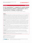
Figure 2 shows an example of successful in vivo
multi-contrast weighted MRI for detection of intraplaque hemorrhage. Table 3 lists technical details of
some studies conducted with respect to in vivo MRI
of human atherosclerotic plaque. The readily available histo-pathological data for comparison of MRI
images after carotid endarterectomy have made in
vivo MRI studies of carotid artery plaque attractive.
Lipid core has traditionally been visualized with T2
weighted spin echo techniques [30]. More recently,
T1 weighted fast spin echo and time-of-flight (TOF)
imaging have gained interest, because these sequences
may lead to better visualization and bright depiction
of lipid core [43, 46, 47]. Multi-contrast weighted
MRI at 3T showed nice depiction of large lipid cores
and an association was reported between large lipid
core and thin or ruptured fibrous cap [48], which is
another marker of plaque vulnerability.
Time-of-flight images were used to investigate the
state of the fibrous cap [49]. Not only thick fibrous
caps were distinguished from intact thin caps, but
Fig. 1 Printed with permission from Larose et al. [39].
Comparison of IVMRI and surface MRI. T1w surface MRI
(a) and IVMRI (b) of a common iliac artery in a subject with
an atheromatous plaque illustrates better image quality with
IVMRI compared with surface MRI. Both images were
produced by use of identical parameters (TR, 500 ms; TE,
13 ms; band width, 16 kHz; field of view, 9 cm; matrix,
256 9 256; no phase wrap) and in-plane resolution of 316 lm,
but the relative SNR is superior for IVMRI. However, in case
of a circumferential plaque the position of the catheter coil
determines the local signal intensity of a plaque component, so
homogeneity of the image SNR is impaired
123
Int J Cardiovasc Imaging (2010) 26:433–445
439
Fig. 2 Printed with permission from Cai et al. [44]. Example
of type VI lesion just distal to carotid bifurcation (acute and
subacute hemorrhages were detected by histology). On
multicontrast-weighted MR images, acute and subacute
hemorrhage had high SI on both TOF and T1WI images, isoSI to slightly high SI on PDWI and T2WI images (arrow).
* indicates lumen. Original magnification9 10
also intact caps could be distinguished from ruptured
ones with support of T1w, proton density (PDw) and
T2w images [49].
Intra-plaque hemorrhage (IPH) has also been
accepted as a marker of plaque vulnerability and is
caused by rupture of fragile neovasculature not
supported with firm connective tissue [50]. A followup MRI study in humans showed that hemorrhage into
the carotid atherosclerotic plaque accelerated plaque
progression in an 18 month period [50]. Erythrocyte
membranes contain more free cholesterol than any
other cell in the body and macrophages surrounding
the bleeding are activated and ingest more oxidized
LDL [50]. Classification of stages of IPH in carotid
arteries of patients scheduled for carotid endarterectomy was performed using multi-contrast weighted
MRI [45]. The classification showed moderate agreement with the classification into fresh, recent and old
categories according to histopathological criteria after
carotid endarterectomy. Another study reported successful discrimination between IPH and luminal
thrombus in human carotid artery lesions with four
different MR weightings [51].
The ability of a non-invasive imaging technique to
discriminate fresh from old luminal thrombus has
obvious clinical relevance. Definitely, recent thrombosis has prognostic implications with regard to
future acute coronary events [7]. Carotid thrombi
were induced in swine by arterial injury and could be
differentiated in recent and old thrombi by assessment of signal intensity on T2w at 1.5 T [52].
In recent years, various mouse models have been
developed in order to study the role of specific genes
in cardiovascular pathophysiology. Several transgenic and knockout models to study vascular biology
and atherosclerosis have been reported: e.g. apolipoprotein E deficient [53], apoE3-Leiden [54], lowdensity lipoprotein receptor deficient [26], and apoE/
eNOS double knockout mice [55].
Successful MRI of aortic plaque in mice at high
field may close the gap between successful in vivo
MRI of human carotid plaque and thus far unsuccessful coronary plaque MRI at low field, because of
similarity of size of human coronary artery and
mouse aorta and the advent of clinical high field
magnets. Wild-type mouse aortic wall thickness has
123
440
123
Table 3 In vivo MRI studies on atherosclerotic plaques: MRI parameters and appearance of plaque components
FA TE
(ms)
Appearance plaque components
Technique
Field
strength
Resolution: in
plane 9 slice
thickness
TR
(ms)
Rabbit/ Abdominal
human aorta/
carotid
artery
1.5 T
500 9 500 lm 9
4 mm
700 ms 90 12
No differentiation between plaque components
90 14
No differentiation between plaque components
90 14
Only peri-adventitial lipid visible
Toussaint
[30]
Human
Carotid
artery
1.5 T
SE T2w
390 9 390 lm 9
5 mm
1 RR
90 20/55
Hypo
Cai [43]
Human
Carotid
artery
1.5 T
DIR FSE PDw
500 9 500 lm 9
1/2 mm
3 RR
90 20
IsoDark Hyper NA
hyper
(Sub)acute: isohyper
DIR FSE T1w
800
90 9.3
Iso
(Sub)acute:
hyper
DIR FSE T2w
3 RR
90 40
IsoDark Iso
hyper
NA
(Sub)acute: isohyper
3D TOF
23
25 3.8
Iso
Dark Hypo
NA
(Sub)acute:
hyper
NA
Author
Vinitski
[28]
Species
Cappendijk Human
[40]
Imaged
vessel
Carotid
artery
SE T1w
CSI FS
CSI WS
1.5 T
FSE PDw
Dark Hyper NA
Hypo
90 20
Hypo
Dark Iso
570
90 14
Iso
Dark Hyper NA
Hyper
390 9 490 lm 9
3 mm
2 RR
90 30/50
Hypo
Dark Hyper NA
Iso
10.3
15 4.0
Hypo
Dark Iso
Hyper
Fresh hem.
Recent hem.
Old hem.
3–4
RR
90 20
Hypo/iso
Hyper
Hypo
FSE T1w
800
90 9.3
Hyper
Hyper
Hypo
FSE T2w
3–4
RR
90 40
Hypo/iso
Hyper
Hypo
3D TOF
23
25 3.5
Hyper
Hyper
Hypo
1.5 T
FSE PDw
625 9 625 lm 9
2 mm
NA
Iso
FA flip angle of excitation pulse, DIR double inversion recovery, EPI echo planar imaging, RR R-peak (ECG) to R-peak interval. For explanation of other abbreviations, see
Table 2
Int J Cardiovasc Imaging (2010) 26:433–445
Carotid
artery
Dark Hyper Fresh: hyper
organising: hypo
Haem.
2 RR
3D EPI T1w
with IR
Human
Calc SMC/ Thr
FT
390 9 390 lm 9
2.5 mm
DIR FSE T1w
FSE T2w
Chu [45]
LC
Author
Mouse strain
Target of imaging
Fayad [54]
ApoE-/-
Field
strength
MRI
technique
TR
(ms)
TE
(ms)
Resolution
9.4 T
SE PDw
2,000
13
97 9 97 9 500 lm/48 9
48 9 500 lm
SE T1w
1,000
13
97 9 97 9 500 lm/48 9
48 9 500 lm
Hist.: 0.300 ± 0.035
SE T2w
2,000
30
97 9 97 9 500/48 9
48 9 500 lm
r = 0.86
SE PDw
2,000
9
109 9 109 9 500 lm
Abdominal aorta,
iliac artery
Choudhury [57]
ApoE-/-, apoE-/-/
apoA-Ia
9.4 T
Correlation between MR and histology
Maximal wall
thickness ± SD, lm
Wall area, mm2
NA
MRI: 0.384 ± 0.046
NA
Int J Cardiovasc Imaging (2010) 26:433–445
Table 4 Mouse MRI studies on atherosclerotic vessel wall: MRI parameters and correlation with histology
MRI: 0.334b
Hist.: 0.126b
Abdominal aorta
r = 0.85
Itskovich [56]
ApoE-/-(/apoA-I)
9.4 T
SE PDw
2,000
9
MRI: 589 ± 164
MRI: 2.09 ± 1.04
SE T1w
500
9
Hist.: 486 ± 155
Hist.: 1.74 ± 0.92
SE T2w
SE T1w
2,000
1,000
30
10
49 9 98 9 300 lm
r [ 0.90
MRI: 238 ± 100
r [ 0.90
MRI: 1.19 ± 0.19
3D FLASH
4.6
1.5
100 9 100 9 39 lm
Hist.: ?
Hist.: 0.96 ± ?
3D FSE ± FS
800
13
140 9 187 9 187 lm
NA
Aortic root
Wiesmann [55]
ApoE-/-
7T
Thoracic aorta
156/78 9 156/78 9
300 lm
r = 0.97
Hockings [26]
LDLR-/-c
7T
Innominate artery
MRI: 0.14 ± 0.086
Hist.: 0.308 ± 0.081
r = 0.8
a
Human apolipoprotein A-I, Choudhury et al. [60]: total study group has a wide range of severity of atherosclerosis
b
Median value
c
Low-density lipoprotein receptor-deficient mice
For explanation of abbreviations, see Table 2
441
123
442
been reported to measure *50 lm at the abdominal
level [56] and *70 lm at the thoracic level [57].
MRI measurements of abdominal aortic wall thickness of ApoE deficient mice revealed doubled wall
thickness when compared to wild-type mice [56]. At
the thoracic and aortic root level the effect of the
gene defect on the aortic wall thickness was even
more conspicuous than at the abdominal level [57,
58]. Aortic wall area/thickness measurements in
atherosclerotic mouse models and correlation coefficients between MRI and histopathology obtained
from various mouse MR studies are listed in Table 4.
MRI showed that increased wall area of the abdominal aorta of apoE deficient mice was completely
compensated by outward remodeling, resulting in a
constant lumen, as could be verified with histopathology [59]. Aortic wall thickness was larger as
assessed by MRI than when assessed by histopathology, which probably is caused by shrinkage through
dehydration by the alcohol.
Imaging of the thoracic aorta in the mouse requires
a nontrivial effort, because of small size and cardiac
and respiratory motions. The heart in an awake mouse
beats 600/min [60, 61]. In vivo assessment of plaque
composition with MRI in the murine aorta has not
been achieved thus far. Nevertheless, wall thickness/
area and plaque area in the murine aortic root and
brachiocephalic artery have been measured with MRI
and compared with histopathology [26, 58]. A
3-dimensional MR technique was used, which offers
the advantage to reconstruct an image in any chosen
orientation after the measurements have taken place
[26]. A very high correlation between MRI measurements and histopathology measurements of aortic wall
area was demonstrated [57]. Probably, the smaller
difference between MRI and histopathology measurements of wall area in the latter study when compared
to Choudhury et al. [59] can be explained by the
smaller pixel dimensions of the images.
In vivo imaging of human coronary arteries
requires cardiac and respiratory gating. Prospective
gating increases measurement time, however retrospective gating and breath-hold techniques increase
time efficiency tremendously. For use as a clinical
screening tool, which scans the whole coronary artery
tree, a sophisticated tracking strategy is required, to
compensate for the motion and tortuosity of the
coronary arteries [62]. Wall thickness and remodelling of coronary arteries have been studied in vivo
123
Int J Cardiovasc Imaging (2010) 26:433–445
with MRI [63, 64]. Until now it has not been possible
to identify different plaque constituents in the coronary arteries in vivo because of earlier mentioned
problems.
Several research groups are involved in the solution
of technical problems, while others are involved in
contrast-enhanced MRI, which allows lower SNR
because of increased contrast between targets of
interest and their background. Equally important, the
possibility to use vehicles carrying not only (super)
paramagnetic agents but also antibodies, peptides or
receptor agonists, provides a technique which is
capable of targeting vulnerable plaque markers which
are more specific and/or sensitive for prediction of
plaque disruption than the classical morphologic
features.
References
1. Fayad ZA, Fuster V (2001) Clinical imaging of the highrisk or vulnerable atherosclerotic plaque. Circ Res 89:
305–316
2. Badimon L (2001) Atherosclerosis and thrombosis: lessons
from animal models. Thromb Haemost 86:356–365
3. Ross R (1999) Atherosclerosis is an inflammatory disease.
Am Heart J 138:S419–S420
4. Mitsumori LM, Hatsukami TS, Ferguson MS, Kerwin WS,
Cai J, Yuan C (2003) In vivo accuracy of multisequence
MR imaging for identifying unstable fibrous caps in
advanced human carotid plaques. J Magn Reson Imaging
17:410–420
5. Leiner T, Gerretsen S, Botnar R, Lutgens E, Cappendijk V,
Kooi E, van EJ (2005) Magnetic resonance imaging of
atherosclerosis. Eur Radiol 15:1087–1099
6. Vink A, Pasterkamp G (2002) Atherosclerotic plaque
burden, plaque vulnerability and arterial remodeling: the
role of inflammation. Minerva Cardioangiol 50:75–83
7. Corti R, Fuster V (2003) New understanding, diagnosis,
and prognosis of atherothrombosis and the role of imaging.
Am J Cardiol 91:17A–26A
8. Ambrose JA, Tannenbaum MA, Alexopoulos D,
Hjemdahl-Monsen CE, Leavy J, Weiss M, Borrico S,
Gorlin R, Fuster V (1988) Angiographic progression of
coronary artery disease and the development of myocardial
infarction. J Am Coll Cardiol 12:56–62
9. de Korte CL, Pasterkamp G, van der Steen AF, Woutman
HA, Bom N (2000) Characterization of plaque components
with intravascular ultrasound elastography in human femoral and coronary arteries in vitro. Circulation 102:617–623
10. Jang IK, Tearney GJ, MacNeill B, Takano M, Moselewski
F, Iftima N, Shishkov M, Houser S, Aretz HT, Halpern EF,
Bouma BE (2005) In vivo characterization of coronary
atherosclerotic plaque by use of optical coherence
tomography. Circulation 111:1551–1555
Int J Cardiovasc Imaging (2010) 26:433–445
11. van de Poll SW, Kastelijn K, Bakker Schut TC, Strijder C,
Pasterkamp G, Puppels GJ, van der LA (2003) On-line
detection of cholesterol and calcification by catheter based
Raman spectroscopy in human atherosclerotic plaque ex
vivo. Heart 89:1078–1082
12. Wang J, Geng YJ, Guo B, Klima T, Lal BN, Willerson JT,
Casscells W (2002) Near-infrared spectroscopic characterization of human advanced atherosclerotic plaques.
J Am Coll Cardiol 39:1305–1313
13. Toutouzas K, Drakopoulou M, Mitropoulos J, Tsiamis E,
Vaina S, Vavuranakis M, Markou V, Bosinakou E,
Stefanadis C (2006) Elevated plaque temperature in nonculprit de novo atheromatous lesions of patients with acute
coronary syndromes. J Am Coll Cardiol 47:301–306
14. Waki H, Masuyama T, Mori H, Maeda T, Kitade K,
Moriyasu K, Tsujimoto M, Fujimoto K, Koshimae N,
Matsuura N (2003) Ultrasonic tissue characterization of the
atherosclerotic carotid artery: histological correlates or
carotid integrated backscatter. Circ J 67:1013–1016
15. Leber AW, Knez A, von ZF, Becker A, Nikolaou K, Paul
S, Wintersperger B, Reiser M, Becker CR, Steinbeck G,
Boekstegers P (2005) Quantification of obstructive and
nonobstructive coronary lesions by 64-slice computed
tomography: a comparative study with quantitative coronary angiography and intravascular ultrasound. J Am Coll
Cardiol 46:147–154
16. Ben-Haim S, Israel O (2006) PET/CT for atherosclerotic
plaque imaging. J Nucl Med Mol Imaging 50:53–60
17. Choudhury RP, Fisher EA (2009) Molecular imaging in
atherosclerosis, thrombosis, and vascular inflammation.
Arterioscler Thromb Vasc Biol 29:983–991
18. Thompson BH, Stanford W (2005) Update on using coronary calcium screening by computed tomography to
measure risk for coronary heart disease. Int J Cardiovasc
Imaging 21:39–53
19. Dey D, Slomka P, Chien D, Fieno D, Abidov A, Saouaf R,
Thomson L, Friedman JD, Berman DS (2006) Direct
quantitative in vivo comparison of calcified atherosclerotic
plaque on vascular MRI and CT by multimodality image
registration. J Magn Reson Imaging 23:345–354
20. Viles-Gonzalez JF, Poon M, Sanz J, Rius T, Nikolaou K,
Fayad ZA, Fuster V, Badimon JJ (2004) In vivo 16-slice,
multidetector-row computed tomography for the assessment of experimental atherosclerosis: comparison with
magnetic resonance imaging and histopathology. Circulation 110:1467–1472
21. Leber AW, Becker A, Knez A, Von ZF, Sirol M, Nikolaou K,
Ohnesorge B, Fayad ZA, Becker CR, Reiser M, Steinbeck G,
Boekstegers P (2006) Accuracy of 64-slice computed
tomography to classify and quantify plaque volumes in the
proximal coronary system: a comparative study using
intravascular ultrasound. J Am Coll Cardiol 47:672–677
22. Kolodgie FD, Petrov A, Virmani R, Narula N, Verjans JW,
Weber DK, Hartung D, Steinmetz N, Vanderheyden JL,
Vannan MA, Gold HK, Reutelingsperger CP, Hofstra L,
Narula J (2003) Targeting of apoptotic macrophages and
experimental atheroma with radiolabeled annexin V: a
technique with potential for noninvasive imaging of vulnerable plaque. Circulation 108:3134–3139
23. Tsimikas S, Shortal BP, Witztum JL, Palinski W (2000) In
vivo uptake of radiolabeled MDA2, an oxidation-specific
443
24.
25.
26.
27.
28.
29.
30.
31.
32.
33.
34.
35.
monoclonal antibody, provides an accurate measure of
atherosclerotic lesions rich in oxidized LDL and is highly
sensitive to their regression. Arterioscler Thromb Vasc
Biol 20:689–697
Rudd JH, Myers KS, Bansilal S, Machac J, Pinto CA, Tong
C, Rafique A, Hargeaves R, Farkouh M, Fuster V, Fayad
ZA (2008) Atherosclerosis inflammation imaging with
18F-FDG PET: carotid, iliac, and femoral uptake reproducibility, quantification methods, and recommendations.
J Nucl Med 49:871–878
Massoud TF, Gambhir SS (2003) Molecular imaging in
living subjects: seeing fundamental biological processes in
a new light. Genes Dev 17:545–580
Hockings PD, Roberts T, Galloway GJ, Reid DG, Harris
DA, Vidgeon-Hart M, Groot PH, Suckling KE, Benson
GM (2002) Repeated three-dimensional magnetic resonance imaging of atherosclerosis development in innominate arteries of low-density lipoprotein receptor-knockout
mice. Circulation 106:1716–1721
Booth RF, Honey AC, Martin JF, Lindon JC, Farrant RD,
Carpenter TA, Hall LD (1990) Lipid characterization in an
animal model of atherosclerosis using NMR spectroscopy
and imaging. NMR Biomed 3:95–100
Vinitski S, Consigny PM, Shapiro MJ, Janes N, Smullens
SN, Rifkin MD (1991) Magnetic resonance chemical shift
imaging and spectroscopy of atherosclerotic plaque. Invest
Radiol 26:703–714
Toussaint JF, Southern JF, Fuster V, Kantor HL (1995)
T2-weighted contrast for NMR characterization of human
atherosclerosis. Arterioscler Thromb Vasc Biol 15:1533–
1542
Toussaint JF, LaMuraglia GM, Southern JF, Fuster V,
Kantor HL (1996) Magnetic resonance images lipid,
fibrous, calcified, hemorrhagic, and thrombotic components of human atherosclerosis in vivo. Circulation
94:932–938
Shinnar M, Fallon JT, Wehrli S, Levin M, Dalmacy D,
Fayad ZA, Badimon JJ, Harrington M, Harrington E,
Fuster V (1999) The diagnostic accuracy of ex vivo
MRI for human atherosclerotic plaque characterization.
Arterioscler Thromb Vasc Biol 19:2756–2761
Toussaint JF, Southern JF, Fuster V, Kantor HL (1997)
Water diffusion properties of human atherosclerosis and
thrombosis measured by pulse field gradient nuclear
magnetic resonance. Arterioscler Thromb Vasc Biol
17:542–546
Rogers WJ, Prichard JW, Hu YL, Olson PR, Benckart DH,
Kramer CM, Vido DA, Reichek N (2000) Characterization
of signal properties in atherosclerotic plaque components
by intravascular MRI. Arterioscler Thromb Vasc Biol
20:1824–1830
Schneider JE, McAteer MA, Tyler DJ, Clarke K, Channon
KM, Choudhury RP, Neubauer S (2004) High-resolution,
multicontrast three-dimensional-MRI characterizes atherosclerotic plaque composition in ApoE-/- mice ex vivo.
J Magn Reson Imaging 20:981–989
Schneiderman J, Wilensky RL, Weiss A, Samouha E,
Muchnik L, Chen-Zion M, Ilovitch M, Golan E, Blank A,
Flugelman M, Rozenman Y, Virmani R (2005) Diagnosis
of thin-cap fibroatheromas by a self-contained intravascular magnetic resonance imaging probe in ex vivo human
123
444
36.
37.
38.
39.
40.
41.
42.
43.
44.
45.
46.
Int J Cardiovasc Imaging (2010) 26:433–445
aortas and in situ coronary arteries. J Am Coll Cardiol
45:1961–1969
MacNeill BD, Lowe HC, Takano M, Fuster V, Jang IK
(2003) Intravascular modalities for detection of vulnerable
plaque: current status. Arterioscler Thromb Vasc Biol 23:
1333–1342
Correia LC, Atalar E, Kelemen MD, Ocali O, Hutchins
GM, Fleg JL, Gerstenblith G, Zerhouni EA, Lima JA
(1997) Intravascular magnetic resonance imaging of aortic
atherosclerotic plaque composition. Arterioscler Thromb
Vasc Biol 17:3626–3632
Larose E, Yeghiazarians Y, Libby P, Yucel EK, Aikawa
M, Kacher DF, Aikawa E, Kinlay S, Schoen FJ, Selwyn
AP, Ganz P (2005) Characterization of human atherosclerotic plaques by intravascular magnetic resonance
imaging. Circulation 112:2324–2331
Larose E, Kinlay S, Selwyn AP, Yeghiazarians Y, Yucel
EK, Kacher DF, Libby P, Ganz P (2008) Improved characterization of atherosclerotic plaques by gadolinium
contrast during intravascular magnetic resonance imaging
of human arteries. Atherosclerosis 196:919–925
Cappendijk VC, Cleutjens KB, Kessels AG, Heeneman S,
Schurink GW, Welten RJ, Mess WH, Daemen MJ, van
Engelshoven JM, Kooi ME (2005) Assessment of human
atherosclerotic carotid plaque components with multisequence MR imaging: initial experience. Radiology 234:
487–492
Cappendijk VC, Heeneman S, Kessels AG, Cleutjens KB,
Schurink GW, Welten RJ, Mess WH, van Suylen RJ,
Leiner T, Daemen MJ, van Engelshoven JM, Kooi ME
(2008) Comparison of single-sequence T1w TFE MRI with
multisequence MRI for the quantification of lipid-rich
necrotic core in atherosclerotic plaque. J Magn Reson
Imaging 27:1347–1355
Luo Y, Polissar N, Han C, Yarnykh V, Kerwin WS,
Hatsukami TS, Yuan C (2003) Accuracy and uniqueness of
three in vivo measurements of atherosclerotic carotid plaque morphology with black blood MRI. Magn Reson Med
50:75–82
Cai JM, Hatsukami TS, Ferguson MS, Small R, Polissar
NL, Yuan C (2002) Classification of human carotid atherosclerotic lesions with in vivo multicontrast magnetic
resonance imaging. Circulation 106:1368–1373
Cai J, Hatsukami TS, Ferguson MS, Kerwin WS, Saam T,
Chu B, Takaya N, Polissar NL, Yuan C (2005) In vivo
quantitative measurement of intact fibrous cap and lipidrich necrotic core size in atherosclerotic carotid plaque:
comparison of high-resolution, contrast-enhanced magnetic resonance imaging and histology. Circulation 112:
3437–3444
Chu B, Kampschulte A, Ferguson MS, Kerwin WS,
Yarnykh VL, O’Brien KD, Polissar NL, Hatsukami TS,
Yuan C (2004) Hemorrhage in the atherosclerotic carotid
plaque: a high-resolution MRI study. Stroke 35:1079–1084
Yuan C, Mitsumori LM, Ferguson MS, Polissar NL,
Echelard D, Ortiz G, Small R, Davies JW, Kerwin WS,
Hatsukami TS (2001) In vivo accuracy of multispectral
magnetic resonance imaging for identifying lipid-rich
necrotic cores and intraplaque hemorrhage in advanced
human carotid plaques. Circulation 104:2051–2056
123
47. Saam T, Hatsukami TS, Takaya N, Chu B, Underhill H,
Kerwin WS, Cai J, Ferguson MS, Yuan C (2007) The
vulnerable, or high-risk, atherosclerotic plaque: noninvasive MR imaging for characterization and assessment.
Radiology 244:64–77
48. Ota H, Yu W, Underhill HR, Oikawa M, Dong L, Zhao X,
Polissar NL, Neradilek B, Gao T, Zhang Z, Yan Z, Guo M,
Zhang Z, Hatsukami TS, Yuan C (2009) Hemorrhage and
large lipid-rich necrotic cores are independently associated
with thin or ruptured fibrous caps: an in vivo 3T MRI
study. Arterioscl Thromb Vasc Biol 29:1696–1701
49. Yuan C, Zhang SX, Polissar NL, Echelard D, Ortiz G,
Davis JW, Ellington E, Ferguson MS, Hatsukami TS
(2002) Identification of fibrous cap rupture with magnetic
resonance imaging is highly associated with recent transient ischemic attack or stroke. Circulation 105:181–185
50. Takaya N, Yuan C, Chu B, Saam T, Polissar NL, Jarvik
GP, Isaac C, McDonough J, Natiello C, Small R, Ferguson
MS, Hatsukami TS (2005) Presence of intraplaque hemorrhage stimulates progression of carotid atherosclerotic
plaques: a high-resolution magnetic resonance imaging
study. Circulation 111:2768–2775
51. Kampschulte A, Ferguson MS, Kerwin WS, Polissar NL,
Chu B, Saam T, Hatsukami TS, Yuan C (2004) Differentiation of intraplaque versus juxtaluminal hemorrhage/
thrombus in advanced human carotid atherosclerotic
lesions by in vivo magnetic resonance imaging. Circulation
110:3239–3244
52. Corti R, Osende JI, Fayad ZA, Fallon JT, Fuster V, Mizsei
G, Dickstein E, Drayer B, Badimon JJ (2002) In vivo
noninvasive detection and age definition of arterial
thrombus by MRI. J Am Coll Cardiol 39:1366–1373
53. Zhang SH, Reddick RL, Piedrahita JA, Maeda N (1992)
Spontaneous hypercholesterolemia and arterial lesions in
mice lacking apolipoprotein E. Science 258:468–471
54. Lutgens E, Daemen M, Kockx M, Doevendans P, Hofker
M, Havekes L, Wellens H, de Muinck ED (1999) Atherosclerosis in APOE*3-Leiden transgenic mice: from proliferative to atheromatous stage. Circulation 99:276–283
55. Kuhlencordt PJ, Gyurko R, Han F, Scherrer-Crosbie M,
Aretz TH, Hajjar R, Picard MH, Huang PL (2001)
Accelerated atherosclerosis, aortic aneurysm formation,
and ischemic heart disease in apolipoprotein E/endothelial
nitric oxide synthase double-knockout mice. Circulation
104:448–454
56. Fayad ZA, Fallon JT, Shinnar M, Wehrli S, Dansky HM,
Poon M, Badimon JJ, Charlton SA, Fisher EA, Breslow JL,
Fuster V (1998) Noninvasive In vivo high-resolution
magnetic resonance imaging of atherosclerotic lesions in
genetically engineered mice. Circulation 98:1541–1547
57. Wiesmann F, Szimtenings M, Frydrychowicz A, Illinger R,
Hunecke A, Rommel E, Neubauer S, Haase A (2003) Highresolution MRI with cardiac and respiratory gating allows
for accurate in vivo atherosclerotic plaque visualization in
the murine aortic arch. Magn Reson Med 50:69–74
58. Itskovich VV, Choudhury RP, Aguinaldo JG, Fallon JT,
Omerhodzic S, Fisher EA, Fayad ZA (2003) Characterization of aortic root atherosclerosis in ApoE knockout
mice: high-resolution in vivo and ex vivo MRM with
histological correlation. Magn Reson Med 49:381–385
Int J Cardiovasc Imaging (2010) 26:433–445
59. Choudhury RP, Aguinaldo JG, Rong JX, Kulak JL, Kulak
AR, Reis ED, Fallon JT, Fuster V, Fisher EA, Fayad ZA
(2002) Atherosclerotic lesions in genetically modified
mice quantified in vivo by non-invasive high-resolution
magnetic resonance microscopy. Atherosclerosis 162:
315–321
60. Weiss RG (2001) Imaging the murine cardiovascular system with magnetic resonance. Circ Res 88:550–551
61. Wehrens XH, Kirchhoff S, Doevendans PA (2000) Mouse
electrocardiography: an interval of 30 years. Cardiovasc
Res 45:231–237
62. Wendt M, Wacker FK (2000) Visualization, tracking, and
navigation of instruments for magnetic resonance imagingguided endovascular procedures. Top Magn Reson Imaging 11:163–172
63. Kim WY, Stuber M, Bornert P, Kissinger KV, Manning
WJ, Botnar RM (2002) Three-dimensional black-blood
cardiac magnetic resonance coronary vessel wall imaging
detects positive arterial remodeling in patients with
445
nonsignificant coronary artery disease. Circulation 106:
296–299
64. Miao C, Chen S, Macedo R, Lai S, Liu K, Li D, Wasserman BA, Vogel-Claussen J, Lima JA, Bluemke DA (2009)
Positive remodeling of the coronary arteries detected by
magnetic resonance imaging in an asymptomatic population: MESA (Multi-Ethnic Study of Atherosclerosis). J Am
Coll Cardiol 53:1708–1715
65. Itskovich VV, Samber DD, Mani V, Aguinaldo JG, Fallon
JT, Tang CY, Fuster V, Fayad ZA (2004) Quantification of
human atherosclerotic plaques using spatially enhanced
cluster analysis of multicontrast-weighted magnetic resonance images. Magn Reson Med 52:515–523
66. Worthley SG, Helft G, Fuster V, Fayad ZA, Fallon JT,
Osende JI, Roqué M, Shinnar M, Zaman AG, Rodriguez
OJ, Verhallen P, Badimon JJ (2000) High resolution ex
vivo magnetic resonance imaging of in situ coronary and
aortic atherosclerotic plaque in a porcine model. Atherosclerosis 150:321–329
123
Academia.edu no longer supports Internet Explorer.
To browse Academia.edu and the wider internet faster and more securely, please take a few seconds to upgrade your browser.
Recent developments and new perspectives on imaging of atherosclerotic plaque: role of anatomical, cellular and molecular MRI Part I and II
The International Journal of Cardiovascular Imaging, 2010
...Read more
Related Papers
Journal of Cardiovascular Magnetic Resonance, 2009
Download

Magnetic Resonance in Medicine, 2012
Download

Current atherosclerosis reports, 2002
Download

Journal of Cardiovascular Magnetic Resonance, 2005
Download

Journal of cardiovascular magnetic resonance : official journal of the Society for Cardiovascular Magnetic Resonance, 2013
Download
European Heart Journal, 2012
Download

Atherosclerosis, 2008
Download

Magnetic Resonance Imaging, 2004
Download

Arteriosclerosis, thrombosis, and vascular biology, 2005
Download

NMR in Biomedicine, 2006
Download

Magnetic Resonance Imaging, 2001
Download

Circulation, 2005
Download

Journal of Cardiovascular Magnetic Resonance, 2013
Download

Arteriosclerosis, Thrombosis, and Vascular Biology, 2004
Download

