Academia.edu no longer supports Internet Explorer.
To browse Academia.edu and the wider internet faster and more securely, please take a few seconds to upgrade your browser.
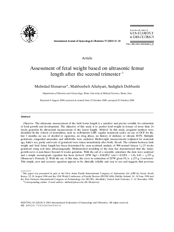
Assessment of fetal weight based on ultrasonic femur length after the second trimester
Assessment of fetal weight based on ultrasonic femur length after the second trimester
2001, International Journal of Gynecology & Obstetrics
Related Papers
Ultrasound in Medicine & Biology
Prediction of birth weight by ultrasound in Turkish population. Which formula should be used in Turkey to estimate fetal weight?2005 •
To determine optimal sonographic fetal weight estimation formula for male and female infants, a prospective study has been performed. Uncomplicated pregnancies and 465 newborns were evaluated. Measurements included birth weight, length and head circumference in addition to fetal head circumference, femur length, abdominal circumference and biparietal diameter. Actual weights were compared with estimated weights calculated by ten different formula. Estimated fetal weight obtained from all formula, except those of Merz, Warsof and Ferrero, tended to be lower than the measured birth weight. The smallest mean differences were obtained with Hadlock 1, Hadlock 2, Hadlock 4 and Shepard formula (19 g-85 g), whereas Merz and Woo produced largest mean differences (110 g-364 g). Intraclass correlation coefficients (ICCs) generated with Hadlock 1 and Hadlock 2 formula were identical (0.90). ICCs obtained with Hadlock 3 and Hadlock 4, Shepard, Merz, Warsof and Campbell formula varied between 0.84 and 0.88. Hadlock 1 and 2 formula gave the closest approximation of birth weight in Turkish population.
International Journal of Gynecology & Obstetrics
Assessment of gestational age based on ultrasonic femur length after the first trimester: a simple mathematical correlation between gestational age (GA) and femur length (FL)2000 •
Clinical and Experimental Obstetrics & Gynecology
Finding the best formulas to estimate fetal weight based on ultrasound for the Turkish population: a comparison of 24 formulas2021 •
American Journal of Obstetrics and Gynecology
Estimation of birth weight by use of ultrasonographic formulas targeted to large-, appropriate-, and small-for-gestational-age fetuses1989 •
Ultrasound in Obstetrics & Gynecology
A new mathematical formula for predicting long bone length in early pregnancy2002 •
ObjectiveTo propose new mathematical formulae to estimate fetal long bone biometry in early pregnancy and to establish their efficacy in comparison to previously constructed mathematical formulae.To propose new mathematical formulae to estimate fetal long bone biometry in early pregnancy and to establish their efficacy in comparison to previously constructed mathematical formulae.MethodsA study population of 1960 singleton euploid fetuses was referred for transvaginal ultrasound examinations between 71 and 112 days of gestation prior to genetic amniocentesis. To determine the relationship between the biparietal diameter and long bone length, a sample group of 400 randomly chosen normal fetuses was evaluated. Regression equations were derived, then tested in the remaining 1560 control fetuses and compared with previously reported mathematical formulae by other authors. Mean absolute error, mean absolute percentage error and mean systematic error with their standard deviations were calculated.A study population of 1960 singleton euploid fetuses was referred for transvaginal ultrasound examinations between 71 and 112 days of gestation prior to genetic amniocentesis. To determine the relationship between the biparietal diameter and long bone length, a sample group of 400 randomly chosen normal fetuses was evaluated. Regression equations were derived, then tested in the remaining 1560 control fetuses and compared with previously reported mathematical formulae by other authors. Mean absolute error, mean absolute percentage error and mean systematic error with their standard deviations were calculated.ResultsThe relationships between femur or humerus length vs. biparietal diameter (BPD) and gestational age (GA) were, respectively: expected femur length = –16.92108 + 0.4569402 × BPD + 0.171617 × GA (P < 0.001) and expected humerus length = –16.28531 + 0.4283019 × BPD + 0.1696017 × GA (P < 0.001). The confidence intervals of the predicted values for different values of biparietal diameter and gestational age and confidence intervals for the regression coefficients, such as the distribution of the residuals, are given. All previous formulae obtained by transabdominal ultrasound demonstrated an overestimation of expected long bones measurements; this was reduced using different formulae obtained in early pregnancy. Using our mathematical formulae, the mean absolute percentage error and the mean systematic error in estimating femur and humerus length were very low (11.15% and −2.02%; 10.59% and –1.74%, respectively).The relationships between femur or humerus length vs. biparietal diameter (BPD) and gestational age (GA) were, respectively: expected femur length = –16.92108 + 0.4569402 × BPD + 0.171617 × GA (P < 0.001) and expected humerus length = –16.28531 + 0.4283019 × BPD + 0.1696017 × GA (P < 0.001). The confidence intervals of the predicted values for different values of biparietal diameter and gestational age and confidence intervals for the regression coefficients, such as the distribution of the residuals, are given. All previous formulae obtained by transabdominal ultrasound demonstrated an overestimation of expected long bones measurements; this was reduced using different formulae obtained in early pregnancy. Using our mathematical formulae, the mean absolute percentage error and the mean systematic error in estimating femur and humerus length were very low (11.15% and −2.02%; 10.59% and –1.74%, respectively).ConclusionsThe new ultrasonographic morphometric models derived from transvaginal measurements in early pregnancy show a good reliability in estimating fetal long bone length. Copyright © 2002 ISUOGThe new ultrasonographic morphometric models derived from transvaginal measurements in early pregnancy show a good reliability in estimating fetal long bone length. Copyright © 2002 ISUOG
International Journal of Gynecology & Obstetrics
A simple estimated fetal weight equation for fetuses between 24 and 34 weeks of gestation1999 •
International Journal of Reproduction, Contraception, Obstetrics and Gynecology
Comparative study between Johnson’s formula and Dare’s formula of fetal weight estimation at term2021 •
Background: Prediction of fetal weight is one of the methods towards effective management of pregnancy and delivery. To assess and compare the accuracy of clinical and sonographic fetal weight estimation in predicting birth weight at term pregnancy, patients who were in latent or in active phase of labour. In the present study, an effort is made to compare two different clinical methods and USG and relate to the actual weight of the baby at birth.Methods: It is a prospective observational study of one hundred pregnant women satisfying the criteria, consenting for the study was recruited. Both USG and clinical methods will be done and compared with estimated the fetal weight. Weight of the baby at birth will be measured.Results: All the three methods had significant relationship with the baby weight. Percentage error was least with USG and the standard deviation of error was lower with Dare’s formula. The standard deviation was minimal for Dare`s formula EFW followed closely by USG.C...
Journal of Ultrasound in Medicine
New formula for estimating fetal weight below 1000 g: comparison with existing formulas1996 •
Integrative Psychological and Behavioral Science
Mindfulness, Phenomenology, and Psychological Science2024 •
Most present-day research on mindfulness treats mindfulness as a variable that is studied in relation to other variables. Although this research may provide us with important knowledge at the population level and mechanism level, it contributes little to our understanding of the phenomenon of mindfulness as it is experienced and enacted at the person level. The present paper takes a person-oriented phenomenological perspective on mindfulness, comparing this perspective with that of von Fircks' (2023). In a first part of the paper, mindfulness is discussed as a phenomenological practice that can be studied by means of experimental phenomenology. It is argued that there is room for the development of an immense variety of personalized mindfulness practices that may serve people's health and well-being. The second part of the paper contains a brief discussion of the possible role of mindful observation and reflection in psychological research. It is argued that mindfulness skills may be important both for improving the quality of phenomenological observation and to facilitate creative thinking in connection with the development of psychological theory. A main implication is that an integration between mindfulness and phenomenology may serve as an important part of this process.
2021 •
Qual imagem faz o juiz italiano do legislador, esse personagem tantas vezes nomeado e tantas vezes evocado ao longo de uma sentença, e em cujo espírito ele busca penetrar, como quer o art. 12 das Disposições sobre a Lei em Geral? A partir de uma pesquisa sobre a argumentação do juiz italiano da Corte de Cassação, busquei traçar em linhas gerais o retrato desse personagem, esforçando-me em determinar os atributos que se lhe reconhecem com mais frequência e que ele deve ter para ser considerado um bom legislador. [...]
RELATED PAPERS
L'immagine sovrana. Urbano VIII e i Barberini, eds. M. Cicconi, F. Gennari Santori, S. Schütze, Roma, Officina Libraria
Di Monte, La scienza moderna e lo specchio del sapere2023 •
Tidsskrift for Den norske legeforening
Behandling av hjerteinfarkt med ST-elevasjon – en observasjonsstudieInnovacion Educativa
Educación media superior, jóvenes y desigualdad de oportunidades2014 •
2023 •
Injury Prevention
Two year active surveillance of injuries in an industrially developing commune in Vietnam2012 •
Social Science Research Network
Why Didn't the Global Financial Crisis Hit Latin America?2011 •
Journal of Manufacturing and Materials Processing
Hydrophobic Material: Effect of Alkyl Chain Length on the Surface RoughnessProceedings of the National Academy of Sciences
Mobile hydrogen carbonate acts as proton acceptor in photosynthetic water oxidation2014 •

 Sedigheh Dehbashi