Academia.edu no longer supports Internet Explorer.
To browse Academia.edu and the wider internet faster and more securely, please take a few seconds to upgrade your browser.
Influence of Ruthenium doping on Structural and Morphological Properties of MoO 3 Thin Films
Influence of Ruthenium doping on Structural and Morphological Properties of MoO 3 Thin Films
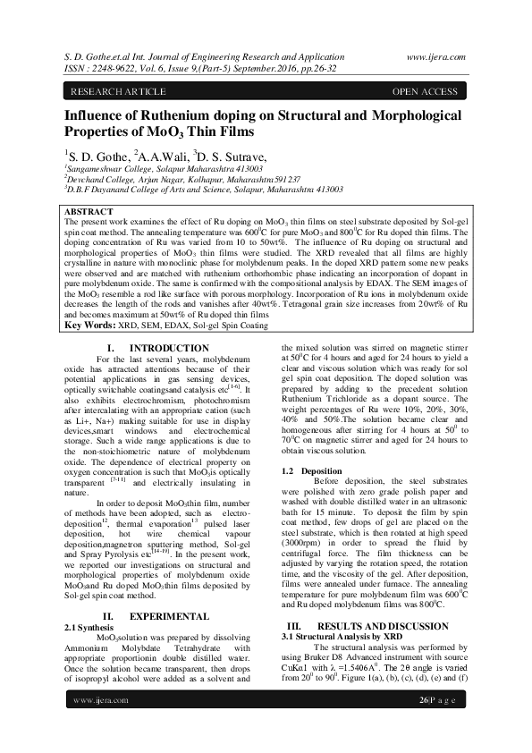
The present work examines the effect of Ru doping on MoO 3 thin films on steel substrate deposited by Sol-gel spin coat method. The annealing temperature was 600 0 C for pure MoO 3 and 800 0 C for Ru doped thin films. The doping concentration of Ru was varied from 10 to 50wt%. The influence of Ru doping on structural and morphological properties of MoO 3 thin films were studied. The XRD revealed that all films are highly crystalline in nature with monoclinic phase for molybdenum peaks. In the doped XRD pattern some new peaks were observed and are matched with ruthenium orthorhombic phase indicating an incorporation of dopant in pure molybdenum oxide. The same is confirmed with the compositional analysis by EDAX. The SEM images of the MoO 3 resemble a rod like surface with porous morphology. Incorporation of Ru ions in molybdenum oxide decreases the length of the rods and vanishes after 40wt%. Tetragonal grain size increases from 20wt% of Ru and becomes maximum at 50wt% of Ru doped thin films
Related Papers
There is a growing necessity of transition metal oxide thin film for many important technological applications such as smart windows gas sensors, solar cells, super capacitors etc. Among the other transition metal oxides, ruthenium oxide is a potential material as it exhibits interesting structural, optical, chemical, electrical properties. In this investigation ruthenium oxide thin films have been synthesized using spin coating technique. Here ruthenium oxide thin films have been deposited on stainless steel substrate by sol-gel spin coating method. Thin film properties of deposited samples were studied by XRD, SEM, FTIR, and EDAX.AFM. The XRD pattern showed sharp intense peaks conform crystalline structure and indicating tetragonal structure of ruthenium oxide. The SEM images of ruthenium oxide thin films showed total coverage of thin films. The thin film had a dense layer covered by agglomeration of particles forming a porous structure. At higher magnification (X 40,000) a porous structure of ruthenium oxide
2018 •
T he molybdenum trioxide MoO 3 thin films were growth on c-Si substrates by magnetron sputtering technique. The structural and morphological properties of MoO 3 thin films were investigated by XRD, RAMAN and SEM analysis. Prior to annealing process, X-ray diffractogram indicated that MoO 3 thin films vere amorphous nature. All of the MoO 3 thin films were applied three different annealing temperature and obtained optimum annealing temperature with 300 0 C. XRD patterns of annealed thin films showed that these MoO 3 thin films have polycrystalline nature with 2 θ peak at 12 ˚ ,23 ˚ , 25 ˚ , 38 ˚ , 55 ˚ and 58 ˚ corresponding to the (020), (110), (040), (060), (112) and (081) planes . RAMAN spectrum of the MoO 3 thin films were determined 1 4 Raman active peaks belong to α -phase MoO 3 . The surface morphology of the MoO 3 thin films as deposited has appeared to be uniform with smaller grains and exhibits a coarse structure. Annealing of the MoO 3 thin films favors growth and agglome...
Journal of Nanoscience and Nanotechnology
Effect of the Growth Temperature and Oxygen Flow Rate on the Properties of MoO3 Thin Films2017 •
Journal of The …
Structural and Optical Properties of Nanocrystalline Er2O3 Thin Films Deposited by a Versatile Low-Pressure MOCVD Approach2008 •
Highly conductive ruthenium metal thin films and ruthenium oxide ones were prepared by a solution process at low temperature (e.g., 6.9 × 10⁻⁵ Ω cm at 300°C for Ru0). Their structure and electric properties depend on the annealing conditions. The process allowed us to fabricate ruthenium electrodes on flexible substrates.
Journal of Vacuum Science & Technology A
MoO3 films grown on polycrystalline Cu: Morphological, structural, and electronic properties2019 •
Academia Engineering
Assessing air pollution emissions vs. abatement costs in agricultural practices2023 •
Agriculture is a vital component of human civilization, providing food, fiber, and fuel for billions of people worldwide. However, the agricultural sector has also been identified as a significant contributor to air pollution. This study aims to comprehensively investigate and analyze the impact of agrofarming activities on air pollution in a very productive area such as Northern Italy. It explores the various sources and mechanisms through which agriculture affects air quality, and the types of pollutants involved, and quantifies the consequences for human health, ecosystems, and the environment. Furthermore, adopting an integrated assessment modelling approach, it highlights the technologies that can mitigate these negative impacts and promote sustainable agriculture. The paper defines policy recommendations for the area at hand analysing the optimal compromise between air quality improvement and pollution abatement costs. It concludes with an outlook of additional options for addressing the air pollution challenges associated with agrofarming activities.
LuanVanCaoHoc.Com
Huong dan su dung phan mem SmartPLS Full Crack Co Ban2020 •
Hướng dẫn sử dụng phần mềm SmartPLS Full Crack 3.2.9 - Sách In Tiếng Việt Cơ Bản - Giá 150.000đ Liên hệ: https://luanvancaohoc.com/smartpls.html
RELATED PAPERS
International Journal of Applied Management Sciences and Engineering
Real Estate Marketing and Factors Impacting Real Estate Purchasing2019 •
Zenodo (CERN European Organization for Nuclear Research)
A 56 Gbaud Reconfigurable FPGA Feed-Forward Equalizer for Optical Datacenter Networks with flexible Baudrate- and Modulation-Format2017 •
SSRN Electronic Journal
Restructuring Security in Africa Beyond the Millennium: Al- Shabaab Menace in Kenya2015 •
Physics Letters B
Measurement of prompt D0 and D‾0 meson azimuthal anisotropy and search for strong electric fields in PbPb collisions at sNN=5.02TeV2021 •
Filosofija. Sociologija
Black Boxes that Curtail Human Flourishing are no Longer Available for Use in Artificial Intelligence (AI) Design2024 •
Journal of Cellular and Molecular Medicine
P60TRP interferes with the GPCR/secretase pathway to mediate neuronal survival and synaptogenesis2011 •
- Find new research papers in:
- Physics
- Chemistry
- Biology
- Health Sciences
- Ecology
- Earth Sciences
- Cognitive Science
- Mathematics
- Computer Science

 IJERA Journal