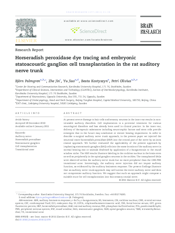
BR A IN RE S E A RCH 1 3 77 ( 20 1 1 ) 4 1 –4 9
available at www.sciencedirect.com
www.elsevier.com/locate/brainres
Research Report
Horseradish peroxidase dye tracing and embryonic
statoacoustic ganglion cell transplantation in the rat auditory
nerve trunk
Björn Palmgrena,b,⁎, Zhe Jinc , Yu Jiaoa,d , Beata Kostyszyna , Petri Oliviusa,b,e
a
Center for Hearing and Communication Research, Karolinska University Hospital, 171 76, Stockholm, Sweden
Department of Clinical Sciences, Intervention and Technology (CLINTEC), Section of Otorhinolaryngology, Karolinska Institutet,
Karolinska University Hospital, 171 76 Stockholm, Sweden
c
Department of Neuroscience, Uppsala University, Box 593, 751 24, Uppsala, Sweden
d
Department of Otolaryngology, Head and Neck Surgery, Beijing TongRen Hospital, Capital Medical University, 100730, Beijing, China
e
ENT clinic, Linköping University hospital, 58185 Linköping, Sweden
b
A R T I C LE I N FO
AB S T R A C T
Article history:
At present severe damage to hair cells and sensory neurons in the inner ear results in non-
Accepted 28 December 2010
treatable auditory disorders. Cell implantation is a potential treatment for various
Available online 6 January 2011
neurological disorders and has already been used in clinical practice. In the inner ear,
delivery of therapeutic substances including neurotrophic factors and stem cells provide
Keywords:
strategies that in the future may ameliorate or restore hearing impairment. In order to
Auditory nerve
describe a surgical auditory nerve trunk approach, in the present paper we injected the
Horseradish peroxidase
neuronal tracer horseradish peroxidase (HRP) into the central part of the nerve by an intra
Statoacoustic ganglion
cranial approach. We further evaluated the applicability of the present approach by
Cell transplantation
implanting statoacoustic ganglion (SAG) cells into the same location of the auditory nerve in
Transitional zone
normal hearing rats or animals deafened by application of β-bungarotoxin to the round
window niche. The HRP results illustrate labeling in the cochlear nucleus in the brain stem
as well as peripherally in the spiral ganglion neurons in the cochlea. The transplanted SAGs
were observed within the auditory nerve trunk but no more peripheral than the CNS-PNS
transitional zone. Interestingly, the auditory nerve injection did not impair auditory
function, as evidenced by the auditory brainstem response. The present findings illustrate
that an auditory nerve trunk approach may well access the entire auditory nerve and does
not compromise auditory function. We suggest that such an approach might compose a
suitable route for cell transplantation into this sensory cranial nerve.
© 2011 Elsevier B.V. All rights reserved.
⁎ Corresponding author. Karolinska University Hospital, 171 76 Stockholm, Sweden. Fax: +46 851774265.
E-mail address: bjorn.palmgren@karolinska.se (B. Palmgren).
Abbreviations: ABR, auditory brainstem response; β-BuTx, β-bungarotoxin; BS, brainstem; CN, cochlear nucleus; CNS, central nervous
system; CSF, cerebrospinal fluid; E13, embryonic day 13; EDTA, ethylenediaminetetraacetic acid; FBS, foetal bovine serum; GFP, green
fluorescent protein; HRP, horseradish peroxidase; IAM, internal auditory meatus; PBS, phosphate-buffered saline; PFA, paraformaldehyde;
PNS, peripheral nervous system; RW, round window; SAG, statoscoustic ganglion; SGN, spiral ganglion neuron; TMB, tetramethylbenzidine; TZ, transitional zone
0006-8993/$ – see front matter © 2011 Elsevier B.V. All rights reserved.
doi:10.1016/j.brainres.2010.12.078
�42
1.
BR A IN RE S EA RCH 1 3 77 ( 20 1 1 ) 4 1 –49
Introduction
Within the peripheral hearing organ, i.e. the cochlea, the
specialized hair cells and neurons are major targets of both
intrinsic genetic changes (e.g. gene mutation) and exogenous
insults (e.g. noise and pharmacological trauma) that consequently result in hearing impairment. In order to restore or even
replace the degenerated cells in the cochlea different substrates
such as pharmacologic agents, viral vectors, mature or immature cells have been delivered into the cochlea using a range of
surgical techniques (Kesser and Lalwani, 2009; Regala et al.,
2005; Richardson et al., 2006; Sekiya et al., 2007b). Several
commonly performed surgical approaches to access the cochlea
(e.g. cochleostomy) may disturb the intracochlear structure and
jeopardize residual hearing. Application of therapeutic substances (e.g. steroids) to the round window membrane presents
a non-traumatic approach in the prevention or treatment of
certain reversible inner ear diseases (Arriaga and Goldman,
1998; Silverstein et al., 1999). The permeability of the human
round window membrane to each therapeutic agent is not yet
fully explored (Carvalho and Lalwani, 1999). In addition, the
majorities of surgical routes to access the cochlea are performed
in smaller experimental animals but are not yet available in
humans. There are also indications that axons from cells
transplanted into the peripheral portion of the cochlear nerve
may be inhibited by the transitional zone (TZ) located between
the central nervous system (CNS) and the peripheral nervous
system (PNS), thereby precluding any further central sprouting
(Fraher, 2000; Sekiya et al., 2007b).
In order to improve the cell delivery process novel
approaches with a potential to counteract irreversible damage
to spiral ganglion neurons (SGNs) including degeneration of the
auditory nerve, might be essential. Only a few experimental
studies have adopted the approach to deliver cells or substances
to the central portion of the auditory nerve (Corrales et al., 2006;
Sekiya et al., 2006, 2007a). In the current paper we describe a
suboccipital approach to initially inject the neuronal tracer
horseradish peroxidase (HRP) into the rat auditory nerve. The
function of the auditory nerve pre- and two weeks postoperatively was monitored by measuring the auditory brainstem
response (ABR). In order to assess the applicability of this
surgical approach, mouse statoacoustic ganglion (SAG) explants
were implanted using the same approach.
Our findings illustrate that the HRP-tracer injected into the
rat auditory nerve trunk by the internal auditory meatus (IAM)
was transported to the central as well as peripheral portions of
the nerve. Furthermore the ABR measurements demonstrated
that the surgical procedure did not compromise auditory
function. We also illustrate that the transplanted SAG
explants can survive in the auditory nerve for up to five
weeks, though in reduced numbers.
2.
Results
2.1.
HRP-tracer distribution after auditory nerve injection
The neuronal tracer HRP was used to verify the precision and
the distribution of the suboccipital approach injection into the
auditory trunk (Table 2). Depending on the volume of the
injected substance there will also be a certain amount of extra
cellular HRP proximal to the injection site as observed by
accumulation of the blue TMB reaction product (Fig. 2). Here,
the accumulation of HRP-tracer around the injection site was
easily identified (Fig. 2A, B). Further, the tracer was transported centrally as well as peripherally from the injection
site (Figs. 2A, B). In the peripheral portion the HRP labeling
was observed in the auditory nerve trunk (Fig. 2a1), in the
Schwann-glial junctional zone of the auditory nerve (Fig. 2a2)
and in the spiral ganglion neurons (Fig. 2b1 and b2). In the
central portion HRP was found in the central SGN terminals in
the cochlear nucleus (Fig. 2b3). No HRP labeling was observed
in the contralateral ear thereby making the possibility of
leakage of HRP tracer from the injection side unlikely.
2.2.
nerve
Transplantation of SAG-GFP cells into the auditory
In both the non-deafened and the β-bungarotoxin-deafened
rats (survival times two to five weeks, groups 2–5, Table 1) we
identified transplanted SAGs at the injection site as well as
Fig. 1 – (A). Experimental setup for intra-auditory nerve trunk
delivery. A Hamilton syringe attached with a 33-gauge
needle was mounted in the clamping device of the
stereotactic frame. (B). The injection site at the auditory nerve
root (arrow) and the injection needle (arrowhead).
�BR A IN RE S E A RCH 1 3 77 ( 20 1 1 ) 4 1 –4 9
43
Fig. 2 – HRP distribution (visualized as blue TMB reaction product) after injection to the rat auditory nerve trunk. The sections A
and B are from different animals. HRP labeling can be found in the auditory nerve trunk (A and a1), in the Schwann-glial
junctional zone (arrowhead) of the auditory nerve (A and a2), in the spiral ganglion neurons (B and b1 and b2). In the central
portion HRP was found in the cochlear nucleus (B and b3). Arrow, blue TMB reaction product; AN, auditory nerve; CN, cochlear
nucleus; SGN, spiral ganglion neuron; PM, peripheral myelin; CM, central myelin. Star, injection site. Scale bar: A, B: 100 μm;
a1–a2, b1–b3: 40 μm.
along the auditory nerve (Fig. 3). In two animals we detected
both GFP- and Tuj1-positive cells with detectable neurites in
the nerve peripherally from the IAM (Figs. 3E and F). We also
observed GFP-positive cell profiles lining the boundary of the
Schwann-glial TZ (Fig. 4C) but we did not observe any cells
passing through the TZ towards the periphery of the auditory nerve. Furthermore there were no transplanted cells
migrating centrally into the cochlear nucleus. In order to
illustrate that our observed GFP-positive profiles (stained with
anti-GFP antibody) are transplanted cells but not artifacts
e.g. autofluorescence from blood or immune cells migrating
from the injection site, we performed sham surgery by
injecting culture medium only by the same approach. In
these control animals we did not observe any cells stained
with anti-GFP antibody (Fig. 4B). In the deafened groups (4 and
5) we found cell profiles in five out of eight animals whereas
among the non deafened animals (groups 2 and 3) cell profiles
were found in one out of nine animals only. In some animals
we observed GFP positive tissue without cell profiles (data not
shown). In four animals we could not detect any GFP-positive
profiles at all. Two animals from the deafened groups had to
be sacrificed due to wound infections.
�44
BR A IN RE S EA RCH 1 3 77 ( 20 1 1 ) 4 1 –49
Table 1 – Groups of rats used either for SAG implantation or HRP injection to the auditory nerve trunk.
Group Number of animals (n) HRP injection to AN SAG transplantation to AN RW application of β-BuTx Survival time
1
2
3
4
5
6
2.3.
6
6
3
4
6
3
+
−
−
−
−
−
−
+
+
+
+
− (only culture medium)
Assessment of auditory function
Immediately before and two weeks after intra auditory nerve
HRP injection ABR was measured at 8, 16 and 40 kHz. The
magnitudes of the ABR threshold shifts pre- as compared to
postoperatively were approximately 5 dB at all frequencies
measured, illustrating no statistically significant differences
between the pre- and post operative ABR values (Fig. 5). In two
out of six animals the ABR thresholds were not altered
postoperatively at all.
3.
Discussion
Studies of transplanted stem cells into the cochlea or the
cochlear nerve have not been able to visualize significant
numbers of newly-formed neuronal connections between the
implant and the cochlear nucleus in the brain stem. In the
present paper, in order to explore differentiation but also
migration of the implanted progenitor cells the aim was to
illustrate a technique for injection of cells into the central
portion of the auditory nerve. Furthermore, by injecting HRP
tracer into the auditory nerve trunk we suggest that the
injection technique allows injected trophic factors or other
substances to reach into the cochlear nucleus region in the
brain stem and also into the SGN in the cochlea. By measuring
the ABR-response we assessed whether the implantation
procedure would have any impact on auditory function.
Finally, by using the same suboccipital approach as for the
HRP injection, we implanted embryonic mouse SAG explants
into the auditory nerve.
Being a widely used tracer for neuronal pathways (van der
Want et al., 1997; Waar et al., 1981) we selected HRP for the
tracing procedure used in this paper. Since the HRP uptake
occurs mainly by passive endocytosis in the axotomized
regions and nerve terminals (van der Want et al., 1997) we
presume that the trauma on the auditory nerve trunk caused
by the injection needle would be permissive for uptake of the
substrate. Seemingly, in accordance with our results other
Table 2 – The distribution of HRP following auditory nerve
trunk injection by the internal auditory meatus.
HRP positive staining area
Number of cochleas
Auditory nerve trunk
Schwann-glial junctional zone
Spiral ganglion neurons
Cochlear nucleus
6
4
5
5
−
−
−
+
+
+
2 days
2 weeks
5 weeks
2 weeks
5 weeks
2 weeks
studies have shown that following pressure injection there are
two different types of HRP uptake into the neurons. Apart from
the local accumulation of HRP there is a diffuse passive uptake
that could be due to HRP pressure in the confined injection
site. The second type of uptake is the physiological incorporation with transportation and diffusion of HRP by the intact
axonal terminals (Leake-Jones and Snyder, 1982). We found
HRP labelings were located by the injection site, in SGN in the
cochlea and in central terminals in the cochlear nucleus (CN)
close to the second order neurons. This verifies the injection
site and illustrates that the injection procedure would
reach into target areas we presume would be relevant for a
successful outcome of an implantation paradigm. We further
speculate that such a paradigm might have a potential to
promote survival of implanted cells to differentiate and send
afferents into the CN in the BS as well as connecting with hair
cells in the cochlea. Locally applied growth factors could also
be distributed to the auditory nerve by diffuse uptake as well
as transported peripherally and centrally. We did not observe
any HRP labeling in the contralateral ear indicating that the
HRP-tracer did neither leak into the CSF nor spread contralaterally via the systemic blood circulation.
Stem cells are present in the rat embryonic inner ear but
decrease in numbers post partum (Oshima et al., 2007). This is
probably one reason for the poor ability of the inner ear to
regenerate damaged spiral ganglion neurons and hair cells.
The SAG explants used in the present experiment were
harvested from the auditory tract in E13 embryos. At this
time period neuronal responses to sound initializes (Friauf,
1992; Uziel et al., 1981). The SAGs contain embryonic progenitor cells responsible for the development of both cochlear
and vestibular neurons (Sher, 1971). Earlier studies have
shown that histological signs of severe rejection appears following transplantation of cells to non-immunosuppressed rats
(Fernandez et al., 2006). All animals in our experiments received daily injections of immunosuppressants and antibiotics
after the cell transplantation during the entire survival time.
In comparison to the distribution of the HRP tracer the
transplanted SAG cells were not observed at longer distances
away from the injection site. Furthermore, although we
observed survival of implanted explants for up to five weeks
these were only found in relatively small numbers. This could
be due to several reasons but we speculate that, in order to
survive in larger numbers, the injected cells might need
exogenous neurotrophic support. Examples of neurotrophic
factors to improve implant survival would be brain-derived
neurotrophic factor (BDNF), glial-derived neurotrophic factor
(GDNF) and neurothrophin-3 (NT-3) that have been shown to
increase SGN survival (Ernfors et al., 1995). In the beginning of
�BR A IN RE S E A RCH 1 3 77 ( 20 1 1 ) 4 1 –4 9
45
Fig. 3 – Survival of SAG cells after transplantation to the rat auditory nerve trunk. Photomicrographs A and B were taken from
non-deafened rats with 5 weeks postoperative survival time, C-F from β-bungarotoxin-deafened rats with 2 weeks
postoperative survival time. Single GFP-positive SAG cells (green color) (A, B and inset in B; arrow) and GFP-positive cell clusters
(C, D; asterisk) were found in the auditory nerve. GFP- and TUJ1- (red color) double stained SAG cells (yellow color) with distinct
neurites (E, F; arrowhead) can also be detected in the auditory nerve. The nuclei were counterstained with DAPI (blue color). AN,
auditory nerve; CN, cochlear nucleus; M, cochlear modiolus. Star, injection site. Scale bar: A, 200 μm; B, 100 μm; C and E
400 μm; D, F and inset in B, 50 μm.
the synaptogenesis it has been shown that the cochlear
neurons are mainly dependant on NT-3 whereas the vestibular neurons are more dependent on BDNF (Fritzsch et al., 1997).
Other studies have shown that, for proper survival, migration
and differentiation, the early SAG neurons are also dependent
on BDNF and fibroblast growth factors (FGFs) (Brumwell et al.,
2000; Hossain et al., 1997). Some technical problems, possibly
precluding implant survival, were encountered involving the
easily disrupted well-vascularized areas by the IAM close to
the auditory nerve. Furthermore, we only examined for any
potential neurite outgrowth during a five week postoperative
period whereas it cannot be excluded that the development of
newly-formed neuritic projections would require a longer
survival time.
We did not observe any SAG cells in the cochlear perilymph
or endolymph indicating that there was no cell leakage via the
CSF or via the canaliculi perforantes in the cochlear modiolus.
In previous studies we have interestingly observed migration
�46
BR A IN RE S EA RCH 1 3 77 ( 20 1 1 ) 4 1 –49
Fig. 5 – ABR threshold in non-deafened rats before and
2 weeks after HRP injection.
Fig. 4 – Migration of transplanted SAG cell profiles to the
Schwann-glial junctional zone. The location of
Schwann-glial junctional zone (A). GFP-positive cells (arrow)
were detected along the boundary of the Schwann-glial
junctional zone (arrowhead) in the animals with SAG
explants transplanted to the auditory nerve (C and inset in C),
but not in the sham-operated animals with culture medium
injection (B). (B) and (C) were shown from the rectangular area
in (A). The nuclei were counterstained with DAPI (blue color).
AN, auditory nerve; PM, peripheral myelin; CM, central
myelin. Scale bar: A, B and C, 50 μm; inset in C, 10 μm.
of embryonic sensory cells from the perilymph to the SGN
in the modiolus following implantation into the inner ear
scala tympani (Hu et al., 2004a; Olivius et al., 2003). We suggested that these migrating cells might utilize the canaliculi
perforantes (Hu et al., 2004b). Furthermore, in the present
paper we did not observe any cells in the contralateral
cochlear specimen even though this does not completely
rule out a possible route for cell migration via the CSF into the
cochlear aqueduct. We speculate that since our SAG cells were
not dispersed in the injected medium but ensheathed with
fibrous tissue in whole explants this may reduce the ability of
the SAGs to migrate and send out neurites. In some specimen
we found GFP-positive tissue without any cells. This could
either be because the transplanted SAG explants did not
contain sufficient numbers of cells or that these did not
survive in sufficient numbers. In terms of SAG cell survival
there was a significant difference between the non-deafened
and β-bungarotoxin-deafened groups. The lower survival-rate
of cells in the non-deafened group could be due to similar
mechanisms indicated by previous studies, e.g. that the
migration of implanted cells in nerves is limited by the
available space in the nerve (Sekiya et al., 2006). Other
explanations for the limited cell migration might be the
CNS-PNS TZ. This border zone between the CNS and PNS,
illustrating a Schwann cell–glial cell barrier, represents a
biological obstacle for various molecules and cells reaching
into the CNS (Fraher, 2000). Subsequently we hypothesize that
the TZ might also hamper the migration of larger molecules
and cells. We are currently investigating the possibility to
make, by injecting selected substances or supporting cells
together with the SAGs, the TZ more permeable and potentially facilitate migration and sprouting of implanted cells.
One possible problem by using an implantation approach
directed towards the auditory nerve is that it might compromise the integrity of the cochlea and the hearing. In the
present study, however, as evaluated from our fairly unaltered
ABR curves the nerve trunk approach does not seem to
significantly impair auditory function.
In summary, the present study illustrates that the surgical
approach presented can be useful in reaching the SGN soma
including their central terminals, e.g. the entire auditory
nerve. The findings also suggest that the approach may hold
the promise to reach regions in the auditory nerve seemingly
relevant for a successful implantation outcome without
compromising hearing. We further suggest that the similar
stereotactic setup may be used for delivery of neurotrophic
factors essential to implant survival and differentiation. Such
studies are under way.
4.
Experimental procedures
All animal experiments followed the national approved
protocol for care and use of animals in Sweden (approval
N58/03, N347/05). Young adult Sprague–Dawley rats (n = 28;
200–250 g) were used in the study. The different animal
groups are presented in Table 1. Preoperative otoscopic
�BR A IN RE S E A RCH 1 3 77 ( 20 1 1 ) 4 1 –4 9
examinations were performed to exclude any visible middle
ear infection.
4.1.
Surgical approach and HRP injection
HRP animals (n = 6) were anaesthetized with an intraperitoneal
(i.p.) injection of a mixture of Ketalar© (50 mg/kg) and
Rompun© (10 mg/kg) and placed in a stereotactic frame. The
skull was put in a fixed position and the skin on the occipital
region shaved and disinfected with 70% ethanol. Under a
surgical microscope a left post-occipital hemi-arcade incision
was made through the skin and underlying soft tissue. By
using a drill a 3 mm diameter hole was made on the
suboccipital bone. By sharp incision the underlying dura was
opened and reflected towards the edge of the hole followed by
drainage of cerebrospinal fluid (CSF). As part of the posterolateral cerebellar hemisphere was gently retracted contralaterally a cotton ball was placed for CSF suction thereby
revealing the auditory nerve trunk between the brainstem and
the internal auditory meatus. A 10 μl Hamilton syringe
attached with a 33-gauge needle was filled with 30% HRP
(Type VI-A, Sigma) and mounted in the clamping device of the
stereotactic frame (Fig. 1A). The needle was positioned above
the auditory nerve trunk with the angle of the tip adjusted
towards the internal auditory meatus. The needle was
lowered into the auditory nerve trunk with a depth of
500 μm by the use of the micromanipulator (Fig. 1B). A total
volume of 4 μl of HRP solution was injected into the nerve
root. After injection the needle was left in place for 10 min
whereafter the wound cavity was filled with sterile saline. A
piece of fascia was used for covering the hole in the dura and
occipital bone. The wound was closed in layers with continuous single sutures. Following removal from the stereotactic
frame the animals were given subcutaneous injections of 3 ml
saline and 0.2 ml Temgesic© (0.3 mg/ml) and placed in a warm
cage to recover before being transferred to the home cage.
4.2.
HRP histochemical staining
Two days following injection the HRP animals were deeply
anaesthetized with an intraperitoneal overdose of pentobarbital (60 mg/ml) and transcardiacally perfused with 0.9% NaCl
followed by 4% of paraformaldehyde (PFA). Following decapitation, the left temporal bone, auditory nerve and adjacent
brainstem were carefully excised in a single tissue block. The
temporal bone was opened and the excess bony tissue removed.
Under a dissecting microscope the cochlea was perfused with
4% PFA in 0.1 M PBS through the round window and a hole was
made in the apical turn. The tissue block was immersed in
fixative for 24 h at 4 °C and washed by PBS. Decalcification with
0.1 M ethylenediaminetetraacetic acid (EDTA) in 0.1 M PBS at
4 °C was carried out on the whole tissue block until the
remaining bony tissue was soft enough for cryostat sectioning.
The tissue block was immersed in 20% sucrose for 24 h,
embedded in optimal cutting temperature (OCT) compound
(Sakura Tissue-Tek) and 12 μm serial cryostat sections were
made. The cryostat sections were processed for HRP using
tetramethylbenzidine (TMB) as the chromagen and sodium
tungstate (ST) as the stabilizer (Gu et al., 1992). In brief, sections
were dried at room temperature (RT) for 2 h, rinsed three times
47
during 10 min in PBS and pre-incubated in reaction medium
(0.5% TMB in ethanol and 1% ST in 0.2 M PBS) at RT for 20 min
while protected from light. The reaction was initiated by adding
0.7 ml of 0.3% hydrogen peroxide every 10 min during the 1 h
incubation period. To terminate the reaction the sections were
rinsed 5 times for 3 min in 0.05 M PBS (pH 5.0–5.4). All sections
with or without eosin counterstaining were dehydrated through
ethanol series, cleared in xylene, mounted with Permount and
photographed using a light microscope (Zeiss) equipped with a
digital camera (Nikon Coolpix 990). Negative controls were made
by omission of TMB in the sections.
4.3.
niche
Application of β-bungarotoxin to the round window
Animals (n = 13) were deafened by application of β-bungarotoxin (β-BuTx) to the round window niche as described
previously (Palmgren et al., 2010). In brief, after i.p. anaesthesia with xylazine (10 mg/kg i.p.) and ketamine (50 mg/kg i.p.)
the round window niche was exposed by a retroauricular
incision. 5 μl of β-BuTx (0.05 μg/ml, Alexis Biochemicals) was
absorbed by gel foam and applied to completely fill the round
window niche. A piece of fascia was placed to cover the hole in
the bulla. The animals were kept for 3 weeks until further
surgical procedures were performed.
4.4.
Transplantation of statoacoustic ganglion explants to
the rat auditory nerve
SAG explants dissection was performed in embryonic day 13
(E13) green fluorescent protein (GFP)-positive BalbC mice in
Hanks Balanced Salt Solution (HBSS) supplemented with
antibiotics. Whole explants were placed into the 4-well cell
culture plates coated with poly-l-lysine and laminin. The SAG
explants were cultured overnight in culture media consisting
of Dulbecco's Modified Eagle's Medium (DMEM)/F12 (Gibco/
Invitrogen) supplemented with 1% Foetal Bovine Serum (FBS),
Insulin-transferrin-sodium selenite supplement (ITSS), 4-(2Hydroxyethyl) piperazine-1ethansulfonic acid (HEPES) and
antibiotics. The explants were removed from the cell culture
plates with a needle and immediately used for implantation.
The host rats had previously been anaesthetized with an
intraperitoneal injection of a mixture of Ketalar© (50 mg/kg)
and Rompun© (10 mg/kg) and the surgery was carried out by
the suboccipital approach described above. The SAG explants
were aspirated from a petri dish with a 10 μl Hamilton syringe.
By using the same needle, the stereotactic frame and a syringe
clamping device (Fig. 1B) the SAG explants were injected
together with 4 μl of medium into the auditory nerve by the
IAM. The needle was kept in place for 10 min after injection
whereafter the wound was closed as above. Sham operated
animals were injected with 5 μl culture medium.
To prevent postoperative infection and immune response
rejection all animals received daily doses of tetracycline
(1.8 mg/ml, i.p.) and cyclosporine (4.2 mg/ml, i.p.). After the
survival period the rats were sacrificed by an overdose of
pentobarbital (60 mg/ml, i.p.), transcardially perfused with
body warm 0.9% saline followed by ice-cold 4% PFA in 0.1 M
PBS. The cochlea, auditory nerve and part of the brainstem
were carefully removed en bloc.
�48
4.5.
BR A IN RE S EA RCH 1 3 77 ( 20 1 1 ) 4 1 –49
Immunohistochemistry
The specimens (cochlea plus auditory nerve including brain
stem) were dissected out and a small hole used for perfusion
with PFA (initially 4% and then 0.5%) was made in the apex.
The cochlea was decalcified in EDTA for 7 days. After 24 h
in 20% sucrose solution the specimens were embedded
and frozen in OCT Compound (Tissue-Tek; Sakura Finetek,
Torrance, CA, USA). The specimens were orientated in the
compound so that mid-modiolar sections would contain the
cochlea, auditory nerve and brain stem (BS) as a continuum.
The 12 μm mid-modiolar cryosections were mounted on glass
slides.
The sections were blocked with 10% goat serum, 5% bovine
serum albumin (BSA) and 0.2% Triton X-100 in 0.1 M PBS for 1 h
at room temperature and incubated for 36 h at 4 °C with
conjugated goat polyclonal to GFP (FITC conjugated) antibody
(1:200; Abcam, Cambridge, UK). Following incubation the
samples were washed in PBS and put into blocking solution
for 1 h at room temperature. For double staining the sections
were incubated for 48 h at 4 °C with rabbit polyclonal β-tubulin
(TUJ1) antibody (1:200; Covance Research Products, Berkeley,
CA, USA). Following incubation the sections were labeled with
goat-anti rabbit-Cy3 (1:2000) for 1 h at room temperature. The
specimens were visualized and photographed using a fluorescence microscope (Zeiss, Stockholm, Sweden) equipped
with a digital camera (Nikon Coolpix 990, Solna, Sweden).
Omission of the primary antibody served as negative control.
Cell nuclei were stained with 4, 6-diamidino-2-phenylindole
(DAPI).
4.6.
Auditory function assessment
The ABR measurements (n = 6) were conducted under general
anaesthesia with ketamine (50 mg/kg, i.p.) and xylazine
(10 mg/kg, i.p.) immediately before and two weeks after
surgery on the left ear in a soundproof booth using a
Tucker-Davis System II (BioSig stimulate/recording system
2.0, Tucker-Davis Technologies, Alachua, FL, USA). The
stimulus intensity was calibrated with a 0.25-inch condenser
microphone (model 4135, Brüel & Kjær, Nærum, Denmark). All
sound pressure levels were expressed in decibel values
relative to 20 μPa. Sound stimulation (tone burst 20 stimuli/
s; single sinusoidal wave) was applied to the left ear using a
high frequency transducer via a flexible tube in the external
auditory meatus. Needle electrodes were placed on the vertex
and below the recorded ear whereas the ground electrode was
placed on the hind leg. The evoked response was amplified
100 000 times and 2048 sweeps were averaged in real time by a
digital signal processor (DSP32C, Lucent Technologies) with a
time-domain artifact rejection. The initial intensity of the
stimulus was 90 dB peak sound pressure level that was
decreased in 5 dB steps until the ABR curves disappeared.
The ABR threshold was defined as the lowest intensity at
which a visible ABR wave was observed in two averaged runs.
Thresholds were measured at three frequencies (8, 16 and
40 kHz). Statistical analysis using student's two-tailed t test
was made from the mean values of the ABR thresholds at
each frequency immediately before and two weeks after
surgery.
REFERENCES
Arriaga, M.A., Goldman, S., 1998. Hearing results of intratympanic
steroid treatment of endolymphatic hydrops. Laryngoscope
108, 1682–1685.
Brumwell, C.L., Hossain, W.A., Morest, D.K., Bernd, P., 2000. Role
for basic fibroblast growth factor (FGF-2) in tyrosine kinase
(TrkB) expression in the early development and innervation of
the auditory receptor: in vitro and in situ studies. Exp. Neurol.
162, 121–145.
Carvalho, G.J., Lalwani, A.K., 1999. The effect of cochleostomy and
intracochlear infusion on auditory brain stem response
threshold in the guinea pig. Am. J. Otol. 20, 87–90.
Corrales, C.E., Pan, L., Li, H., Liberman, M.C., Heller, S., Edge, A.S.,
2006. Engraftment and differentiation of embryonic stem
cell-derived neural progenitor cells in the cochlear nerve trunk:
growth of processes into the organ of Corti. J. Neurobiol. 66,
1489–1500.
Ernfors, P., Van De Water, T., Loring, J., Jaenisch, R., 1995.
Complementary roles of BDNF and NT-3 in vestibular and
auditory development. Neuron 14, 1153–1164.
Fernandez, E., Mannino, S., Tufo, T., Pallini, R., Lauretti, L.,
Albanese, A., Denaro, L., 2006. The adult “paraplegic” rat:
treatment with cell graftings. Surg. Neurol. 65, 223–237.
Fraher, J.P., 2000. The transitional zone and CNS regeneration.
J. Anat. 196 (Pt 1), 137–158.
Friauf, E., 1992. Tonotopic order in the adult and developing
auditory system of the rat as shown by c-fos
immunocytochemistry. Eur. J. Neurosci. 4, 798–812.
Fritzsch, B., Silos-Santiago, I., Bianchi, L.M., Farinas, I., 1997. The
role of neurotrophic factors in regulating the development of
inner ear innervation. Trends Neurosci. 20, 159–164.
Gu, Y., Chen, Y., Ye, L., 1992. Electron microscopical
demonstration of horseradish peroxidase by use of
tetramethylbenzidine as chromogen and sodium tungstate as
stabilizer (TMB-ST method): a tracing method with high
sensitivity and well preserved ultrastructural tissue.
J. Neurosci. Methods 42, 1–10.
Hossain, W.A., Rutledge, A., Morest, D.K., 1997. Critical periods of
basic fibroblast growth factor and brain-derived neurotrophic
factor in the development of the chicken cochleovestibular
ganglion in vitro. Exp. Neurol. 147, 437–451.
Hu, Z., Ulfendahl, M., Olivius, N.P., 2004a. Central migration of
neuronal tissue and embryonic stem cells following
transplantation along the adult auditory nerve. Brain Res. 1026,
68–73.
Hu, Z., Ulfendahl, M., Olivius, N.P., 2004b. Survival of neuronal
tissue following xenograft implantation into the adult rat inner
ear. Exp. Neurol. 185, 7–14.
Kesser, B.W., Lalwani, A.K., 2009. Gene therapy and stem cell
transplantation: strategies for hearing restoration. Adv.
Otorhinolaryngol. 66, 64–86.
Leake-Jones, P.A., Snyder, R.L., 1982. Uptake and transport of
horseradish peroxidase by cochlear spiral ganglion neurons.
Hear. Res. 8, 199–223.
Olivius, P., Alexandrov, L., Miller, J., Ulfendahl, M.,
Bagger-Sjoback, D., Kozlova, E.N., 2003. Allografted fetal dorsal
root ganglion neuronal survival in the guinea pig cochlea.
Brain Res. 979, 1–6.
Oshima, K., Grimm, C.M., Corrales, C.E., Senn, P., Martinez
Monedero, R., Geleoc, G.S., Edge, A., Holt, J.R., Heller, S., 2007.
Differential distribution of stem cells in the auditory and
vestibular organs of the inner ear. J. Assoc. Res. Otolaryngol. 8,
18–31.
Palmgren, B., Jin, Z., Ma, H., Jiao, Y., Olivius, P., 2010.
Beta-bungarotoxin application to the round window: an in
vivo deafferentation model of the inner ear. Hear. Res. 265,
70–76.
�BR A IN RE S E A RCH 1 3 77 ( 20 1 1 ) 4 1 –4 9
Regala, C., Duan, M., Zou, J., Salminen, M., Olivius, P., 2005.
Xenografted fetal dorsal root ganglion, embryonic stem cell
and adult neural stem cell survival following implantation
into the adult vestibulocochlear nerve. Exp. Neurol. 193,
326–333.
Richardson, R.T., Noushi, F., O'Leary, S., 2006. Inner ear therapy for
neural preservation. Audiol. Neurootol. 11, 343–356.
Sekiya, T., Kojima, K., Matsumoto, M., Kim, T.S., Tamura, T., Ito, J.,
2006. Cell transplantation to the auditory nerve and cochlear
duct. Exp. Neurol. 198, 12–24.
Sekiya, T., Holley, M.C., Kojima, K., Matsumoto, M., Helyer, R., Ito,
J., 2007a. Transplantation of conditionally immortal auditory
neuroblasts to the auditory nerve. Eur. J. Neurosci. 25,
2307–2318.
Sekiya, T., Kojima, K., Matsumoto, M., Holley, M.C., Ito, J., 2007b.
Rebuilding lost hearing using cell transplantation.
Neurosurgery 60, 417–433 discussion 433.
49
Sher, A.E., 1971. The embryonic and postnatal development of the
inner ear of the mouse. Acta Otolaryngol. Suppl. 285, 1–77.
Silverstein, H., Arruda, J., Rosenberg, S.I., Deems, D., Hester, T.O.,
1999. Direct round window membrane application of
gentamicin in the treatment of Meniere's disease. Otolaryngol.
Head Neck Surg. 120, 649–655.
Uziel, A., Romand, R., Marot, M., 1981. Development of cochlear
potentials in rats. Audiology 20, 89–100.
van der Want, J.J., Klooster, J., Cardozo, B.N., de Weerd, H.,
Liem, R.S., 1997. Tract-tracing in the nervous system of
vertebrates using horseradish peroxidase and its conjugates:
tracers, chromogens and stabilization for light and electron
microscopy. Brain Res. Brain Res. Protoc. 1, 269–279.
Waar WB, de Olmos, J.S., Heimer L., 1981. Horseradish peroxidase:
the basic procedure. In: Heimer L, Robards MJ, (Eds.),
Neuroanatomical Tract Tracing Methods. New York, Plenum
Press, pp. 207–262.
�
 Björn Palmgren