IMMUNOLOGY AND PATHOGENESIS OF VIRAL HEMORRHAGIC FEVERS
A Mouse Model for Studying Dengue Virus
Pathogenesis and Immune Response
Katherine L. Williams,a Simona Zompi,a P. Robert Beatty,b
and Eva Harrisa
a
b
Division of Infectious Diseases and Vaccinology, School of Public Health, and
Department of Molecular and Cellular Biology, University of California Berkeley,
Berkeley, California, USA
A small animal model for studying dengue disease is of critical importance to furthering
many areas of dengue research, including host immunity, disease pathogenesis, and
drug and vaccine development. Recent characterization of the AG129 mouse model has
demonstrated it to be one of the only models at this time that permits infection by all
four serotypes of dengue virus (DENV), supports replication in relevant cell and tissue
types comparable to human infection, and allows antibody-mediated protection and
enhancement of DENV infection. Thus, this model enables testing hypotheses arising
from epidemiological observations and in vitro experiments in an in vivo system with
a functional adaptive immune response. This review provides a brief overview of the
development of a mouse model of DENV infection, describes the work completed to date
characterizing the AG129 model, and examines several of the unanswered questions
remaining in the field.
Key words: dengue; mouse model; antibody-dependent enhancement; AG129 mice
Introduction
Dengue virus (DENV) is a mosquito-borne
virus of the Flaviviridae family and is related to
yellow fever, West Nile (WNV), and Japanese
Encephalitis viruses.1 Endemic to tropical and
subtropical regions of the world, DENV is
the most medically important arthropod-borne
virus worldwide and a major public health
challenge. Three billion people are at risk for
DENV infection, with an estimated 50 million cases of dengue fever (DF) annually and
250,000–500,000 cases of the potentially fatal
dengue hemorrhagic fever/dengue shock syndrome (DHF/DSS), characterized by vascular
leak leading to hypotensive shock.2 The four
DENV serotypes (DENV1-4) are transmitted
to humans primarily by the mosquitoes Aedes
Address for correspondence: Eva Harris, Division of Infectious Diseases and Vaccinology, School of Public Health, University of California,
Berkeley, 1 Barker Hall, Berkeley, CA 94720-7354. Voice: 510-642-4845;
fax: 510-642-6350. eharris@berkeley.edu
aegypti and Ae. albopictus. The clinical course
of DHF/DSS is initially quite similar to DF;
however, at defervescence, DHF/DSS patients
rapidly deteriorate into life-threatening conditions characterized by vascular leak, hemorrhagic manifestations, and thrombocytopenia
with or without shock.3 The inability to differentiate between DF and DHF/DSS at early
stages of illness contributes to the difficulty in
treating the disease. Further, individuals previously infected with DENV are at increased risk
of severe disease upon secondary infection with
a different (heterologous) serotype.4 At present,
no effective antiviral therapy or vaccine exists,
and treatment is largely supportive in nature.
Role of the Adaptive Immune
Response in Modulating Secondary
DENV Infection
Host factors, including human leukocyte
antigen (HLA) polymorphisms and prior
Immunology and Pathogenesis of Viral Hemorrhagic Fevers: Ann. N.Y. Acad. Sci. 1171: E12–E23 (2009).
c 2009 New York Academy of Sciences.
doi: 10.1111/j.1749-6632.2009.05057.x �
E12
�Williams et al.: Mouse Model for Studying Dengue
T and B cell immunity, are key determinants
of susceptibility to severe dengue disease.5 Interferon (IFN) α/β, and γ are necessary for resistance to DENV-induced disease,6 and both
arms of the adaptive immune response have
been demonstrated to play an important role
in both protection and enhancement of dengue
disease. In general, most individuals infected
with DENV for the first time (primary infection) experience inapparent infection, undifferentiated fever, or DF. In contrast, epidemiological data and experimental human studies suggest that the greatest risk factor for development
of DHF/DSS is prior infection with a different
DENV serotype4 ; Halstead7 estimated the rate
of DHF/DSS to be 40 times more frequent
during secondary than primary infections.
Two nonmutually exclusive hypotheses have
been proposed to explain the role of the adaptive immune response in the development of
DHF/DSS. The first implicates antibodies in
mediating enhanced disease. In hallmark studies, Halstead and O’Rourke8 demonstrated
that DENV did not replicate in peripheral
blood leukocytes (PBL) of nonimmune primates
but did replicate in PBL isolated from immune
primates; however, nonimmune PBL could be
infected by adding anti-DENV antisera. This
illustrates a phenomenon termed “antibodydependent enhancement” (ADE),7 in which
non-neutralizing anti-DENV antibodies facilitate entry of the virus into Fcγ receptor (FcγR)bearing cells.7,9 This increase in infected cells
directly contributes to the higher viremia levels
associated with DHF/DSS,7 and the infection
of target monocyte/macrophage cells leads to
activation, ultimately promoting the “cytokine
storm” that characterizes DHF/DSS.10 Additional evidence for ADE in vivo is derived
from the observation that most severe cases
in primary DENV infections occur in infants
in endemic areas, where DENV-specific antibodies are transferred transplacentally to infants from their dengue-immune mothers.11
These antibodies wane over time until they
can enhance DENV infection,11,12 suggesting
that pre-existing antibodies alone are sufficient
E13
to promote severe DENV illness.13 The second hypothesis explaining the immunopathology underlying severe secondary DENV infection implicates T cells. Specifically, low-affinity,
cross-reactive CD4+ and CD8+ memory T
cells resulting from a primary infection are
over-activated during secondary DENV infection with a different serotype, and these
serotype cross-reactive T cells produce high levels of cytokines, such as tumor necrosis factor-α
(TNF)-α, which contribute to increased vascular permeability.5,14,15 While in vitro and ex
vivo experiments provide insight into individual
contributions of the humoral and cell-mediated
immune response, an in vivo model is required
to mechanistically define their combined contribution to protection and enhancement of
DENV infection and disease.
Development of a Mouse Model
for DENV Infection and Disease
A small animal model for studying dengue
disease is of critical importance to furthering
many areas of dengue research, including host
immunity, disease pathogenesis, and drug and
vaccine development. Although many attempts
were made over the past several decades to develop an animal model for studying DENV,
most animal species proved to be resistant to
DENV infection.16 Over the past 7 years, our
group and others17,18 have developed and characterized the AG129 mouse (IFN-α/β, and -γ
receptor-deficient) as a tool for studying dengue
pathogenesis and immunology. This review will
briefly outline the development of a small animal model of DENV infection and disease, discuss the contributions of the AG129 model to
date, and elaborate upon its utility in studying
aspects of DENV pathogenesis, immunity, and
therapeutic drug development. For more extensive recent reviews of dengue mouse models, see
Bente and Rico-Hesse,19 Balsitis and Harris,16
and Yauch and Shresta.20
Initial attempts to develop a mouse model
for dengue in immunocompetent mice required
�E14
high doses of virus and were hindered by the
inability to recapitulate several important aspects of human DENV infection, including
replication in peripheral tissues and development of the hallmark symptoms of DENV disease.16 As another approach, humanized mice
developed by engrafting severe combined immunodeficient (SCID) mice with either human
peripheral blood lymphocytes or a variety of
DENV-susceptible tumor cells, including human K562 erythroleukemic cells and human
liver HepG2 and Huh-7 cells, were explored
as potential DENV mouse models.16 Improvement of the humanized mouse model has included the development and characterization
of nonobese diabetic (NOD)/SCID mice21 and
RAG2−/− γ−/− mice22 engrafted with CD34+
human cord blood hematopoietic stem cells. Interestingly, the reconstituted NOD/SCID mice
developed viremia as well as clinical signs of
dengue, including fever, rash, and thrombocytopenia following a subcutaneous (sc) infection with DENV2. However, the interaction
between human cells and the mouse immune
system may not completely reproduce a functional immune system, limiting the study of the
adaptive immune response to DENV infection.
Additionally, these mice are difficult and expensive to generate, thus hindering large-scale
studies.
Severely immunocompromised strains,
BALB/c athymic nu/nu mice23 and RAG1−/−
mice,6 both succumb to infection with DENV,
but death results from paralysis instead of a
defined vascular leak phenotype. Given the importance of IFNs in controlling viral infections,
studies were conducted in IFN-α/β, and -γ
receptor-6,24 and STAT1-deficient25,26 mice.
Johnson and Roehrig24 initially established the
utility of AG129 mice to study primary DENV
infection and vaccine challenge. Comparison
between mice lacking either the IFN-α/β or
IFN-γ receptor or both suggested that both
knockouts are required for early viral replication in peripheral tissues and subsequent
disease.6 Despite the immunodeficiency, the
AG129 mouse model for dengue allows for
Annals of the New York Academy of Sciences
investigation of tropism and pathogenesis
in context of a functional adaptive immune
system.
Characterization of the AG129
Mouse Model
Over the past several years, multiple studies have characterized the tissue and cellular
tropism of DENV in the AG129 model,27,28
demonstrating significant parallels with human
infection.27,29,30 Specifically, initial tropism
studies using the AG129 model demonstrated
that clinical isolates from all four DENV
serotypes replicate efficiently in spleen, lymph
node, bone marrow, and muscle. Negativestrand viral RNA was detected in dendritic
cells and macrophages of the lymph node and
spleen.28 Similarly, antibodies directed against
the nonstructural NS3 DENV protein indicated active viral replication in macrophages,
dendritic cells, hepatocytes, and bone marrowderived myeloid cells in infected AG129 mice.27
Both of these studies coincide with tropism
data from human autopsy studies27,30 and flow
cytometry analysis of infected human peripheral blood mononuclear cells,29 where the infected cells were predominantly of the myeloid
lineage. Importantly, the sc route of infection and 102 –105 pfu inoculum used in these
murine tropism studies are compatible with the
natural route and viral inoculum found in a
mosquito bite.31 In addition, AG129 mice exhibit thrombocytopenia inversely related to viral load and develop high levels of soluble NS1
during DENV infection comparable to levels
in humans18 (data not shown).
Characterization of the AG129 immune response revealed a functional adaptive immune
response to DENV infection. Specifically, antibodies elicited by infection are a mixture
of serotype-specific and serotype–cross-reactive
antibodies, including long-lasting neutralizing
antibodies,32 and the distribution of IgG isotypes among DENV-specific antibodies include
IgG1, IgG2a, and IgG2b in ratios similar to the
ratios of isotypes elicited by viral infection of
�Williams et al.: Mouse Model for Studying Dengue
wild-type 129 mice33 (data not shown). Sequential infections of one DENV serotype followed
4–52 weeks later by another serotype demonstrated reduction in viral load in the second
infection as compared to naive mice experiencing a primary infection.32 Passive transfer studies demonstrated that transfer of high doses of
monoclonal antibodies (mAbs) directed against
different epitopes on the envelope [E] protein
(domain II fusion loop or domain III lateral
ridge; see below) or anti-DENV polyclonal sera
24 h prior to DENV infection protect against
viral challenge with either the same or a different serotype as measured by a reduction in
viral load in spleen, lymph node, and serum32
(data not shown). Additionally, studies examining the T cell response to DENV infection
in AG129 mice determined that CD8+ T cells
release IFN-γ and TNF-α and have cytotoxic
effects in vivo.17 Mapping studies identified 12
immunodominant epitopes that mapped to six
DENV proteins, and vaccination with four of
these epitopes supported clearance of viral proteins.17 Taken together, these results support
the role of the mouse model in studying both
serotype-specific and cross-reactive antibodymediated protection.
To further investigate the role of dengue
pathogenesis in vivo, a virulent, mouse peripherally adapted strain of DENV2, D2S10, was
generated by alternately passaging the virus
through mice and mosquito cells 10 times, harvesting mouse serum in each cycle.34 A high inoculum of D2S10 administered to AG129 mice
results in a lethal “vascular-leak” syndrome
within 4–5 days. D2S10 has only two mutations, N124D and K128E, in the E protein that
differentiate it from the parental clinical isolate PL046. These mutations decrease heparan
sulfate binding and consequently reduce clearance of the virus, thus increasing viremia and
contributing to the lethal disease phenotype.35
A triple plaque-purified clone of D2S10, S221,
has an additional mutation in NS1 and causes
the same phenotype as D2S10.17
To study the role of antibodies in mediating
enhancement of DENV disease, Balsitis et al.
E15
(manuscript submitted) tested whether mAbs
or polyclonal sera raised in AG129 mice could
enhance a sublethal dose of DENV2 strain
D2S10. Passive transfer of both cross-reactive
mAbs and heterotypic sera was found to be
capable of lethal enhancement. Interestingly,
although a higher dose of homotypic serum
was protective, enhancement was observed following transfer of lower doses of homotypic
sera, implying that once the dilution of serum
falls below the threshold required for neutralization, the serum can become enhancing regardless of serotype specificity. This is consistent with in vitro observations of neutralization
and enhancement.36 Additionally, mice that
succumbed to lethal disease had significantly
increased levels of TNF-α and IL-10 and reduction in platelets, characteristics of human
DHF/DSS. Viral tropism data indicated a significant increase in viral load in bone marrow,
serum, white blood cells, lymph node, liver, and
small and large intestine between mice infected
with a sublethal DENV2 dose and mice experiencing lethal ADE of DENV2 infection. Interestingly, this increased viral load was not significantly different than that caused by a lethal
dose of D2S10. Cellular tropism data supported
an increase in infection in macrophages and
dendritic cells in numerous organs as well as
sinusoidal endothelial cells in the liver. Taken
together, the viral and cellular tropism data imply that the pathogenesis of an enhanced infection is fundamentally comparable to that of a
lethal, non-enhanced infection, where the main
outcome is increased infection of targeted cell
types resulting in elevated viral load. Again, the
cell types targeted are similar to those infected
by DENV in humans. In this model, infection
with human DENV1 and DENV2 clinical isolates was similarly enhanced with pretransfer of
either heterotypic anti-DENV sera or mAbs. In
summary, the AG129 mouse model is currently
the only model of DENV infection that supports replication of all four serotypes, demonstrates infection in relevant cell types and
tissues, succumbs to a fatal vascular leak syndrome from a lethal dose of a mouse-adapted
�E16
strain of DENV, and can reproduce both protection and ADE. Together these data indicate
that the AG129 mouse model can be used to
study DENV pathogenesis and answer pertinent questions regarding the roles of both humoral and cell-mediated immune responses in
contributing to protection and enhancement of
DENV disease.
Unanswered Questions about
Dengue Pathogenesis
and Immune Response
To date, a majority of observations regarding
factors contributing to the severity of DENV
disease have resulted from epidemiological observations and in vitro analysis. As most aspects
of the immune system are difficult to duplicate in vitro, many questions regarding the contribution of the adaptive immune response to
modulating secondary DENV infection still remain. With the development of a small animal model to study DENV disease, we can
now test hypotheses generated from epidemiological and in vitro data in an in vivo system.
Specifically, what components of the immune
response contribute to protection and enhancement? Are both the humoral and cell-mediated
components necessary for protection? Dissecting the humoral immune response to DENV
requires consideration of multiple contributing factors, including serotype-, domain-, and
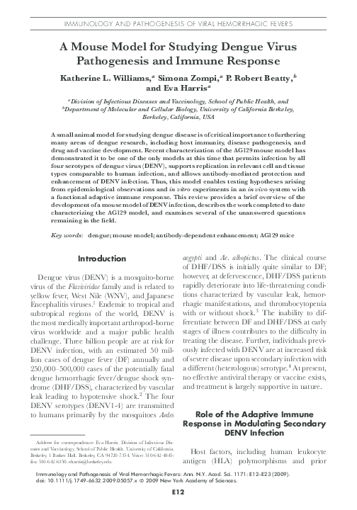
epitope-specificity of antibodies, stoichiometry and mechanism of neutralization, interaction between specific isotypes, complement and
FcγR subtypes, and maturation state of the
virus37 (Fig. 1). Which antibody characteristics are associated with protection versus enhancement in vivo? How does the ratio of mature to immature virus particles contribute to
antibody-mediated neutralization or enhancement? What candidate antivirals are protective
against DENV infection and disease in vivo?
Last, identification of a functional in vitro correlate of in vivo protection is critical for vaccine
development. These are some of the issues that
Annals of the New York Academy of Sciences
can now be addressed using the AG129 dengue
mouse model.
Understanding the Role of Serotype-,
Domain-, and Epitope-Specificity in
Mediating Protection or Enhancement
during a Secondary DENV Infection
One important question that remains unanswered is which characteristics of the antibody
response contribute to modulation of infection outcome. For instance, to what degree
are cross-reactive antibodies protective? To answer this question in an in vivo system, we
are directly comparing the ability of serotypespecific versus cross-reactive antibodies to protect/neutralize or enhance DENV infection
by diluting sera derived against each of the
four serotypes to the same neutralizing titer
(NT50 ) against DENV2, inoculating mice with
this sera followed by infection with DENV2
D2S10, and then measuring viral load and
morbidity/mortality. This should address the
contribution of quantity (titer) versus quality
(serotype-specificity). The role of antibodies directed against different domains of the DENV
E protein is also unclear. DENV E consists of
three domains (EDI, II, III),38 and antibodies targeted to EDI/II are often cross-reactive
among the four DENV serotypes and other flaviviruses,39,40 while antibodies against EDIII
are more likely to be neutralizing and DENV
serotype-specific.39,41 Further, in mice, the immunodominant and most neutralizing antibodies are directed against EDIII, while in humans,
the immunodominant response to WNV42 and
DENV43 (A. deSilva, personal communication) appears to be directed against EDI/II,
though strongly neutralizing anti-EDIII antibodies exist43 (F. Sallusto, personal communication). By depleting mouse and human sera of
EDIII-specific antibodies, measuring neutralization capacity in vitro, and inoculating mice
with depleted and nondepleted sera followed by
DENV2 challenge, we can directly determine
how domain-specific antibodies contribute to
neutralization in vitro and protection in vivo.
�E17
Williams et al.: Mouse Model for Studying Dengue
Figure 1. Factors influencing function of anti-dengue virus (DENV) antibodies. (A) Cryoelectron microscopy reconstruction of a mature DENV virion, with individual domains EDI,
EDII, and EDIII indicated by red, yellow, and blue, respectively. Reprinted with permission
from Kuhn et al.56 (B) Ribbon diagram of the antiparallel DENV E protein homodimer viewed
from the top. The fusion peptide and individual domains (EDI, red; EDII, yellow; EDIII blue) are
indicated. Arrows indicated well-characterized epitopes. Modified from Modis et al.57 (C)
Additional viral and host factors that may modulate protection and enhancement of secondary
DENV infection in vivo.
Throughout these experiments, the correlation
between in vitro assays and in vivo outcomes will
be addressed.
Likewise, the role of specific epitopes in protection/enhancement is not well-understood.
At least eight epitopes on the flavivirus E
protein with distinct biologic activities have
been defined by antibody mapping,44 including two strongly neutralizing epitopes (the
FG loop of DENV E, analogous to the lateral ridge in WNV45 and the A strand of
EDIII46 ) and a cross-reactive epitope in the fusion loop in EDII.44 Using Reporter Viral Particles (RVPs)47 and infectious DENV2 D2S10
clones ablated for particular epitopes, the role
of these epitopes in neutralization in vitro and
protection in vivo can be assessed. Conversely,
alphavirus/DENV virus replicon particles expressing particular B cell epitopes can be used
to immunize mice and then test the contribu-
tion of these epitopes to protection upon subsequent viral challenge. Little is known about
prM/M antibodies; this can be addressed directly by comparing the ability of anti-prM/M
mAbs and anti-E mAbs to protect or enhance
in the mice. Since anti-prM/M antibodies interact with immature/partially immature virions, these experiments can be combined with
questions of virion maturation, which can be
experimentally manipulated by preparing virions that are fully mature, partially immature, or
fully immature.48,49 By ablating the site of FcγR
or C1q interaction in recombinant mAbs (e.g.,
mAbs containing the N297Q mutation in the
heavy chain constrant region), the role of antibody effector functions in enhancement can be
determined (Balsitis et al., manuscript submitted). We have shown that N297Q-containing
mAbs added after infection can protect against
ADE elicited by priming with the same mAb
�E18
prior to infection (see below). This then allows investigation of whether ADE triggered by
priming mice with mAbs directed to one epitope can be prevented by treatment with mAbs
directed against another epitope or domain.
Thus, many aspects of the humoral response to
DENV infection can be examined mechanistically using this mouse model.
Establishing the Role of the B Cell
Response in Modulating Immune
Response to Secondary DENV Infection
While the memory immune response is
clearly involved in both protection and enhancement of secondary DENV infection in
humans,5 little mechanistic data dissecting the
roles of individual subsets of the memory response currently exists. Specifically, what components of the memory response—long-lived
plasma cells (LPCs) or memory B and T cells—
are important in controlling disease of a secondary DENV infection? Additionally, there
is little data identifying and characterizing
the epitope-specific memory B cells that contribute to protection or enhancement of DENV
infection.
We have previously reported protection
against secondary heterologous DENV infection between 4 and 52 weeks after primary infection, using sequential DENV1-DENV2 and
DENV2-DENV4 infections in AG129 mice.32
This in vivo protection correlated with detectable titers of heterologous neutralizing antibodies. Moreover, prior infection with DENV1
protects mice against both a sublethal (105 pfu)
and lethal (107 pfu) secondary infection with
DENV2 D2S10 8 weeks later (data not shown).
Passive transfer of high-titer polyclonal sera or
mAbs against E provided protection against
secondary infection, whereas adoptive transfer
of DENV1-immune splenocytes to naı̈ve recipients provided only partial protection against
secondary DENV2 infection.32 These data underscore the importance of preformed antibodies for protection against DENV in vivo dur-
Annals of the New York Academy of Sciences
ing sequential infections and imply a role for
DENV-immune cells in the spleen.
The importance of the cellular immune response has been further illustrated by recent
experiments using cyclophosphamide (CP). CP
is an alkylating agent and immunosuppressive drug that primarily affects proliferating
lymphocytes. CP-treated mice show a marked
decrease in CD4+ and CD8+ T cell subsets, antibody-forming cells, and antibody levels.50,51 CP has previously been shown to decrease survival of mice infected intracerebrally
with DENV, and passive transfer of immune
sera after CP treatment protected mice against
fatal DENV infection.52 Using the AG129
model, we showed that immune mice treated
with CP prior to secondary DENV infection
died on day 7.5–8.5, demonstrating a role for
the cellular memory response in protection
(Fig. 2A). Furthermore, CP-treated naı̈ve mice
experiencing a primary DENV infection died
on day 4 (with no antibodies or cellular immune response), in contrast to the delayed
death of CP-treated immune mice with a secondary DENV infection (containing LPC and
preformed antibodies), supporting the contribution of preformed antibodies to protection
(Fig. 2B). Antibody levels did not change significantly between days -1 and day 6 p.i., but
viremia increased on average 2.5 logs between
days 4 and 6.5 p.i. only in CP-treated mice
(Fig. 2C). At day 6.5, or 24–48 h prior to death,
viral load in CP-treated mice was ∼1 log lower
than that of mice with lethal ADE 10 h prior to
death (data not shown). This indicates an initial role for antibodies in early protection and
a later role for the cellular immune response.
Together, these data imply the importance of a
functional B cell response and LPCs in protection against secondary DENV infection.
The partial protection provided by the cellular immune response may have been induced
either by memory CD4+ cells, memory CD8+
cells, or by memory B cells that rapidly differentiated into antibody-producing cells upon secondary DENV infection, thus producing high
titers of neutralizing antibody. Depletion of the
�E19
Williams et al.: Mouse Model for Studying Dengue
Figure 2. Role of memory immune response in dengue virus (DENV)-infected AG129 mice. (A) Mice
(n = 3–4/group) were infected with 105 pfu sc of DENV1 or DENV4 and 6 weeks later were infected with
105 pfu of DENV2 D2S10 intravenously (iv). On days −1, 0, 1, 2, 3, and 4, mice were treated with 50
mg/kg of cyclophosphamide (CP) or PBS (no CP). Survival was monitored for 10 days. (B) As in A, mice
(n = 3–4/group) were infected with 105 pfu sc of DENV1 or DENV4 or C6/36 cell medium and 6 weeks
later infected with 105 pfu iv of DENV2 D2S10. On days −1, 0, 1, 2, 3, and 4, mice were treated with 50
mg/kg of CP. (C) DENV1- and DENV4-immune mice treated or not treated with CP were infected with 105
DENV2 D2S10 iv 6 weeks after 1◦ infection. Retro-orbital eye bleeds were performed on days 4 and 6.5,
and DENV viremia was quantified by qRT-PCR. Significant differences were measured by Wilcoxon rank sum
analysis; ∗ P < 0.05.
different cellular components using mABS and
adoptive transfer of presorted T and B cells will
allow us to differentiate which component(s) are
necessary and/or sufficient to induce protection during secondary DENV infection, as well
as to investigate memory B cells and LPCs and
the specific contributions of individual B cell
epitopes.
Development of Therapeutics for
DENV Infection
While no antivirals for dengue currently
exist, there has recently been an upsurge in
interest in development of anti-dengue therapeutic agents.18,53 While in vitro analysis of
different compounds is necessary for screening
�E20
purposes, preclinical testing using a small animal model is crucial to further development
of candidate therapeutic agents. Several classes
of potential anti-dengue drugs currently exist:
antivirals that reduce viral load by targeting either the virus or host processes that are critical
for the virus, compounds that interfere with the
antibody-FcγR interaction and prevent ADE,
and drugs that prevent severe systemic inflammatory disease (e.g., vascular leak). The rationale for the first class of drugs that target
a reduction in viral load arises from clinical
studies showing that DHF/DSS patients have
1- to 2-log higher viremia than DF cases.54,55
This data suggests that compounds that target virus replication early in disease may lower
viral load and prevent progress to severe disease. One such class of reagents are iminosugars
designed to inhibit host cell proteins required
for E glycoprotein folding and maturation.
One iminosugar, N -nonyl-deoxynojirimycin
(N N-DNJ) tested in AG129 mice reduced
viremia and splenomegaly.18 Consistent with
these results, we have obtained preliminary
data demonstrating efficacy for additional iminosugars in our AG129 mouse model of
ADE.
The second class of drugs involves a novel
therapeutic approach that derives from the critical role of the antibody–FcγR interaction for
ADE in vitro,9 which we have recently demonstrated is essential for ADE in vivo (Balsitis et al.,
manuscript submitted). Interestingly, recombinant mAbs lacking the binding site for FcγR
(N297Q) transferred either concurrently with
heterotypic sera or enhancing mAbs in the
AG129 ADE model, or 24 h after antibodyenhanced lethal DENV infection completely
prevented mortality. Treatment with this mAb
variant significantly decreased viremia, tissue
viral burden, and serum TNF-α levels as compared to lethal infection. Moreover, when transferred 48 h post infection, 80% survival was observed with higher concentrations of mAb and
40% survival with lower doses. In addition to
therapeutic use, mAbs lacking the FcγR binding site can be used to ask fundamental ques-
Annals of the New York Academy of Sciences
tions regarding the mechanism of ADE (see
above).
The third class of drugs are antiinflammatory compounds designed to reduce
the pathogenic response to DENV infection.
We tested an anti-inflammatory peptide and
found it to modestly but significantly delay mortality in our mouse model of ADE (data not
shown). For all three classes of drugs, in vivo
analysis in a small animal model is essential for
further consideration of these candidate therapeutic agents in treating the clinical manifestations of DENV disease. Wide-scale use of
dengue antivirals is much more feasible now,
after development and increasingly widespread
distribution of an acute-phase diagnostic based
on detection of NS1, a viral protein that is secreted from DENV-infected cells whose antigenemia correlates with DENV viremia.54 As
dengue continues to spread worldwide and increase in both incidence and severity, there is
a heightened sense of urgency to move animal
testing of lead antiviral compounds forward.
In conclusion, the AG129 mouse model is
now positioned as the first robust small animal
model to study the role of the adaptive immune
system in modulating protection and enhancement of DENV infection. Epidemiological data
has implicated the adaptive immune response
in mediating severe secondary infections; however, crucial questions regarding the specific
role of the humoral and cell-mediated immune
response currently remain unanswered. With
the development of the AG129 mouse model,
necessary experimental tools now exist to further examine the role of antibody repertoire
and function in mediating DENV protection
and enhancement as well as to unravel the contribution of LPCs and B and T memory cells
in modulating secondary DENV infection. Finally, this model can be used to evaluate therapeutic drug candidates to treat DENV disease.
Conflicts of Interest
The authors declare no conflicts of interest.
�E21
Williams et al.: Mouse Model for Studying Dengue
References
1. Burke, D.S. & T.P. Monath. 2001. Flaviviruses.
In Fields Virology. D.M. Knipe & P.M. Howley,
Eds.: 1043–1126. Lippincott Williams and Wilkins.
Philadelphia.
2. Gibbons, R.V. & D.W. Vaughn. 2002. Dengue: an
escalating problem. BMJ 324: 1563–1566.
3. Gibbons, R.V., S. Kalanarooj, R.G. Jarman, et al.
2007. Analysis of repeat hospital admissions for
dengue to estimate the frequency of third or fourth
dengue infections resulting in admissions and dengue
hemorrhagic fever, and serotype sequences. Am. J.
Trop. Med. Hyg. 77: 910–913.
4. Kyle, J.L. & E. Harris. 2008. Global persistence and
spread of dengue. Annu. Rev. Microbiol. 62: 71–92.
5. Green, S. & A. Rothman. 2006. Immunopathological mechanisms in dengue and dengue hemorrhagic
fever. Curr. Opin. Infect. Dis. 19: 429–436.
6. Shresta, S., J.L. Kyle, H.M. Snider, et al. 2004.
Interferon-dependent immunity is essential for resistance to primary dengue virus infection in mice,
whereas T and B cell-dependent immunity is less
critical. J. Virol. 78: 2701–2710.
7. Halstead, S.B. 2003. Neutralization and antibodydependent enhancement of dengue viruses. Adv. Virus
Res. 60: 421–469.
8. Halstead, S.B. & E.J. O’Rourke. 1977. Antibodyenhanced dengue virus infection in primate leukocytes. Nature 265: 739–741.
9. Boonnak, K., B.M. Slike, T.H. Burgess, et al. 2008.
Role of dendritic cells in antibody-dependent enhancement of dengue virus infection. J. Virol. 82:
3939–3951.
10. Kurane, I. & F. Ennis. 1997. Immunopathogenesis of
dengue virus infections. In Dengue and Dengue Hemorrhagic Fever. D.J. Gubler & G. Kuno, Eds.: 273–290.
CAB International. New York.
11. Kliks, S.C., S. Nimmanitya, A. Nisalak, et al. 1988.
Evidence that maternal dengue antibodies are important in the development of dengue hemorrhagic
fever in infants. Am. J. Trop. Med. Hyg. 38: 411–419.
12. Chau, T.N., N.T. Quyen, T.T. Thuy, et al. 2008.
Dengue in Vietnamese infants–results of infectionenhancement assays correlate with age-related disease epidemiology, and cellular immune responses
correlate with disease severity. J. Infect. Dis. 198: 516–
524.
13. Halstead, S.B., N.T. Lan, T.T. Myint, et al. 2002.
Dengue hemorrhagic fever in infants: research opportunities ignored. Emerg. Infect. Dis. 8: 1474–
1479.
14. Mongkolsapaya, J., W. Dejnirattisai, X.N. Xu, et al.
2003. Original antigenic sin and apoptosis in the
15.
16.
17.
18.
19.
20.
21.
22.
23.
24.
25.
26.
27.
28.
pathogenesis of dengue hemorrhagic fever. Nat. Med.
9: 921–927.
Beaumier, C.M., A. Mathew, H.S. Bashyam, et al.
2008. Cross-reactive memory CD8(+) T cells alter the immune response to heterologous secondary
dengue virus infections in mice in a sequence-specific
manner. J. Infect. Dis. 197: 608–617.
Balsitis, S.J. & E. Harris. 2009. Animal models of
dengue virus infection: applications, insights, and
frontiers. In Frontiers in Dengue Virus Research, K.A.
Hanley & S.C. Weaver, Eds. Horizon Scientific Press.
Norwich, UK. In press.
Yauch, L.E., R.M. Zellweger, M.F. Kotturi, et al.
2009. A protective role for dengue virus-specific
CD8+ T cells. J. Immunol. 182: 4865–4873.
Schul, W., W. Liu, H.Y. Xu, et al. 2007. A dengue
fever viremia model in mice shows reduction in viral
replication and suppression of the inflammatory response after treatment with antiviral drugs. J. Infect.
Dis. 195: 665–674.
Bente, D.A. & R. Rico-Hesse. 2006. Models of
dengue virus infection. Drug Discov. Today Dis. Models 3: 97–103.
Yauch, L.E. & S. Shresta. 2008. Mouse models of
dengue virus infection and disease. Antiviral Res. 80:
87–93.
Bente, D.A., M.W. Melkus, J.V. Garcia, et al. 2005.
Dengue fever in humanized NOD/SCID mice. J.
Virol. 79: 13797–13799.
Kuruvilla, J.G., R.M. Troyer, S. Devi, et al. 2007.
Dengue virus infection and immune response in humanized RAG2(-/-)gamma(c)(-/-) (RAG-hu) mice.
Virology 369: 143–152.
Hotta, H., I. Murakami, K. Miyasaki, et al. 1981.
Inoculation of dengue virus into nude mice. J. Gen.
Virol. 52: 71–76.
Johnson, A.J. & J.T. Roehrig. 1999. New mouse
model for dengue virus vaccine testing. J. Virol. 73:
783–786.
Chen, S.T., Y.L. Lin, M.T. Huang, et al. 2008.
CLEC5A is critical for dengue-virus-induced lethal
disease. Nature 453: 672–676.
Shresta, S., K.L. Sharar, D.M. Prigozhin, et al.
2005. Critical roles for both STAT1-dependent and
STAT1-independent pathways in the control of primary dengue virus infection in mice. J. Immunol. 175:
3946–3954.
Balsitis, S.J., J. Coloma, G. Castro, et al. 2009.
Tropism of dengue virus in mice and humans defined
by viral nonstructural protein 3-specific immunostaining. Am. J. Trop. Med. Hyg. 80: 416–424.
Kyle, J.L., P.R. Beatty & E. Harris. 2007. Dengue
virus infects macrophages and dendritic cells in a
mouse model of infection. J. Infect. Dis. 195: 1808–
1817.
�E22
Annals of the New York Academy of Sciences
29. Durbin, A., M.J. Vargas, B. Thumar, et al. 2008.
Phenotyping of peripheral blood mononuclear cells
during acute dengue illness demonstrates infection
and increased activation of monocytes in severe cases
compared to classic dengue fever. Virology 376: 429–
435.
30. Jessie, K., M.Y. Fong, S. Devi, et al. 2004. Localization
of dengue virus in naturally infected human tissues,
by immunohistochemistry and in situ hybridization.
J. Infect. Dis. 189: 1411–1418.
31. Styer, L.M., K.A. Kent, R.G. Albright, et al. 2007.
Mosquitoes inoculate high doses of West Nile virus
as they probe and feed on live hosts. PLoS Pathog. 3:
1262–1270.
32. Kyle, J.L., S.B. Balsitis, L. Zhang, et al. 2008. Antibodies play a greater role than immune cells in
heterologous protection against secondary dengue
virus infection in a mouse model. Virology 380: 296–
303.
33. Van Den Broek, M.F., U. Muller, S. Huang, et al.
1995. Immune defence in mice lacking type I and/or
type II interferon receptors. Immunol. Rev. 148: 5–
18.
34. Shresta, S., K.L. Sharar, D.M. Prigozhin, et al. 2006.
A murine model for dengue lethal disease with increased vascular permeability. J. Virol. 80: 10208–
10217.
35. Prestwood, T.R., D.M. Prigozhin, K.L. Sharar, et al.
2008. A mouse-passaged dengue virus strain with
reduced affinity for heparan sulfate causes severe disease in mice by establishing increased systemic viral
loads. J. Virol. 82: 8411–8421.
36. Pierson, T.C. & M.S. Diamond. 2008. Molecular
mechanisms of antibody-mediated neutralisation of
flavivirus infection. Expert Rev. Mol. Med. 10: e12.
37. Pierson, T.C., D.H. Fremont, R.J. Kuhn, et al. 2008.
Structural insights into the mechanisms of antibodymediated neutralization of flavivirus infection: implications for vaccine development. Cell Host Microbe 4:
229–238.
38. Modis, Y., S. Ogata, D. Clements, et al. 2005. Variable
surface epitopes in the crystal structure of dengue
virus type 3 envelope glycoprotein. J. Virol. 79: 1223–
1231.
39. Crill, W.D. & G.J. Chang. 2004. Localization and
characterization of flavivirus envelope glycoprotein
cross-reactive epitopes. J. Virol. 78: 13975–13986.
40. Goncalvez, A.P., R.H. Purcell & C.-J. Lai. 2004. Epitope determinants of a chimpanzee Fab antibody that
efficiently cross-neutralizes dengue type 1 and type 2
viruses map to inside and in close proximity to fusion
loop of the dengue type 2 virus envelope protein. J.
Virol. 78: 12919–12928.
41. Lisova, O., F. Hardy, B. Petit, et al. 2007. Mapping to completeness and transplantation of a group-
specific, discontinuous, neutralizing envelope protein
of dengue virus. J. Gen. Virol. 88: 2387–2397.
Oliphant, T., G.E. Nybakken, S.K. Austin, et al.
2007. Induction of epitope-specific neutralizing antibodies against West Nile virus. J. Virol. 81: 11828–
11839.
Crill, W.D., H.R. Hughes, M.J. Delorey, et al. 2009.
Humoral immune responses of dengue fever patients
using epitope-specific serotype-2 virus-like particle
antigens. PLoS ONE 4: e4991.
Roehrig, J.T. 2003. Antigenic structure of flavivirus
proteins. Adv. Virus Res. 59: 141–175.
Nybakken, G.E., T. Oliphant, S. Johnson, et al.
2005. Structural basis of West Nile virus neutralization by a therapeutic antibody. Nature 437: 764–
769.
Sukupolvi-Petty, S., S.K. Asutin, W. Purtha, et al.
2007. Type and sub-complex-specific neutralizing
antibodies against domain III of dengue virus type-2
envelope protein recognize adjacent epitopes. J. Virol.
81: 11828–11839.
Ansarah-Sobrinho, C., S. Nelson, C.A. Jost, et al.
2008. Temperature-dependent production of pseudoinfectious dengue reporter virus particles by complementation. Virology 10: 67–74.
Zybert, I.A., H. Van Der Ende-Metselaar, J.
Wilschut, et al. 2008. Functional importance of
dengue virus maturation: infectious properties
of immature virions. J. Gen. Virol. 89: 3047–
3051.
Nelson, S., C.A. Jost, Q. Xu, et al. 2008. Maturation
of West Nile virus modulates sensitivity to antibodymediated neutralization. PLoS Pathog. 4: e1000060.
Loveless, S.E., G.S. Ladics, C. Smith, et al. 2007. Interlaboratory study of the primary antibody response
to sheep red blood cells in outbred rodents following
exposure to cyclophosphamide or dexamethasone. J.
Immunotoxicol. 4: 233–238.
Miyauchi, A., C. Hiramine, S. Tanaka, et al. 1990.
Differential effects of a single dose of cyclophosphamide on T cell subsets of the thymus and spleen
in mice: flow cytofluorometry analysis. Tohoku J. Exp.
Med. 162: 147–167.
Chaturvedi, U.C., P. Tandon, A. Mathur, et al. 1978.
Host defence mechanisms against dengue virus infection of mice. J. Gen. Virol. 39: 293–302.
Patkar, C.G. & R.J. Kuhn. 2006. Development of
novel antivirals against flaviviruses. Novartis Foundation Symposium 277: 41–52; discussion 52–46, 71–43,
251–253.
Libraty, D.H., P.R. Young, D. Pickering, et al. 2002.
High circulating levels of the dengue virus nonstructural protein NS1 early in dengue illness correlate
with the development of dengue hemorrhagic fever.
J. Infect. Dis. 186: 1165–1168.
42.
43.
44.
45.
46.
47.
48.
49.
50.
51.
52.
53.
54.
�Williams et al.: Mouse Model for Studying Dengue
55. Vaughn, D.W., S. Green, S. Kalayanarooj, et al. 2000.
Dengue viremia titer, antibody response pattern, and
virus serotype correlate with disease severity. J. Infect.
Dis. 181: 2–9.
56. Kuhn, R.J., W. Zhang, M.G. Rossmann, et al. 2002.
Structure of dengue virus: implications for flavivirus
View publication stats
E23
organization, maturation, and fusion. Cell 108: 717–
725.
57. Modis, Y., S. Ogata, D. Clements, et al. 2003. A
ligand-binding pocket in the dengue virus envelope
glycoprotein. Proc. Natl. Acad. Sci. USA 100: 6986–
6991.
�
 P. Beatty
 Simona Zompi