Neuroscience 167 (2010) 1168 –1174
ACETYL-L-CARNITINE INCREASES ARTEMIN LEVEL AND PREVENTS
NEUROTROPHIC FACTOR ALTERATIONS DURING NEUROPATHY
E. VIVOLI,a L. DI CESARE MANNELLI,a* A. SALVICCHI,a
A. BARTOLINI,a A. KOVERECH,b R. NICOLAI,b
P. BENATTIb AND C. GHELARDINIa
allodynia or ongoing pain can persist in the absence of visible
injury or clinically measurable inflammation (Woolf and Mannion, 1999; Gilron et al., 2006; Baron, 2006). Actual therapies
for pain improvement are not able to revert the nervous
alteration or to induce tissue regeneration. The most clinically used compound gabapentin is active on pain but it is
ineffective on neuroprotection or neuroregeneration. On
the other hand, many growth factors for the nervous system do not relieve pain. Nerve growth factor (NGF), the
prototypical neurotrophic factor, maintains the survival of
sympathetic and sensory neurons as well as neurite outgrowth but it also exerts profound biological effect on nociceptors that express high-affinity NGF receptors. NGF is
upregulated by inflammatory states and by peripheral
nerve injury with the ensuing Wallerian degeneration
(Ramer et al., 1997); it is able to sensitize peripheral
nociceptive terminals inducing nociceptor gene expression. Moreover, NGF is retrogradely transported to sensory neuron soma and regulates genes involved in pain
processing (Sah et al., 2003; Pezet and McMahon, 2006).
Therefore NGF acts as pain mediator and its administration in rats results in pronounced mechanical and thermal
hyperalgesia (Levin et al., 1994). Similar hyperalgesic effects are referable also to BDNF and, at a lesser extent, to
NT3, other members of the neurotrophin family (Sah et al.,
2003).
A different profile has been described for the glial cell
line-derived neurotrophic factor (GDNF)-related family, in
particular for GDNF and Artemin (ARTN). These factors
are notable for their ability to promote growth and survival
of neurons, are neuroprotective and support regeneration
after nervous tissue damage (Chen et al., 2001; Wang et
al., 2008); nevertheless, they are also able to normalize
pain threshold. GDNF has been reported to reduce mechanical hyperalgesia and ectopic discharges within sensory neurons after nerve injury (Boucher and McMahon,
2001). Porreca and co-workers (Gardell et al., 2003; Wang
et al., 2008) described that a repeated ARTN administration prevented pain behaviour after spinal nerve ligation.
The acetyl ester of L-Carnitine isomer (ALCAR) is able
to raise the pain threshold, showing an analgesic effect in
acute pain conditions (Ghelardini et al., 2002; Galeotti et al.,
2004) and an anti-hyperalgesic effect both in animal and
human neuropathic conditions (Di Cesare Mannelli et al.,
2009; Osio et al., 2006). Moreover, ALCAR shows a protective and regenerative action profile on the nervous tissue after toxic (Pisano et al., 2003) or traumatic injuries
(Fernandez et al., 1989; Hart et al., 2002; McKay-Hart et
al., 2002; Di Cesare Mannelli et al., 2007).
a
Department of Preclinical and Clinical Pharmacology, University of
Florence, Viale Pieraccini 6, 50139, Florence, Italy
b
Sigma-Tau Industrie Farmaceutiche Riunite S.p.A., Via Pontina km
30,400, I-00040 Pomezia, Rome, Italy
Abstract—Damages to the nervous system are the primarily
cause of neuropathy and chronic pain. Current pharmacological treatments for neuropathic pain are not able to prevent or
revert morphological and molecular consequences of tissue
injury. On the other hand, many neurotrophins, like nerve
growth factor (NGF), paired off restorative effects with hyperalgesia. Interestingly, the glial cell line– derived neurotrophic
factors GDNF and Artemin (ARTN) seem to support neuron
survival and to normalize abnormal pain behaviour. In the
present research protein levels of NGF, GDNF and ARTN
were evaluated in a rat model of peripheral neuropathy, the
chronic constriction injury (CCI). NGF was increased by CCI
in the ipsilateral dorsal root ganglia (DRG), in the spinal cord
and in the periaqueductal grey matter (PAG). On the contrary,
ARTN was decreased bilaterally in DRG, spinal cord and
PAG. GDNF levels decreased in ipsilateral DRG, whereas the
constriction did not modify its expression in the central nervous system districts. Repeated treatments with the antihyperalgesic and neuroregenerative compound acetyl-L-carnitine (ALCAR; 100 mgkgⴚ1 i.p. twice daily for 15 days) was
able to prevent the increase of NGF levels. In conditions of
pain relief ALCAR normalized peripheral and central alterations of GDNF and ARTN levels. Characteristically, sham
animals that underwent the same ALCAR treatment, showed
increased levels of ARTN both in the DRG and in the spinal
cord. These data offer a new point of view on the mechanism
of the antihyperalgesic as well as the neuroprotective effect
of ALCAR. © 2010 IBRO. Published by Elsevier Ltd. All rights
reserved.
Key words: artemin, GDNF, NGF, pain, neurorestoration.
Neuropathic pain is a chronic algic sensation, a characteristic symptom of neuropathies along altered sensibility and
loss of functionality. Lesions to the central or peripheral
nervous system are the causes of this syndrome, which
may result from traumatic events or metabolic or toxic
insults. In these conditions the protective role of pain is
lost, it does not offer biological advantage and cause suffering and distress; the signalling is altered and hyperalgesia,
*Corresponding author. Tel: ⫹39-0554271316; fax: ⫹39-0554271280.
E-mail address: lorenzo.mannelli@unifi.it (L. Di Cesare Mannelli).
Abbreviations: ALCAR, acetyl-L-carnitine; ARTN, artemin; CCI,
chronic constriction injury; DRG, dorsal root ganglia; GDNF, glial cell
line-derived neurotrophic factor; NGF, nerve growth factor; PAG, periaqueductal grey matter.
0306-4522/10 $ - see front matter © 2010 IBRO. Published by Elsevier Ltd. All rights reserved.
doi:10.1016/j.neuroscience.2010.03.017
1168
�E. Vivoli et al. / Neuroscience 167 (2010) 1168 –1174
Aimed to study the complex relationship between pain
relieve and neuroprotection and to explore the pharmacodynamic profile of ALCAR, we characterized a rat model of
chronic constriction injury in terms of ARTN levels in comparison with GDNF and NGF levels, both in the peripheral
and central nervous system. The effect of repeated administration of ALCAR on pain and on neurotrophic factor
expressions was analyzed.
EXPERIMENTAL PROCEDURES
Animals
Male Sprague–Dawley rats from Harlan-Italia (Varese, Italy) were
used. Four rats were housed per cage (size 26⫻41 cm2) and
placed in the experimental room for acclimatization 24 h before
the test. The animals were fed with standard laboratory diet and
with tap water ad libitum, and kept at 23⫾1 °C with a 12 h
light/dark cycle, light at 7 AM. All animal manipulations were carried
out according to the European Community guidelines for animal
care (DL 116/92, application of the European Communities Council Directive of November 24, 1986 (86/609/EEC)). Ethical policy
of the University of Florence complies with the Guide for the Care
and Use of Laboratory Animals of the US National Institutes of
Health (NIH Publication No. 85-23, revised 1996; University of
Florence assurance number: A5278-01). All efforts were made to
minimize animal suffering and to reduce the number of animals
used.
Peripheral rat mononeuropathy
Neuropathy was induced according to the procedure described by
Bennett and Xie (1988). Briefly, rats were anaesthetized with 400
mg/kg i.p. chloral hydrate (Merck, Darmstadt, Germany). Under
aseptic conditions, the right common sciatic nerve was exposed at
the level of the middle thigh by blunt dissection. Proximal to the
trifurcation, the nerve was carefully freed from the surrounding
connective tissue and four chromic cat gut ligatures (4 – 0, Ethicon, Norderstedt, Germany) were tied loosely around it with about
1 mm spacing. After hemostasis was confirmed, the incision was
closed in layers. The animals were allowed to recover from surgery and then housed one per cage with free access to water and
standard laboratory chow. Another group of rats were subjected to
sham surgery in which the sciatic nerve was only exposed but not
ligated.
Drug administration
ALCAR was provided from Sigma-Tau (Pomezia, Italy) and the
administration was performed i.p. twice daily for 15 consecutive
days.
Paw pressure test
The nociceptive threshold in the rat was determined with an
analgesimeter (Ugo Basile, Varese, Italy), according to the
method described by Leighton et al. (1988). Briefly, a constantly
increasing pressure was applied to a small area of the dorsal
surface of the paw using a blunt conical probe by a mechanical
device to a small area of the paw. Mechanical pressure was
increased until vocalization or a withdrawal reflex occurred while
rats were lightly restrained. Vocalization or withdrawal reflex
thresholds were expressed in grams. Rats scoring below 40 g or
over 75 g during the test before drug administration (25%) were
rejected. An arbitrary cut-off value of 250 g was adopted. The data
were collected by an observer who was blinded to the protocol.
1169
Tissue explants
After treatment, animals were sacrificed by decapitation and the
ipsilateral (right) and contralateral (left) dorsal root ganglia (DRG),
the entire spinal cord and periaqueductal grey matter (PAG) were
explanted. Tissue extracted was approximately 10, 400 and 100
mg respectively.
Western blot
Tissue was mechanically homogenized on ice with lysis buffer
containing 1 M Tris–HCl pH 7.5, 10% sodium dodecyl sulfate
(SDS), 40 mM p-Nitrophenyl Phosphate, 57 mM Phenylmethylsulfonyl fluoride, 100 mM Sodium Orthovanadate, 100 mM Sodium
Pyrophosphate, 1.4 mg/ml Aprotinin, 2 mg/ml Leupeptine, 1 M
NaCl, 100 mM EGTA and 50 mM EDTA. The samples were
centrifuged at 10000⫻g for 15 min at 4 °C. The supernatant was
collected and stored at ⫺80 °C. Protein concentrations in the
supernatant were measured by Bradford’s method (Protein assay
kit, Bio-Rad Laboratories, Milan, Italy). Protein homogenates were
separated on a 10% SDS-polyacrylamide gel by electrophoresis
and transferred onto nitrocellulose membranes (Bio-Rad Laboratories, Milan, Italy). Membranes were blocked with 5% non-fat dry
milk in phosphate-buffered saline (PBS) containing 0.1% Tween
20 (PBST) and then probed overnight with primary antibodies
specific versus NGF, GDNF, ARTN (Santa Cruz Biotechnology
Inc, CA, USA) and used in PBST/5% non fat dry milk at concentration 1:1000 for NGF and GDNF and 1:500 for ARTN. After
washing with PBST, the membranes were incubated for 2 h in
PBST/milk containing the appropriate horse radish peroxidaseconjugated secondary antibody (1:5000). Blots were then extensively washed according to the manufacture’s instruction and
developed using a colorimetric method (OPTI 4 CN substrate kit,
Bio-Rad Laboratories, Milan, Italy). Densitometric analysis was
performed on a Macintosh Imac computer. Measurements in control samples (sham, saline) were assigned a relative value of
100%. -actin was used as loading control.
Statistical analysis
All experimental results are given as mean⫾SEM analysis of
variance ANOVA, followed by Fisher’s post hoc comparison, was
used to verify significance between two means. Data were analyzed with StatView software for the Macintosh (1992). P-values of
less than 0.05 were considered significant.
RESULTS
The chronic constriction injury (CCI) model induced a pain
syndrome characterized by hyperalgesia and nerve tissue
alterations well demonstrated 15 days after nerve injury.
Following this period the hyperalgesic behaviour of rats
that underwent the right sciatic nerve ligation were confirmed by Paw pressure test. As shown in Table 1 repeated
treatments with ALCAR (100 mgkg⫺1 i.p. twice per day) for
Table 1. Paw-Pressure test—15 d after nerve ligation
ALCAR (100 mg kg⫺1 s.c. twice
daily)
Saline
Left paw
Right paw
Left paw
Right paw
60.9⫾4.7
34.1⫾4.3*
118.4⫾6.7**
58.9⫾6.2**
Each value represents the mean of two experiments with 12 rats per
group.
* P⬍0.01 versus Saline left nerve.
** P⬍0.01 versus Saline.
�1170
E. Vivoli et al. / Neuroscience 167 (2010) 1168 –1174
15 days starting on the day of the operation, was able to
induce an antihyperalgesic effect, evaluated on the right
paw. The same treatment was able to induce an analgesic
effect evaluated on the left unoperated paw. At day 15,
after the measurement of the pain threshold, rats were
sacrified and the expression levels of NGF, GDNF and
ARTN were analyzed by Western blot in the left (contralateral) and in the right (ipsilateral) DRG, in the spinal cord
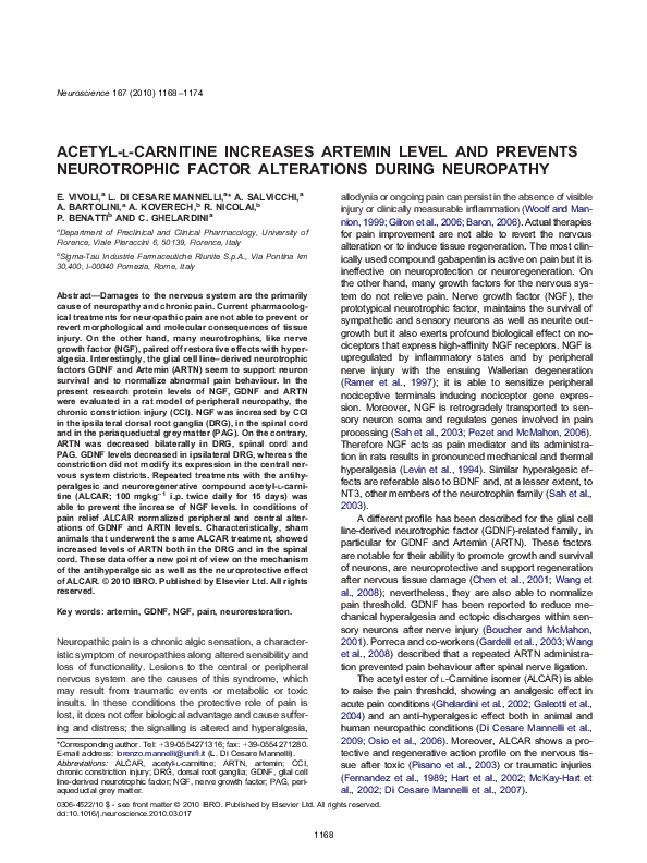
and in the PAG. In CCI saline-treated animals NGF was
increased in the ipsilateral DRG (150%), in the spinal cord
(350%) and in the PAG (180%) in respect to the sham
saline-treated group arbitrarily assigned a relative value of
100% (Fig. 1–CCI, saline). In all districts ALCAR administration was able to prevent NGF increase (Fig. 1–CCI,
ALCAR) in CCI-treated rats whereas it did not modify NGF
expression profile of sham rats (Fig. 1—sham, ALCAR).
In Fig. 2—panels A–D the expression of GDNF was
shown. The loose ligation of the sciatic nerve decreased
GDNF expression in the DRG of the ipsilateral side in
respect to the sham saline-treated group (70%; Fig. 2
panel A–CCI, saline). Repeated treatment with ALCAR
restored the control level (110%; Fig. 2 panel A–CCI,
ALCAR). No alterations were observed in the contralateral
DRG as well as in the spinal cord and in the PAG (Fig. 2,
panels B–D). ALCAR did not has a per se effect on GDNF
expression in sham animals (Fig. 2—sham, ALCAR).
The nerve injury was able to induce a decrease of
ARTN expression bilaterally in the DRG of CCI, salinetreated rats, up to 50% in the ipsilateral and at a lesser
extent in the contralateral (75%) (Fig. 3, panel A–CCI,
saline). Analogous reduction was measured in the spinal
cord (60%) (Fig. 3, panel B–CCI, saline). Both in DRG and
spinal cord of CCI the effects of ALCAR administration
consisted of a complete prevention in the ARTN decrease
(the expression level in DRG was bilaterally about 120%
and 150% in the spinal cord; Fig. 3, panels A, B–CCI,
ALCAR). ALCAR treatment was also able to increase
ARTN levels in DRG and spinal cord of sham animals
(160% and 240% respectively; Fig. 3, panel A, B—sham,
ALCAR). Fig. 3 panel C shows the pattern of expression of
Fig. 1. NGF protein expression levels. Rats underwent to surgical process with (CCI) or without (sham) the loose ligation of the sciatic nerve; the
administration of saline or ALCAR (100 mgkg⫺1 i.p., twice per day) was started on the day of the operation and continued for 15 days. (A) The right
(ipsilateral) and the left (contralateral) DRG were analyzed by Western blot: a densitometric evaluation is shown. (B) NGF levels in the total spinal cord
and (C) in the PAG. (D) Representative Western blots in respect to -actin expression. Results are expressed as the mean⫾SEM of five different
animals. Measurements in control samples (sham, saline) were assigned a relative value of 100%. * P⬍0.05, significantly different from sham, saline;
^ P⬍0.05 significantly different from CCI, saline.
�E. Vivoli et al. / Neuroscience 167 (2010) 1168 –1174
1171
Fig. 2. GDNF, protein expression levels. (A) contralateral and ipsilateral DRG were analyzed using a specific antibody. Effects of saline or ALCAR
treatment (100 mgkg⫺1 i.p., twice per day) were also evaluated in (B) spinal cord and (C) PAG. Western blot was performed 15 days after the
operation, (D) representative results are shown. Results are expressed as the mean⫾SEM of five different animals; -actin normalization was
performed for each sample. Measurements in sham, saline samples were assigned a relative value of 100%. * P⬍0.05, significantly different from
sham, saline; ^ P⬍0.05 significantly different from CCI, saline. For interpretation of the references to color in this figure legend, the reader is referred
to the Web version of this article.
ARTN in PAG: animals suffered a decrease up to 60% in
consequence of CCI also in this area (CCI, saline). Conversely, in the ALCAR-treated group a 190% expression
level of ARTN was measurable (Fig. 3, panel C–CCI,
ALCAR). ALCAR did not increase ARTN in PAG of sham
rats (Fig. 3C—sham, ALCAR).
DISCUSSION
Chronic pain developed in consequence of lesions to the
nervous system is the most observable phenomena of a
complex syndrome founded on dysfunctional signalling.
Pathological responses include neurotrophic factor expression. The role of NGF, GDNF and ARTN on pain has
been extensively studied but the relationship between their
expression levels and pain is not well established. The
gray matter located in the midbrain around the cerebral
aqueduct plays a key role in nociceptive processing (Behbehani, 1995). It receives afferent projections from a
number of brainstem and spinal areas which are known to
be involved in the modulation and conduction of nociception. The PAG is also a major component of the endogenous pain control system that is able to inhibit and facilitate
pain processing. The present research describes an increase in NGF expression levels in DRG during a painful
neuropathy induced by traumatic injury to the sciatic nerve.
This alteration dramatically raises in the central nervous
system, in particular in the spinal cord as well as in the
PAG. Blocking the action of NGF provides highly effective
pain relief in many animal models of acute and chronic
pain (Li et al., 2003; Gwak et al., 2003; Zahn et al., 2004;
Hefti et al., 2006). Current options about NGF-related therapies being explored involve the use of protein such as
antibodies, peptibodies and small protein domains, which
either sequester NGF or prevent the binding to its receptor
TrkA (Watson et al., 2008). On the contrary, GDNF and
ARTN, members of another family of growth factors primarily related to glia cells, have an antihyperalgesic effect.
Their characteristic actions on pain and neuron survival
�1172
E. Vivoli et al. / Neuroscience 167 (2010) 1168 –1174
Fig. 3. ARTN. Protein expression levels were analyzed in CCI rats in respect to sham and the effect of ALCAR (100 mgkg⫺1 i.p. twice per day) was
evaluated after administrations repeated for 15 days. Densitometric analysis of (A) contralateral and ipsilateral DRG; (B) total spinal cord; (C) PAG.
Panel (D) shows representative Western blot performed in the different areas with an ARTN specific antibody; a colorimetric method was used to
visualize the peroxidase-coated bands. Results are expressed as the mean⫾SEM of five different animals; -actin normalization was performed for
each sample. Measurements in control samples (sham, saline) were assigned a relative value of 100%. * P⬍0.05, significantly different from sham,
saline; ^ P⬍0.05 significantly different from CCI, saline.
strongly suggest these factors as therapeutic agents in
neuropathy. Our data showed a GDNF decrease in the
peripheral (DRG) but not in the CNS of CCI rats. On the
other hand, in the same rat model Nagano et al. (2003)
showed that the GDNF content in DRG was markedly
decreased at day 7 after the operation and stayed at low
levels at day 14; comparable reductions of GDNF levels
were observed in DRG on the injured side at 14 postoperative days in a model of spinal nerve ligation. GDNF decrease seems to be not specifically related to neuropathic
pain since GDNF was down-regulated in both dorsal root
ganglia and spinal cord of rats with chronic inflammation
induced by injection of complete Freund’s adjuvant (Fang
et al., 2003). Spinal infusion of GDNF prevents abnormal
pain behaviour in the L5 spinal nerve ligation and partial
sciatic nerve ligation models in the rat (Boucher et al.,
2000; Boucher and McMahon, 2001). McMahon and coworkers (Pezet et al., 2006) also evaluated the possibility
to administer GDNF by the use of lentiviral vectors. The
GDNF-expressing vectors were injected unilaterally into
the spinal dorsal horn 5 weeks before a spinal nerve
ligation producing a partial but significant reversal of thermal and mechanical hyperalgesia. Despite these effects of
GDNF on experimentally induced neuropathic pain, an
important caveat regarding GDNF therapy is that it elicits
clinically unacceptable side effects in vivo, such as weight
loss and allodynia (Nutt et al., 2003; Kordower et al.,
1999). On the other hand this strategy might increase the
risk of cancer because constant activation of its functional
receptor Ret by continuously GDNF can lead to malignancy (Bespalov and Saarma, 2007).
Along GDFN, ARTN is the neurotrophic factor mainly
involved in the strategy of pain treatment. In 2003, Porreca
and coworkers demonstrated that systematically administration of ARTN normalized the behavioural hypersensitivity to mechanical and thermal stimuli in rat that underwent
to spinal nerve ligation (Gardell et al., 2003). ARTN treatment was able to restore sensorimotor functions and improve morphological and neurochemical features of the
injury state induced by spinal nerve axotomy (Bennett et
�E. Vivoli et al. / Neuroscience 167 (2010) 1168 –1174
al., 2006) and dorsal root crush (Wang et al., 2008). Trophic effect of ARTN seems orientated to sensory neurons
where its receptor GFR␣3 is mainly expressed. Thus
ARTN could be a valuable tool to affect neuropathic pain
without having a broader effects on other organs and
tissues (Sah et al., 2003). For the first time, at our knowledge, in the present article a measure of ARTN levels was
performed in DRG, spinal cord and PAG. In respect to
GDNF, ARTN levels was more involved in the CCI-induced
alterations. ARTN expression was reduced in DRG, not
only ipsilaterally but also in the contralateral part. On the
other hand the peripheral injury was able to induce ARTN
decrease at spinal and supraspinal level. A mirror effect in
respect to NGF increase.
In this pathological condition ALCAR repeated treatments displayed an antihyperalgesic effect. Contrary to the
great part of the compounds clinically-used to treat neuropathic pain ALCAR joints this effect with neurorestorative
properties and a good safety profile. On the other hand,
ALCAR shows any analgesic or antihyperalgesic effect
after a single administration (data not shown) allowing to
hypothesize a relationship among the efficacy on pain
relief and the neuroprotective profile observed after a repeated treatment.
Clinical studies on diabetic peripheral neuropathy
show that ALCAR reduces pain sensation and improves
nerve conduction velocities and regeneration (Evans et al.,
2008). Open studies involving HIV-positive patients have
shown that a chronic treatment with ALCAR ameliorates
pain symptoms related to peripheral polyneuropathy (Scarpini et al., 1997). Hart and colleagues (2002) report that 6
months of oral ALCAR treatment results in peripheral
nerve regeneration of small sensory fibers as observed
from skin biopsies in patients with distal symmetrical polyneuropathy. Finally, in our laboratory it has been highlighted that the repeated treatment with ALCAR prevents
the apoptotic cascade in the peripheral nerve after CCI (Di
Cesare Mannelli et al., 2007, 2009).
Only the antiapoptotic effect has been related to a
specific pharmacodynamic property of ALCAR (Di Cesare
Mannelli et al., 2009) while the neuroregenerative profile
has been associated to the important ALCAR role for
energetics in the brain. ALCAR transports fatty acids from
the cell cytoplasm into the mitochondria where they provide a substrate for ATP generation via oxidative phosphorylation (Kidd, 2008).
The evidences shown in the present research suggest
the capability of ALCAR to restore altered levels of all the
evaluated neurotrophic factors: it prevents NGF increase
as well as GDNF and ARTN decrease. At least for NGF
and GDNF this effect seems strictly related to the injury
since ALCAR did not increase these factors in sham rats.
On the other hand an ALCAR normalizing effect on altered
neurotrophic factors has been described. In condition of
NGF depletion, as aged rats or rats subjected to total
fimbria–fornix transection, ALCAR upregulated the expression of the NGF and of its receptor, p75NGFR, in the CNS
(Piovesan et al., 1994; Foreman et al., 1995).
1173
Characteristically, ALCAR is able to increase ARTN
levels in a pathology-independent manner since it increases ARTN levels both in the peripheral and CNS of
sham animals in respect to saline-treated group. These
findings suggest that the neuroprotective profile of ALCAR
could be mediated, at least in part, by a signalling pathway
that target ARTN expression levels. Topic of debate is
whether the effect on NGF and GDNF are due to the ARTN
increase and which mechanism rules these molecular
events.
REFERENCES
Baron R (2006) Mechanisms of disease: neuropathic pain-a clinical
perspective. Nat Clin Pract Neurol 2:95–106.
Behbehani MM (1995) Functional characteristics of the midbrain periaqueductal gray. Prog Neurobiol 46:575– 605.
Bennett DLH, Boucher TJ, Michael GJ, Popat RJ, Malcangio M, Averill
SA, Poulsen K, Priestley JV, Shelton DL, McMahon SB (2006)
Artemin has potent neurotrophic actions on injured C-fibres. J
Peripher Nerv Syst 11:330 –345.
Bennett GJ, Xie YK (1988) A peripheral mononeuropathy in rat that
produces disorders of pain sensation like those seen in man. Pain
33:87–107.
Bespalov MM, Saarma M (2007) GDNF family receptor complexes are
emerging drug targets. Trends Pharmacol Sci 28:68 –74.
Boucher TJ, Okuse K, Bennett DLH, Munson JB, Wood JN, McMahon
SB (2000) Potent analgesic effects of GDNF in neuropathic pain
states. Science 290:124 –127.
Boucher TJ, McMahon SB (2001) Neurotrophic factors and neuropathic pain. Curr Opin Pharmacol 1:66 –72.
Chen ZY, Chai YF, Cao L, Lu CL, He C (2001) Glial cell line-derived
neurotrophic factor enhances axonal regeneration following sciatic
nerve transection in adult rats. Brain Res 902:272–276.
Di Cesare Mannelli L, Ghelardini C, Calvani M, Nicolai R, Mosconi L,
Vivoli E, Pacini A, Bartolini A (2007) Protective effect of acetyl-Lcarnitine on the apoptotic pathway of peripheral neuropathy. Eur
J Neurosci 26:820 – 827.
Di Cesare Mannelli L, Ghelardini C, Calvani M, Nicolai R, Mosconi L,
Toscano A, Pacini A, Bartolini A (2009) Neuroprotective effects of
acetyl-L-carnitine on neuropathic pain and apoptosis: a role for the
nicotinic receptor. J Neurosci Res 87:200 –207.
Evans JD, Jacobs TF, Evans EW (2008) Role of acetyl-L-carnitine in
the treatment of diabetic peripheral neuropathy. Ann Pharmacother 42:1686 –1691.
Fang M, Wang Y, He QH, Sun YX, Deng LB, Wang XM, Han JS (2003)
Glial cell line-derived neurotrophic factor contributes to delayed
inflammatory hyperalgesia in adjuvant rat pain model. Neuroscience 117:503–512.
Fernandez E, Pallini PR, Gangitano C, Aurora DF, Sangiacomo CO,
Sbricioli A, Ricoy J, Rossi GF (1989) Effects of L-carnitine, Lacetylcarnitine and gangliosides on the regeneration of the
transected sciatic nerve in rats. Neurol Res 11:57– 62.
Foreman PJ, Perez-Polo JR, Angelucci L, Ramacci MT, Taglialatela G
(1995) Effect of acetyl-L-carnitine treatment and stress exposure
on the nerve growth factor receptor (p75NGFR) mRNA level in the
central nervous system of aged rats. Prog Neuropsychopharmacol
Biol Psychiatry 19:117–133.
Galeotti N, Bartolini A, Calvani M, Nicolai R, Ghelardini C (2004)
Acetyl-L-carnitine requires phospholipase C-IP3 pathway activation to induce antinociception. Neuropharmacology 472:286 –294.
Ghelardini C, Galeotti N, Calvani M, Mosconi L, Nicolai R, Bartolini A
(2002) Acetyl-L-carnitine induces muscarinic antinocieption in
mice and rats. Neuropharmacology 43:1180 –1187.
Gardell LR, Wang R, Ehrenfels C, Ossipov NH, Rossomando AJ,
Miller S, Buckley C, Cai AK, Tse A, Foley SF, Gong B, Walus L,
Carmillo P, Warley D, Huang C, Engber T, Pepinsky B, Cate RL,
�1174
E. Vivoli et al. / Neuroscience 167 (2010) 1168 –1174
Vanderah TW, Lai J, Sah DW, Porreca F (2003) Multiple actions
of systemic artemin in experimental neuropathy. Nat Med 9:
1383–1389.
Gilron I, Watson PN, Cahill CM, Moulin DE (2006) Neuropathic pain: a
pratical guide for the clinician. CMAJ 175:265–275.
Gwak YS, Nam TS, Paik KS, Hulsebosch CE, Leem JW (2003) Attenuation of mechanical hyperalgesia following spinal cord injury by
administration of antibodies to nerve growth factor in the rat. Neurosci Lett 336:117–120.
Hart AM, Wiberg M, Youle M, Terenghi G (2002) Systemic acetyl-Lcarnitine eliminates sensory neuronal loss after peripheral axotomy: a new clinical approach in the management of peripheral
nerve trauma. Exp Brain Res 145:182–189.
Hefti FF, Rosenthal A, Walicke PA, Wyatt S, Vergara G, Shelton DL,
Davies AM (2006) Novel class of pain drugs based on antagonism
of NGF. Trends Pharmacol Sci 27:85–91.
Kidd PM (2008) Alzheimer’s disease, amnestic mild cognitive impairment, and age-associated memory impairment: current understanding and progress toward integrative prevention. Altern Med
Rev 13:85–115.
Kordower JH, Palfi S, Chen EY, Ma SY, Sendera T, Cochran EJ,
Cochran EJ, Mufson EJ, Penn R, Goetz CG, Comella CD (1999)
Clinicopathological findings following intraventricular glial-derived
neurotrophic factor treatment in a patient with Parkinson’s disease.
Ann Neurol 46:419 – 424.
Leighton GE, Rodriguez RE, Hill RG, Hughes J (1988) kappa-opioid
agonist produce antinociception after i.v. and i.c.v. but not intrathecal administration in the rat. Br J Pharmacol 93:553–560.
Levin GR, Rueff A, Mendell LM (1994) Peripheral and central mechanisms of NGF-induced hyperalgesia. Eur J Neurosci 6:1903–1912.
Li L, Xian CJ, Zhong JH, Zhou XF (2003) Lumbar 5 ventral root
transection-induced upregulation of nerve growth factor in sensory
neurons and their target tissues: a mechanism in neuropathic pain.
Mol Cell Neurosci 23:232–250.
McKay-Hart A, Wiberg M, Terenghi G (2002) Pharmacological enhancement of peripheral nerve regeneration in the rat by systemic
acetyl-L-carnitine treatment. Neurosci Lett 334:181–185.
Nagano M, Sakai A, Takahashi N, Umino M, Yoshioka K, Suzuki H
(2003) Decreased expression of glial cell line-derived neurotrophic
factor signaling in rat models of neuropathic pain. Br J Pharmacol
140:1252–1260.
Nutt JG, Burchiel KJ, Comella CL, Jankovic J, Lang AE, Laws ER Jr,
Lozano AM, Penn RD, Simpson RK Jr, Stacy M, Wooten GF
(2003) Randomized double-blind trial of glial cell line-derived
neurotrophic factor (GDNF) in PD. Neurology 60:69 –73.
Osio M, Muscia F, Zampini L, Nascimbene C, Mailland E, Cargnel A,
Mariani C (2006) Acetyl-L-carnitine in the treatment of painful
antiretroviral toxic neuropathy in human immunodeficiency virus
patients: an open label study. J Peripher Nerv Syst 11:72–76.
Pezet S, Krzyzanowska A, Wong LF, Grist J, Mazarakis ND, Georgievska B, McMahon SB (2006) Reversal of neurochemical
changes and pain-related behavior in a model of neuropathic pain
using modified lentiviral vectors expressing GDNF. Mol Ther 13:
1101–1109.
Pezet S, McMahon SB (2006) Neurotrophins: mediators and modulators of pain. Annu Rev Neurosci 29:507–538.
Piovesan P, Pacifici L, Taglialatela G, Ramacci MT, Angelucci L
(1994) Acetyl-L-carnitine treatment increases choline acetyltransferase activity and NGF levels in the CNS of adult rats following
total fimbria-fornix transection. Brain Res 633:77– 82.
Pisano C, Pratesi G, Laccabue D, Zumino F, Lo Giudice P, Bellocci A,
Pacifici L, Camerini B, Vesci L, Castorina M, Cocuzza S, Tredici G,
Marmiroli P, Nicolini G, Galbiati S, Calvani M, Carminati P, Cavaletti G (2003) Paclitaxel and cisplatin-induced neurotoxicity. A protective role of acetyl-L-carnitine. Clin Cancer Res 9:5756 –5767.
Ramer MS, French GD, Bisby MA (1997) Wallerian degeneration is
required for both neuropathic pain and sympatethetic sprouting
into the DRG. Pain 72:71–78.
Sah D, Ossipov M, Porreca F (2003) Neurotrophic factors as novel
therapeutics for neuropathic pain. Nat Rev Drug Discov 2:460 – 472.
Scarpini E, Sacilotto G, Baron P, Cusini M, Scarlato G (1997) Effect of
acetyl-L-carnitine in the treatment of painful peripheral neuropathies in HIV⫹ patients. J Peripher Nerv Syst 2:250 –252.
Wang R, King T, Ossipov MH, Rossomando AJ, Vanderah TW, Harvey P, Cariani P, Frank E, Sah DWY, Porreca F (2008) Persistent
restoration of sensory function by immediate or delayed systemic
artemin after dorsal root injury. Nat Neurosci 11:488 – 496.
Watson JJ, Allen SJ, Dawbarn D (2008) Targeting nerve growth factor
in pain. What is the therapeutic potential? BioDrugs 22:349 –359.
Woolf CJ, Mannion RJ (1999) Neuropathic pain: aetiology, symptoms,
mechanisms, and management. Lancet 353:1959 –1964.
Zahn PK, Subieta A, Park SS, Brennan TJ (2004) Effect of blockade of
nerve growth factor and tumor necrosis factor on pain behaviors
after plantar incision. J Pain 5:157–163.
(Accepted 8 March 2010)
(Available online 16 March 2010)
�
 Aleardo Koverech