zyxwvutsrqpo
Eur J. Biochem. 109, 285-290 (1980)
T> by FEBS 1980
Recovery of Pure Ribosomal Proteins from Stained Gels
A Fast Method of Purification of Active Proteins
Carmelo BERNABEU, Francisco SANCHEZ-MADRID, and Ricardo AMlLS
Instituto de Bioquimica de Macromoleculas, Centro de Biologia Molecular,
Consejo Superior de Investigaciones Cientificas y Universidad Autonoma de Madrid
(Received February 181May 19, 1980)
A simple technique has been developed for eluting ribosomal proteins from stained gels in the
presence of an acetic acid solution. The ribosomal proteins are then separated from the dye by
anion-exchange chromatography under dissociating conditions.
Ribosomal proteins purified by these methods give total cross-reaction with proteins obtained
by standard procedures, when tested by immunodiffusion against their corresponding antibodies,
and show the same electrophoretic mobility as standard proteins in bidimensional polyacrylamide
gel systems.
Ribosomal proteins L7/L12, recovered from stained gels and purified by these methods, are able
to reconstitute the elongation-factor-G-dependent GTPase activity of ribosomal particles deprived
of these proteins. Radioactive protein L1, recovered in the same way, is incorporated into a total
reconstituted 50-S subunit, competing with an excess of standard L1 present in the pool of total
proteins from 50-S subunits used for reconstitution.
These results suggest that bidimensional electrophoresis can be considered an alternative system
of purification of active proteins from complex mixtures.
zyx
zyxwvutsr
zyxwvutsrqpo
zyx
Ribosomal proteins are usually purified by a combination of ion-exchange chromatography and gel
filtration in the presence of urea or high ionic strength
to avoid formation of protein complexes (for reviews,
see [1,2]).
Monodimensional gel electrophoresis has also
been used to prepare ribosomal proteins, either by
elution of migrating proteins from the end of gel
columns during electrophoresis [3,4] or by extraction
of proteins from slices cut from gel columns after
electrophoresis [5 - 121. Bidimensional polyacrylamide
gel electrophoresis provides better resolution of complex protein mixtures than either of the two procedures
mentioned. It has, however, rarely been used for preparative purposes [13,14], because resolved components can be easily detected in gel slabs only by staining and must, therefore, be separated from the dye
after extraction from the gel.
We have previously shown [14] that dye can be
separated from proteins eluted from bidimensional
gels by filtration on Sephadex G-25. In this report we
show that this separation can be achieved more
rapidly and conveniently by anion-exchange chromatography.
~-
MATERIALS A ND METHODS
5 0 3 subunits were obtained from Eschcrichia coli
MRE600 70-S ribosomes [15]. 80-S ribosomes from
Succlzurom,yces cerevisiue Y166 [I 61. Total proteins
from 50-S subunits and 80-S ribosomes were extracted
following Barritault et al. [17].
Chemical labelling of total proteins from the 50-S
subunit with '''I was performed basically as described
by Greenwood et al. [I81 in the presence of 8 M urea.
Separation of free ' 'I was carried out in a Sephadex
G-25 column using 5 % (v/v) acetic acid as eluant.
The protein peaks were lyophilized. All the acetic acid
solutions were vacuum degassed for 10 min before use.
DEAE-cellulose (DE-23), BD-cellulose and Dowex-1 (dry mesh 200-400) were purchased from
Whatman, Serva and Sigma respectively, 12'1 (15 mCi/
pg) from Amersham.
Bidimensionul Polyacrylumide Gel Electroplzort~sis
0.7 mg total proteins from 50-S subunits and 1 mg
total proteins from 80-S ribosomes were dissolved in
buffer A (8 M urea, 20 mM NH4HC03, 10% sucrose
and 0.1 M 2-mercaptoethanol), and electrophoresed
under the conditions of Kaltschmidt and Wittmann
zyxwvu
Ahhrrvzution. EF-G, elongation factor G.
�zyxwvutsrqpo
zyxwvutsrq
zyxwvutsrq
zyxwvutsr
286
Purification of Proteins from Gels
[19] as modified by Howard and Traut [20]. After
electrophoresis the gel slabs were stained for 2 h at
4 ' C with 0.18 % Coomassie blue, 50% methanol and
5 %acetic acid. Destaining was performed with methanol 50% and 7.5% acetic acid at 4'C.
loot
Extraction of Rihosomul ProtPins
und Coonzussie Blue
Spots were cut out and processed essentially as
described [14]. Macerated gels were extracted in the
presence of 1.5 ml 66'x) (v/v) acetic acid, except as
otherwise indicated. After 12 h of extraction at 4 ' C ,
the supernatant was separated from the gel with a
pasteur pipette.
Separation of Ribosomal Proteins
.from Coonzussie Blue
Supernatants containing the ribosomal proteins
and Coomassie blue were applied to a pasteur pipette
plugged with G FjC fiber filter, which contained
approximately 0.7 ml packed resin, previously washed
with acetic acid and equilibrated with distilled water.
When needed, a modified column was prepared by
placing 0.7 ml Dowex-I on the top of a Sephadex G-25
column (0.6 x 12 cm); it was washed with acetic acid
and equilibrated with distilled water. After the chromatography ihe eluted volumes were diluted with
distilled water to a 20% (v/v) acetic acid and lyophilized.
zyxwv
Acetic acid (%)
zyx
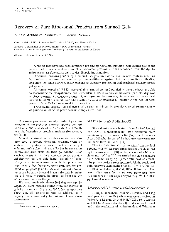
Fig. 1. Extraction qf rihosomul proteins jrom gels at dif;frrent acetic.
acid concentrations. Proteins L2 (O), L3 (0),and LIY ( A ) were
homogenized and 1.5 ml acetic acid at the indicated concentration
was added. After 12 h extraction at 4 'C the supernatant was separated from the gel and the radioactivity measured in a scintillation
gamma counter
RESULTS
To determine the recovery in the various steps of
purification, ribosomal proteins were chemically labeled with 1251.These labeled proteins were resolved
by bidimensional polyacrylamide gel electrophoresis.
After staining, the spots were cut out and ribosomal
proteins and Coomassie blue were extracted from the
gels at different acetic acid concentrations. As shown
in Fig. 1, 40 % (v/v) acetic acid is sufficient to give
maximum protein release after 12 h of incubation
at 4°C.
Knowing the highly acidic character of Coomassie
blue (three sulfonic groups) and the basic or weak
acidic character of the ribosomal proteins, we saw
the possibility of using ion-exchange chromatography
to separate the proteins from the dye. Table 1 shows
the percentages of protein and dye recovered from the
eluant after the chromatography of stained proteins in
small columns of various exchange resins.
Anion exchangers retained the dye while permitting
the elution of most of the radioactive proteins applied
to the column. Amberlite showed some deviation from
this behavior, probably because of its chemical alteration at the concentration of acetic acid used in the
experiment.
As expected, cation exchangers produced the opposite effect; however, the recovery of radioactive proteins from the columns, by means of changes in pH,
high ionic strength and denaturing agents, was extremely difficult. Anion exchangers were chosen as
they permitted adequate separation of proteins from
dye without any other manipulation of the sample.
Dowex-1, BD-cellulose and DEAE-cellulose were
selected for further studies because the diffusion of
the dye on the column was minimal.
zyxwvutsrqpo
Preparation of Spec~ficAntisern
and Inzmunodijjusion Tests
Antisera were raised in rabbits against proteins
L7/L12 from E. coli and L15 from S. cerevisiue. Processing of antisera is described elsewhere [21]. Immunodiffusion tests were carried out on 1 agar
plates in 10 mM Tris-HC1 pH 7.4, 150 mM NaCl.
The proteins were resuspended in 10 mM Tris-HCI
p H 7.4, 2 mM 2-mercaptoethanol, 6 M urea. The
amount of protein in the different samples was estimated by radial immunodiffusion [22].
Otizer Metizods
Reconstitution of 50-S ribosomal particles was
carried out as described [23] and the reconstituted
particles were treated with NHeCl/ethanol to obtain
Po cores [24]. EF-G-dependent GTPase assays were
performed according Modolell and Vazquez [25] and
the poly(Phe) synthesis directed by poly(U) was done
as described elsewhere [15]. The purification of proteins L7/L12 from E. coli following the procedure of
F. Sanchez-Madrid et al. [21], the purification of L15
from S. cerevisiae will be described elsewhere.
�zyxwvutsrqponm
zyxwvuts
C . Bernabeu, F. Sanchez-Madrid, and R. Amils
287
The influence of acetic acid concentration on protein recovery is similar in the three systems (Fig. 2).
DEAE-cellulose columns can be used with the widest
range of acetic acid concentrations, 15 - 66'%,,avoiding
dilution o f the sample when 66% acetic acid is used
for extraction.
The rates of recovery achieved with eight SO-S
ribosomal proteins eluted through DEAE-cellulose
after extraction with 66% acetic acid (Table 2) show
that an average of 64% of the protein present in a
spot is recoverable in its pure form.
Previous work with Sephadex G-25 [14] demonstrated that this method of extraction and purification
did not change the protein's electrophoretic mobilities. Proteins purified by anion-exchange chromatography also exhibit the same electrophoretic mobility
as standard ones (data not shown).
To test whether these methods of protein purification were capable of maintaining antigenic determinants, several ribosomal proteins from Escherichiu
coli and Succlzuromyces cerevisiue were extracted from
stained gels and the dye was separated on DEAE-
Table 1. Sepurution o j radiouctive proteins and Coomussie blue b.y
ion-excliunge chromutograpliy
Samples of 1 ml each containing '251-labelled total proteins from
50-S subunits (70 pg/ml) and Coomassie blue ( 5 0 pM) in 33% acetic
acid were applied, as described in Materials and Methods, to small
columns of different ion exchangers, previously equilibrated with
distilled water. After the elution the columns were washed with
1 ml 33% acetic acid. The radioactivity and the absorbance at
605 nm were determined
Table 2. Ribosomal protein recovery ufier extraction and anionrxckanRe chromutogruphy in DEAE-cellulose
Stained 'ZsI-labelled ribosomal proteins separated by bidimensional
polyacrylamide gel electrophoresis were cut, macerated and extracted at 4'-C with 1.5 ml 66'j: acetic acid for 12 h. The supernatants were applied to small DEAE-cellulose columns, as described in Materials and Methods. The protein radioactivity in the
gel after the extraction, bound to the column and eluted, was
estimated. The percentages refer t o the radioactivity originally in
the spots, which ranged from 1 x l o 5 to 8 x l o 5 counts/min
zyxwvutsrq
Ion exchangers
Coomassie
blue eluted
Protein
eluted
Protein
Radioactivity
in the gel after
extraction
zyxw
Radioactivity
in the column
Radioactivity
in the eluant
zyxwvutsrq
Cation exchangers
Dowex-SOX2 (Serva)
Cellex-P (BioRad)
Phosphocellulose, PI 1 (Whatman)
CM-cellulose, C M 52 (Whatman)
Amberlite, CG-120 (Prolabo)
Anion exchangers
1
10
89
94
17
70
79
79
84
78
57
0
0
0
0
21
2
2
2
22
Dowex-l (Sigma)
Dowex-1x2 (Serva)
DEAE-cellulose, DE-23 (Whatman)
BD-cellulose (Serva)
Amberlite, CG-4B (Prolabo)
L5
L10
L13
L14
L15
L16
L23
L25
Mean
30
49
27
29
31
29
40
21
32
69
45
69
70
68
65
50
72
64
1
7
5
2
2
6
10
7
9
5 1 3
10
zyxwvutsrq
100
1.0
A
0.8
0.6
8
T
zyx
zyxwvuts
zyxwv
T
0.4
9
0.2
- - - - - -- - - - - - _
I
0
20
40
60
0
20
40
60
0
20
40
60
0
Acetic acid ( m l / 100rnl)
Fig. 2. Separation o j Coomussie hluefrom ribosomal proteins in anion-exckunge resins. 1.5-ml samples containing 'Z51-labelledtotal proteins
from 50-S subunits (0.2 mg/ml) and Coomassie blue G-250 (50 pM), at the indicated acetic acid concentrations, were applied, as described
in Materials and Methods, to small columns of Dowex-1 (A), BD-cellulose (B) and DEAE-cellulose (C). Protein radioactivity in the eluted
volumes and in the resins was estimated. Absorbance at 605 n m was measured t o determine the amount of Coomassie blue present in the
eluant (100% dye = 0.8 unit absorbance)
�Purification of Proteins from Gels
28K
by the addition of purified L7/L12. We chose this
reconstitution experiment to test the ability of proteins
L7/L12, purified from gels, to restore the GTPase
activity of the cores Po.
Proteins L7/L12, purified by anion exchangers,
showed in preliminary experiments that the above
conditions did not separate the proteins from a contaminant that has a strong inhibitory effect on the
reconstitution of the GTPase activity (Table 3, compare lines 5 and 6 with 3) and poly(Phe) synthesis
(data not shown). This contaminant, presumably small
polymers of acrylamide released from the gels during
the acidic extraction, is not retained on the resins
under these conditions.
In order to separate this contaminant, a chromatographic system combining anion exchange and
gel filtration was devised. Compared to the same
amount of standard L7/L12, the correspondent proteins extracted from gels and purified by this modified
column restored up to 70% of the cores Po EF-Gdependent GTPase activity (Table 3, lines 7 and 8).
When a control (material extracted from a piece of
gel without protein) was run in the same column,
fractions with an inhibitory effect were obtained in
positions corresponding to a smaller molecular weight
than that of the proteins. Similar results were obtained
in the poly(Phe) synthesis assay.
To test the ability of proteins prepared by this
method to compete with proteins prepared by standard
ones, the total reconstitution system of the 50-S ribosomal subunit was chosen. We selected ribosomal
proteins able to reconstitute 50-S particles whcn
labelled with iodine. '231-labelled ribosomal protein
L1 from E. coli purified from gels was mixed with
total proteins from the 50-S subunit and total rRNA
in the conditions already described for total reconstitution. After incubation, the particles were precipitated with ethanol in the presence of 1 M NH4CI to
eliminate non-specific interactions, resuspended and
run through a sucrose gradient. Fig.4 shows that a
significant amount of radioactive L1, 287; of the
maximum expected, had been incorporated into the
reconstituted 50-S subunits, competing successfully
with an excess of untreated L1.
zyxwvutsrqponm
zyxwvutsrqpo
zyxwvutsrqp
zyxwvutsrq
Fig. 3. Double-rmnzunodifftrsion test. (A) Well 1, antiserum to E. coli
L71L12; well 2, proteins L71L12 obtained by standard procedures
( 3 pg); well 3, proteins L7/L12 recovered from gel slab and purified
with Dowex-I. (B) well 1, antiserum to S. cerevisiue L15; well 2,
protein L15 obtained chromatographically (2 pg), well 3, L15 recovered from gel slab and purified with DEAE-cellulose
cellulose and Dowex-I. Fig. 3 shows the total crossreaction of proteins L7/L12 from E. coli and L15
from S . cerevisiue, with their counterparts purified by
standard procedures when tested against their corresponding antibodies. Similar results have been obtained with other ribosomal proteins.
We tried to compare the activity of proteins purified by this method with those prepared by the
standard systems. In E. coli ribosomes it is difficult
to assign specific roles to any of the proteins, with
the exception of the acidic proteins L7/L12. When
these proteins are removed by ethanol precipitation
under high ionic strength, the resulting cores (Po)
are deficicnt in GTPase activity, which can be restored
DISCUSSION
Bidimensional polyacrylamide gel electrophoresis
provides a versatile, gentle and high-resolution method
for fractionation of complex protein mixtures based
on size and net charge. We intend to use this method
purification of active ribosomal proteins. The KaltSchmidt and Wittmann system was chosen because
it permits an adequate resolution of all the proteins
of the ribosomal particle [19]. Staining the proteins
simplifies their location and identification.
�zyxwvutsrqponm
zy
zyxwvuts
289
C. Bernabeu, F. Sanchez-Madrid, and R. Amils
Table 3. EF-G dependent GTPuse activity
The ashay was carried out as described in Materials and Methods. All the components of the reaction, with the exception of the GTP,
were preincubated for 10 min at 37°C. The concentration of the proteins was determined by radial immunodiffusion. The activity is given
as pmol GTP hydrolysed/pmol particles
Additions
Activity
-
standard
L7/L12
L7lL12 from
gel\ + anion
exchange
control froin
gels
L7/L12 from
gels modified
procedure
+
pmol/pmol
704
Po
Po
Po
PO
Po
Po
Po
a
zyxw
zy
400C
Y)
c
C
3
8
zyxwvutsrq
I
H
UI
-0 200c
N
1
at 4°C with acetic acid solution above 40% (vlv)
(Fig. 1). These extraction conditions do no affect the
ribosomal proteins activity. On the contrary, proteins
exposed to long incubations (24 h or more) with 66 %
acetic acid at 4 "C give higher yields of total reconstitution of the 50-S ribosomal subunit than proteins exposed to standard incubations (1 h) (unpublished
work).
The most important advantage of anion-exchange
chromatography over gel filtration in separating proteins from Coomassie blue is the large volume of
samples that can be processed, resulting in greater
recovery in the extraction and in the chromatography.
Subsequent applications of the purified proteins
will determine when simple anion-exchange cliromatography is sufficient. If active proteins are required,
the separation of the contaminant released from the
gels is needed. This can be achieved by the modified
system that combines anion exchange and gel filtration.
In addition to the good recovery obtained with
this technique, the electrophoretic mobilities, the antigenic determinants, the reconstitution ability and the
activity of the proteins are preserved.
The results suggest that this method can be considered an alternative system of purifying protein
mixtures, with these advantages over standard chromatographic methods : selectivity, high yields and
small time consumption, permitting a wide spectrum
of possible applications. Using this approach our
laboratory is currcntly producing antisera against
specific ribosomal proteins; which, while being an
important tool in the ribosome field, has been available up to now only to groups with a large purification
capacity.
zyxwvutsrq
.
C
(100)
(6)
(103)
(26)
(29)
(15)
(60)
(70)
zyxwvut
1
1.5
Volumes equivalent to the protein extracted froin gels were used.
.-E
42.5
2.6
44.0
11.0
12.3
6.4
25.5
29.6
(7;)
zyxwvut
zyxwvut
zyxwvutsrqponm
C
5
10
15
20
Fraction number
Z
Fig. 4. I n c ~ q ~ o r u f i OoJn E. coli ribosomul prort.in L1 e.utrc~reciJi-om
grls into 50-S particles by tofu1 reconstitution, '2'1-L3, recovered
from a stained gel and purified as described in Materials and
Methods, was lyophilized and then mixed with total proteins from
50-S subunits and r R N A in the conditions described for total reconstitution. Aftcr the incubation the reconstituted particles were
precipitated with ethanol in the presence of 1 M NH&I and the
resuspended particles run in a sucrose gra d lent
'
Previously we reported that 66 7(:(v/v) acetic acid
efficiently extracted stained proteins from gels aftcr
6 h incubation at room temperature [14]. This extraction system was intended for use in studies of the
structure and function of the ribosome, thus optimal
conditions causing the least possible damage were
sought. Optimal extraction of ribosomal proteins
from gels can be achieved by overnight incubation
�290
zyxwvutsr
zyxwvutsrqpon
zyxwvutsrqpo
zyxwvutsrqpon
zyxwvutsrqp
zyxw
zyxwvutsrq
C. Bernabeu, F. Sanchez-Madrid. and R. Amils: Purification ol' Proteins from Gels
This work was supported by an Institutional Grant to the
Centro de Biologiu Mulecirlur ,fi.om Comision Administradoru del
Desc'uenlo Complementurio (Instituto Nacionul de Previsihn) and
personal grants from Essex Laboratories and Lilly Indian of Spain.
We thank to D r J. P. G. Ballesta for encouraging us and for
helpful discussions, and Drs S. Ochoa and C. Cantor for their
critical evaluation of the manuscript.
12. Mardian, J. K. W. & Isenberg, I. (1978) Anul. Biocliem. 91,
1-12.
13. Goerl, M., Wellle, H . & Bielka, H. (1978) Biochim. 5ioplzj.s.
Actu, 519, 418-427.
14. Bernabeu, C., Conde, F. P. & Vazquez, D. (1978) Anal. Bioelinn. 84, 97- 102.
15. Amils, R., Matthcws, E. & Cantor, C. R. (1978) M e r h d s
Enzyniol. 59, 449 461.
16. Battaner, E. & Vazquez, D . (1971) Biocliim. Biopli.~~.~.
Actri,
254, 316-330.
17. Barritault, D., Expert-Bezanqon, A,, Guerin, M . F. & Hayes,
D. (1976) Eur. J . Bioclzem. 63, 131-135.
18. Greenwood, F. C., Hunter, W. M . & Glover, J. S. (1963) Biochern. J. 89, 114- 123.
19. Kaltschmidt, E. & Wittmann, H . G . (1970) Pruc. Nut1 Acud.
S1.i. USA, 67, 1276-1282.
20. Howard, G . A. & Traut, R. R. (1974) Metliuds Enzymol. 30,
526-539.
21. Sanchez-Madrid, F., Reyes, R., Conde, P. & Ballesta, J. P. G.
(1979) Eur. J . Biocliem. Y8, 409-416.
22. Mancini, G., Carbonara, A. 0. & Heremans, J. F. (1965) Inirnunochernistry, 2, 235 -254.
23. Amils, R., Matthews, E. A. & Cantor, C. (1978) Nuc,leic Acid,
Rcs. 5, 2455 - 2470.
24. Hamcl, E., Koka, M. & Nakamoto, T. (1972) J . Biol. Cliem.
247, 805-814.
25. Modolell. J. & Vazquez. D. (1973) J. Biol. ( % m z . 248. 488493.
-
REFERENCES
1 . Wittmann, H. G . (1974) in Rilwsonie.~(Nomura, M., Tissieres,
A. & Lengyel, R., eds) pp. 93- 114, Cold Spring Harbor,
New York.
2. Dijk, J. & Littlechild, J. (1978) Methuds Enzyniol. 59, 481 -502.
3. Racusen, D. & Calvanico, N . (1964) Anal. Biocliem. 7 , 62-66.
4. Shuster, L. (1971) Metlzotls Emyniol. 22, 412-433.
5. Lewis, U . J . &Clark, M. 0. (1963) A m / . Biocliem. 6, 303-315.
6. Martinage, A,, Sautiere, P., Kerckaert, J. P. & Biserte, G. (1976)
Biochim. Biophys. Acta, 420, 37-41.
7. Sulitzeanu, D., Slavin, M . & Yecheskeli, E. (1967) Anal. Bioc l u m 21, 57-67.
8. Kohen, A. L. & Shaw, C. R. (1964) Anul. Riocliern. 9, 495498.
9. Ziola, B. R. & Scraba, D. (1976) AiiaI.Biochem. 72. 366-371.
10. Hjerten, S. (1 973) in Metliotlologicul Developments in Biochcmistry (Reid, E., ed.) vol. 2, pp. 39-47, Longman, London.
1I . Stephens, R. E. (1975) Ancrl. Bioclieni. 65, 369- 379.
C . Bernabeu, F. Sanchez-Madrid, and R . A i d s , Instituto de Bioquimica de Macromoleculas, Centro de Biologia Molecular,
Consejo Superior de Investigaciones Cientificas y Universidad Autbnoma de Madrid, Facultad de Ciencias,
Universidad Autonoma de Madrid, lnstituto de Biologia del Desarrollo, Canto Blanco, Madrid-34, Spain
�
 Carmelo Bernabeu