review
review
Tyrosine phosphorylation in semaphorin signalling:
shifting into overdrive
Mélanie Franco & Luca Tamagnone+
Institute for Cancer Research and Treatment (IRCC), University of Torino, Candiolo, Italy
The semaphorins constitute a large family of molecular signals with
regulatory functions in neuronal development, angiogenesis, cancer progression and immune responses. Accumulating data indicate that semaphorins might trigger multiple signalling pathways,
and mediate different and sometimes opposing effects, depending
on the cellular context and the particular plexin-associated subunits of the receptor complex, which can include receptor-type or
cytoplasmic tyrosine kinases such as MET, ERBB2, VEGFR2, FYN,
FES, PYK2 and SRC. It has also been shown that a specific plexin
can alternatively associate with different tyrosine kinase receptors,
eliciting divergent signalling pathways and functional outcomes.
Tyrosine phosphorylation is a pivotal post-translational protein
modification that regulates intracellular signalling. Therefore,
phosphorylation of tyrosines in the intracellular domain of plexins could determine or modify their interactions with additional
signal transducers. Here, we discuss the potential relevance of
tyrosine phosphorylation in semaphorin-induced signalling, with
an emphasis on its probable role in dictating the choice between
multiple pathways and functional outcomes. The identification of
implicated tyrosine kinases will pave the way to target individual
semaphorin-mediated functions.
Keywords: kinase; plexin; receptor; semaphorin; tyrosine
EMBO reports (2008) 9, 865–871. doi:10.1038/embor.2008.139
See Glossary for abbreviations used in this article.
Introduction
The semaphorins constitute a wide family of membrane-bound and
secreted proteins that provide guidance cues for axon pathfinding and cell migration (Tamagnone & Comoglio, 2000; Zhou et al,
2008). Moreover, semaphorins are implicated in the regulation of
many biological processes, such as neural development and organ
morphogenesis in the embryo, and immune response, angiogenesis
and invasive tumour growth in the adult. Semaphorin signalling
affects cytoskeletal remodelling and integrin-dependent adhesion,
Division of Molecular Oncology, Institute for Cancer Research and Treatment (IRCC),
University of Torino, S.P. 142, 10060 Candiolo, Torino, Italy
*Corresponding author. Tel: +39 (0)11 993 3204; Fax: +39 (0)11 993 3225;
E-mail: luca.tamagnone@ircc.it
Submitted 25 January 2008; accepted 24 June 2008; published online 25 July 2008
©2008 EUROPEAN MOLECULAR BIOLOGY ORGANIZATION
consequently impinging on cell migration; however, it has also been
implicated in the regulation of cell proliferation and apoptosis, and,
recently, in cell differentiation (for reviews, see Casazza et al, 2007;
Kruger et al, 2005; Tamagnone & Giordano, 2006).
The main functional receptors for semaphorins are members of the
plexin family (Takahashi et al, 1999; Tamagnone et al, 1999); notably,
a subset of secreted semaphorins require obligate co-receptors associated with the plexins—which are known as neuropilins—although
their potential role in intracellular signal transduction remains controversial. In mammals, nine plexins have been identified and divided
into four subfamilies: plexin A1 to plexin A4, plexin B1 to plexin
B3, plexin C1 and plexin D1. Plexins are large single-pass transmembrane molecules, the extracellular moiety of which contains
conserved protein motifs, such as the ‘sema domain’ that presents
a ‘β-propeller’ structure (for a comprehensive review, see Gherardi
et al, 2004), the cysteine-rich MET-related-sequences or PSI motifs,
and the IPT domains (Artigiani et al, 1999). These structural domains
are thought to mediate protein–protein interactions, although their
specific functional relevance has not yet been elucidated.
The cytoplasmic region of plexins is highly conserved among
the family members, and yet it is unique, owing to its lack of
apparent homology with other proteins (Maestrini et al, 1996).
This domain has been shown to associate with several intracellular signal transducers, eliciting multiple signalling pathways
in response to semaphorin stimulation (Kruger et al, 2005; Fig 1).
For instance, the intracellular domains of plexins contain GTPaseactivating protein (GAP)-like motifs that are able to interact with—
and downregulate—the monomeric G-protein R-Ras (Oinuma
et al, 2004). Another region in the intracellular domain of plexins
has been shown to interact with Rho family GTPases such as Rnd1
and Rac (Rohm et al, 2000; Tong et al, 2007). Current data indicate
that the association of these monomeric G-proteins with plexins
could induce the conformational changes required to elicit their
GAP activity or to allow for the recruitment of additional signal
transducers. Plexins have also been found to interact with GTPaseexchanger factors (GEFs), such as the Rac-GEF FARP2 (Toyofuku
et al, 2005) and PDZ-Rho-GEF (Driessens et al, 2002; Perrot et al,
2002; Swiercz et al, 2002). By contrast, the cytoplasmic domain of
plexin A1 and plexin B1 can interact with p190 Rho-GAP (Barberis
et al, 2005); therefore, regulation of RhoA might vary in response
to semaphorin signals in different cells (Swiercz et al, 2008). This
is in agreement with the common observation that plexins have
EMBO reports
VOL 9 | NO 9 | 2008 8 6 5
reviews
Tyrosine phosphorylation in semaphorin signalling
M. Franco & L. Tamagnone
Glossary
CDK
CRMP
DAP12
EGF
ERBB2
FAK
FES
MAPK
PI(3)K
PLCγ
PTB
PYK2
RhoA
Rnd1
Sema4A
SH2
TREM2
VEGFR2
cyclin-dependent kinase
collapsin response-mediator protein
DNAX-activating protein of 12 kDa
epidermal growth factor
erythroblastic leukaemia viral oncogene homologue 2
focal adhesion kinase
feline sarcoma oncogene
mitogen-activated protein kinase
phosphatidylinositol-3-kinase
phospholipase-Cγ
phosphotyrosine-binding
proline-rich tyrosine kinase 2
Ras homologue gene family member A
Rho family GTPase 1
semaphorin 4A
SRC homology 2
triggering receptor expressed on myeloid cells 2
vascular endothelial growth factor receptor 2
A
been found to trigger multiple intracellular pathways, sometimes
leading to opposing functional effects.
In fact, semaphorin signals are often mediated by multimeric
receptor complexes that contain additional transmembrane subunits
associated with plexins. In particular, receptor tyrosine kinases (RTKs)
might associate with plexins on the cell surface, and have been shown
to have a pivotal role in semaphorin signalling pathways (Conrotto
et al, 2004; Giordano et al, 2002; Swiercz et al, 2004, 2008;
Toyofuku et al, 2004a; Winberg et al, 2001). Moreover, in dendritic
cells and osteoclast precursors, plexin A1 can be found in a complex
with the transmembrane protein known as TREM2, which is, in turn,
associated with the immunoreceptor tyrosine-based activation motif
(ITAM) transducer, DAP12 (Takegahara et al, 2006), responsible for
activating the intracellular tyrosine kinase SYK. Notably, certain semaphorins might also signal through receptors that are distinct from
plexins. For instance, in the immune system, Sema4A and Sema4D
might use the alternative low-affinity receptors TIM2 and CD72,
respectively (Kikutani & Kumanogoh, 2003; Kumanogoh & Kikutani,
2001). CD72 controls tyrosine phosphorylation cascades in B cells
B
Semaphorin
Semaphorin
Plexin
Plexin
RTK
RTK
Integrin
Integrin
α
β
TK
β
R-Ras
P
P
GA
TK
α
β
R-Ras
P
Integrin
TK
P
P
-GT
Rac -GTP
Rnd
GTPase
regulators
GTPase
regulators
P
GA
α
P
-GT
Rac -GTP
Rnd
P
TK
CDK5
PI(3)K
PI(3)K
PI(3)K
AKT
AKT
AKT
Rho/Rac
PI(3)K
AKT
Rho/Rac
CRMP
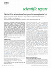
Fig 1 | Tyrosine kinases at the crossroads of semaphorin signalling pathways. Semaphorin receptors can elicit multiple intracellular signalling cascades to control
axon guidance, cell migration and invasive growth (reviewed by Kruger et al, 2005; Zhou et al, 2008). Notably, the cytoplasmic tail of plexins might become tyrosine
phosphorylated by either RTKs or cytoplasmic tyrosine kinases, indicating further regulatory mechanisms that have not been characterized (see text and Table 1 for details
and specific references). (A) Semaphorin receptor complexes often include plexin-associated RTKs. For example, plexin B1 and plexin A1 have been found in association
with tyrosine kinases such as MET, RON, ERBB2, VEGFR2 or OTK (off-track kinase) in a cell-specific manner. On semaphorin binding, these RTKs become activated,
resulting in a differential regulation of cell migration, invasive growth and morphogenesis. (B) The intracellular domain of plexins might also associate with cytoplasmic
tyrosine kinases implicated in signal transduction. For example, on Sema4D stimulation, endothelial cell chemotaxis might require integrin-dependent activation of the
kinases PYK2 and SRC, triggering the PI(3)K/AKT pathway. Moreover, in different neuronal populations, the tyrosine kinases FAK, FYN and FER/FPS regulate neurite
outgrowth in response to Sema3A and Sema3B. Therefore, semaphorin signals control cytoskeletal dynamics, integrin function, axon guidance, cell migration and invasive
growth, leading sometimes to opposing functional effects, due to the activation of distinct pathways in a cell-specific manner. CDK5, cyclin-dependent kinase 5;
CRMP, collapsin response-mediator protein; ERBB2, erythroblastic leukaemia viral oncogene homologue 2; FAK, focal adhesion kinase; FER, feline sarcoma oncogene;
GAP, GTPase-activating protein; PI(3)K, phosphatidylinositol-3-kinase; PYK2, proline-rich tyrosine kinase 2; RND, Rho family GTPase 1; RTK, receptor tyrosine kinase;
Sema4D, semaphorin 4D; SRC, sarcoma viral oncogene homologue; TK, tyrosine kinase; VEGFR2, vascular endothelial growth factor receptor 2.
8 6 6 EMBO reports
VOL 9 | NO 9 | 2008
©2008 EUROPEAN MOLECULAR BIOLOGY ORGANIZATION
reviews
Tyrosine phosphorylation in semaphorin signalling
M. Franco & L. Tamagnone
Table 1 | Semaphorin signalling pathways that implicate associated tyrosine kinases
Semaphorin
Receptor complex
Cell type
Activity
References
Sema3A
Plexin A1*/NRP1/FES-FPS or FER
DRG neurons, COS
Growth-cone collapse
Mitsui et al, 2002;
Shapovalova et al,
2007
Plexin A2*/NRP1/SRC family kinases
DRG neurons,
HEK293 cells
Growth-cone collapse
Sasaki et al, 2002
Plexin/SRC family kinases
Cortical neurons
Neurite formation,
dendritic branching
Morita et al, 2006
NRP1/L1/FAK kinases
Cortical neurons
Growth-cone collapse
Bechara et al, 2008
Sema3B
NRP2/NrCAM (?)/FAK–SRC family
kinases
Commissural neurons
Axon outgrowth attraction
(or repulsion/collapse)
Falk et al, 2005
Sema4D
Plexin B1*/MET or RON
Epithelial cells and
carcinoma cells, COS,
NIH3T3
Increased cell migration, invasive
growth
Giordano et al, 2002;
Conrotto et al, 2004
Plexin B1*/SRC/PYK2
HUVEC
Increased migration
Basile et al, 2005
Plexin B1/MET
HUVEC
Increased migration, angiogenesis
Conrotto et al, 2005
Plexin B1*/MET
MDA-MB 468
RhoA inhibition,
inhibited migration
Swiercz et al, 2008
Plexin B1*/ERBB2
MCF-7
RhoA activation,
increased migration
Swiercz et al, 2008
Plexin B1*/ERBB2
HEK293 cells, PC12,
hippocampal neurons
RhoA activation,
growth-cone collapse
Swiercz et al, 2004
Sema5A
Plexin B3/MET
NIH3T3, HUVEC
Increased cell migration
(or cell collapse)
Artigiani et al, 2004
Sema6B
Reverse signalling through Sema6B
cyto-tail/SRC
COS
Unknown
Eckhardt et al, 1997
Sema6D
Plexin A1/OTK
Endocardiac cells of the
ventricle region
Inhibition of migration,
cell repulsion
Toyofuku et al, 2004a
Plexin A1/VEGFR2
Endocardiac cells of the CT
segment
Increased cell migration, invasive
growth
Toyofuku et al, 2004a
Plexin A1–TREM2–DAP12/SYK
Dendritic cells
Cell differentiation
Takegahara et al, 2006
Reverse signalling through Sema6D
cyto-tail/ABL (plexin A1 ectodomain)
Myocardiac cells
Increased cell migration, invasive
growth
Toyofuku et al, 2004b
Asterisks indicate plexins reported to become tyrosine phosphorylated.
CT, conotruncal; DAP12, DNAX-activating protein of 12 kDa; DRG, dorsal root ganglion; ERBB2, erythroblastic leukaemia viral oncogene homologue 2; FAK, focal adhesion kinase;
Fes, feline sarcoma oncogene; HUVEC, human umbilical vein endothelial cell; NrCAM, neuron-glia-related cell-adhesion molecule; NRP, neuropilin; OTK, off-track kinase; PYK2,
proline-rich tyrosine kinase 2; RhoA, Ras homologue gene family member A; Sema, semaphorin; Trem2, triggering receptor expressed on myeloid cells 2; Src, sarcoma viral oncogene
homologue; VEGFR2, vascular endothelial growth factor receptor 2.
through the regulation of the associated tyrosine phosphatase SHP1.
Finally, Sema7A might interact in trans with β1-integrin and trigger
the activation of FAK and MAPK signalling cascades (Pasterkamp
et al, 2003; Suzuki et al, 2007).
Tyrosine kinases at the crossroads of semaphorin pathways
Tyrosine phosphorylation is a pivotal post-translational protein modification that regulates intracellular signalling in response to several
extracellular signals, which are either receptor ligands or extracellular matrix components. It is mediated by tyrosine kinases, which can
be subdivided into receptor type (transmembrane) and non-receptor
type (cytoplasmic and commonly membrane associated). Numerous
cytoplasmic and receptor-type tyrosine kinases are implicated in
controlling integrin-mediated adhesion, cytoskeletal dynamics, cell
©2008 EUROPEAN MOLECULAR BIOLOGY ORGANIZATION
migration and axon guidance, which are the main processes regulated by semaphorins. Notably, tyrosine kinase inhibitors have been
shown to inhibit Sema3A-mediated and Sema3B-mediated functions
in neurons (Falk et al, 2005; Morita et al, 2006; Sasaki et al, 2002).
Moreover, the crucial involvement of tyrosine kinases in the signalling pathways mediated by Sema4D and Sema6D in non-neuronal
cells has been shown using small-molecule inhibitors, the expression
of dominant-negative constructs and RNA interference-based genesilencing approaches (Conrotto et al, 2004, 2005; Giordano et al,
2002; Swiercz et al, 2004, 2008; Toyofuku et al, 2004a,b; Table 1).
As mentioned above, semaphorins might be able to trigger
the activation of RTKs associated with plexins in receptor complexes on the cell surface (Fig 1A). For example, Sema4D stimulation can activate and induce tyrosine phosphorylation of MET,
EMBO reports
VOL 9 | NO 9 | 2008 8 6 7
reviews
Sidebar A | In need of answers
(i) How is the association of tyrosine kinases with plexins regulated in
different cells?
(ii) What are the relevant tyrosine phosphorylation sites in the cytoplasmic
domain of plexins?
(iii) What are the functional role(s) of plexin tyrosine phosphorylation—
for example, to regulate protein conformation, as docking sites for adaptors/
transducers, or in receptor trafficking?
RON and ERBB2 RTKs in different cell types (Conrotto et al,
2004; Giordano et al, 2002; Swiercz et al, 2008). Notably, it has
been shown that the pattern of ERBB2 tyrosine phosphorylation
induced by Sema4D is not the same as that seen upon EGF stimulation, which could point to the specific recruitment of different
downstream effectors in response to these signals (Swiercz et al,
2004). Moreover, this cross-talk might be responsible for switching between different signalling pathways. For instance, Swiercz
and co-workers found that the pro-migratory and anti-migratory
effects observed in response to Sema4D in different epithelial
cells seem to correlate with the ability of distinctive receptor
complexes to regulate RhoA activity (Swiercz et al, 2008). In this
regard, the activation of the plexin B1–ERBB2 receptor complex
elicits RhoA activation and directional cell migration through the
involvement of a plexin-associated PDZ-Rho-GEF, whereas the
plexin B1–MET complex seems to mediate Rho inhibition and
migration block. However, considering that MET activation in
response to Sema4D might also lead to increased migration and
invasive growth in other cell types (Giordano et al, 2002), the role
of MET signalling in response to semaphorins remains controversial, and might be strongly dependent on the cellular context. As
another example, Sema6D-mediated signals in different developing cardiac cells can induce opposing migratory effects, due to
the involvement of different receptor complexes. In particular,
in endocardiac cells of the conotruncal segment, the plexin A1–
VEGFR2 complex mediates cell migration and invasive growth
in response to Sema6D. By contrast, the migration of cardiac
cells of the ventricle region, which express the plexin A1–OTK
(off-track kinase) receptor complex, is inhibited in response to
Sema6D (Toyofuku et al, 2004a). Notably, although OTK is an
unusual RTK that is devoid of catalytic activity, VEGFR2 is known
to activate intracellular signalling pathways, including PLCγ and
PI(3)K/AKT (Olsson et al, 2006).
The mechanisms that mediate the activation of plexin-associated
tyrosine kinases in response to semaphorin stimulation have not
been clearly elucidated (Sidebar A). As the association of RTKs with
plexins seems to pre-date ligand binding, it has been proposed—
although still not shown—that semaphorins can cluster in large
receptor complexes on the cell surface, which is a process that is
known to activate RTKs.
Cytoplasmic tyrosine kinases have also been implicated in
semaphorin signalling, and have been found to be associated
with semaphorin receptors (Fig 1B). The mechanism mediating the
recruitment and functional activation of these plexin-associated
non-receptor tyrosine kinases is less obvious, and might be direct
or indirect. The intracellular domain of plexins might contain
motifs that recognize conserved protein domains such as SH2 and
SH3, which are frequently found in cytoplasmic tyrosine kinases;
8 6 8 EMBO reports
VOL 9 | NO 9 | 2008
Tyrosine phosphorylation in semaphorin signalling
M. Franco & L. Tamagnone
the conformational change after recruitment to the plexin might
be sufficient to disturb the auto-inhibitory intramolecular interaction, leading to its functional activation (Moarefi et al, 1997). For
example, plexin A1 and plexin A2 include proline-rich putative
SH3-domain-binding sequences (Sasaki et al, 2002; M.F., unpublished observations). Notably, independent studies have shown
that the intracellular tyrosine kinases FYN and FES/FPS can associate with plexins of the A-subfamily in a ligand-independent manner (Mitsui et al, 2002; Sasaki et al, 2002), and that PYK2 and SRC
are recruited to the plexin B1 receptor complex, and are functionally activated in endothelial cells on Sema4D stimulation (Basile
et al, 2005). Moreover, additional transmembrane molecules
within semaphorin-receptor complexes might control the association and activation of intracellular tyrosine kinases; for example,
the Sema3A co-receptor neuropilin 1 has been implicated in the
recruitment of FES and FAK tyrosine kinases (Bechara et al, 2008;
Mitsui et al, 2002).
The functional role of FYN in response to Sema3A in neurons seems to implicate the activation of the CDK5 phosphorylation cascade—known to regulate cytoskeletal rearrangement and
membrane endocytosis (Sasaki et al, 2002; Yamashita et al, 2007).
Moreover, FES and the related kinase FER were reported to phosphorylate CRMP-associated molecules, which are substrates of CDK5
(Mitsui et al, 2002; Yamashita et al, 2007). It was shown recently that
Sema3A stimulation of neurons expressing neuropilin-1/L1 receptor complexes also leads to the recruitment and activation of FAK,
which is a pathway that is implicated in the turnover of integrinbased adhesions and the inhibition of axonal outgrowth (Bechara
et al, 2008). Intriguingly, the same group had shown previously that
FAK and SRC kinases are activated in response to Sema3B in anterior commissural neurons, the axons of which are attracted by the
semaphorin (Falk et al, 2005). By contrast, FAK and SRC activation
are not observed in posterior commissural axons, which are repelled
by Sema3B. It seems that the addition of a specific SRC family kinase
inhibitor not only abolishes the attractive response to Sema3B, but
also converts it into axon repulsion (Falk et al, 2005). Therefore,
FAK/SRC signalling induced by Sema3B is a crucial component
of the attractive response in commissural neurons, and the kinase
activity seems to tightly control the switch between opposing outcomes. Sema7A-dependent axonal outgrowth was further explained
by integrin engagement, as well as activation of the FAK and MAPK
signalling cascades (Pasterkamp et al, 2003). In endothelial cells,
Sema4D stimulation leads to the recruitment and activation of the
integrin-associated kinase PYK2, thereby eliciting the PI(3)K/AKT
pathway, which is implicated in cell motility and angiogenesis
(Basile et al, 2005, 2007). Therefore, the association of semaphorin
signals with the activation of tyrosine kinases that regulate integrin
functions, such as SRC-family kinases FAK and PYK2, as well as MET
and ERBB2 (Guo et al, 2006; Trusolino et al, 2001), might represent
another mechanism of controlling integrin-mediated adhesion, in
addition to the inactivation of monomeric GTPases.
Cytoplasmic tyrosine kinases have also been implicated in the
so-called ‘reverse’ signalling pathways mediated by the intracellular
domain of transmembrane semaphorins. In particular, Toyofuku and
co-workers used the ABL kinase inhibitor STI-571 to show that this
tyrosine kinase is required for Sema6D/plexin A1-induced migration of endocardiac cells during heart development (Toyofuku et al,
2004b). The intracellular domain of Sema6D associates with the ABL
substrate Enabled, the tyrosine phosphorylation of which is required
©2008 EUROPEAN MOLECULAR BIOLOGY ORGANIZATION
reviews
Tyrosine phosphorylation in semaphorin signalling
M. Franco & L. Tamagnone
3OH[LQ%
3OH[LQ%
3OH[LQ%
3OH[LQ$
3OH[LQ$
3OH[LQ$
3OH[LQ$
3OH[LQ'
3OH[LQ&
<
<55.6.4$/5'<..94,4/(1/(6695'5&..()7'/07(07'/76'//*6*,3)/'<.9<$(5,))3*+5(63/+5'/*93(655379(4*/
<5+.6.4$/5'<4.9/94/(6/(7*9*'4&5.()7'/07(07'/66'/(*6*,3)/'<57<$(5$))3*+**&3/43.3(*3*('*+&$7954*/
<:5.644$(5(<(.,.64/(*/((695'5&..()7'/0,(0('471'9+($*,39/'<.7<7'59))/36.'*'.'90,7*./',3(355399(4$/
<.5.65'$'57/.5/4/40'1/(659$/(&.($)$(/47',+(/71'/'*$*,3)/'<57<$059/)3*,('+39/.(0(94$19(.6/
<.5.74'$'57/.5/4/40'1/(659$/(&.($)$(/47',1(/71+0'(94,3)/'<57<$959/)3*,($+39/.(/'73319(.$/
<.5.65(1'/7/.5/4040'1/(659$/(&.($)$(/47',1(/76'/'56*,3</'<57<$059/)3*,('+39/5(/(94*1*44+9(.$/
<.5.65(6'/7/.5/4040'1/(659$/(&.($)$(/47',+(/76'/'*$*,3)/'<57<7059/)3*,('+39/5'/(93*<54(59(.*/
7.655$(5<:4.7//40((0(64,5((,5.*)$(/47'07'/7.(/1564*,3)/(<.+)9757))3.&66/<((5<9/3647/164*664$4(7+3//*(:.,3(6&5310((*,
75+.6.(/65.4644/(//(6(/5.(,5'*)$(/40'./'99'6)*793)/'<.+)$/57))3(6**)7+,)7('0+15'$1'.1(6/
3OH[LQ%
3OH[LQ%
3OH[LQ%
3OH[LQ$
3OH[LQ$
3OH[LQ$
3OH[LQ$
3OH[LQ'
3OH[LQ&
<
*4/61//16./)/7.),+7/(6457)6$5'5$<9$6//79$/+*./(<)7',/57//6'/9$4<9$.13./0/557(799(.//71:06,&/<7)95'69*(3/<0/)
74/61//16./)//7/,+7/((436)645'5&+9$6//6/$/+*./(</7',057//*'/$$+<9+513./0/55/$/7:75:5*7(709(.//71:/6,&/<$)/5(9$*(3/<0/)
<4)61//16.6)/,1),+7/(145()6$5$.9<)$6//79$/+*./(<<7',0+7/)/(//(4<99$.13./0/556(799(50/61:06,&/<4</.'6$*(3/<./)
7/)*4//7..+)//7),57/($456)605'5*19$6/,07$/4*(0(<$7*9/.4//6'/,(.1/(6.1+3.///557(69$(.0/71:)7)//<.)/.(&$*(3/)0/<
5/)*4//+65$)9/7),+7/($466)605'5*79$6/709$/465/'<$7*//.4//$'/,(.1/(6.1+3.///557(69$(.0/71:)7)//+.)/.(&$*(3/)//<
./)$4/,11.9)//7),57/(/456)605'5*19$6/,07*/4*5/(<$7'9/.4//6'/,'.1/(1.1+3.///557(69$(.0/71:)$)//+.)/.(&$*(3/)0/<
./)$4/,11.9)//6),57/(6456)605'5*19$6/,079/46./(<$7'9/.4//$'/,'.1/(6.1+3.///557(69$(.0/71:)7)//<.)/.(&$*(3/)6/)
6/)66//11.+)/,9)9+$/(44.')$95'5&6/$6//7,$/+*./(<<76,0.(//9'/,'$6$$.13./0/557(699(.0/71:06,&0<6&/5(79*(3))///
7$/'$/,&1.6)/979,+7/(.4.1)69.'5&/)$6)/7,$/47./9</76,/(9/75'/0(4&61043./0/557(699(.//71:069&/6*)/5(79*(3)<//9
3OH[LQ%
3OH[LQ%
3OH[LQ%
3OH[LQ$
3OH[LQ$
3OH[LQ$
3OH[LQ$
3OH[LQ'
3OH[LQ&
<
5*,.+49'.*39'697*.$.<7/1'15//5('9(<53/7/1$//$9*3*$*($4*939.9/'&'7,64$.(.0/'4/<.*93/7453'357/'9(:56*9$*+/,/6'('976(94
5$,4<49'.*39'$97*.$.57/1'65//5('9()43/7/09/9*3*$**$$*66(04593$59/'7'7,749.(.9/'49<.*73)645369+$/'/(:56*/$*+/7/6'('/76974
.$,.+49(.*39'$94..$.<7/1'7*//*''9(<$3/7969,94'(*9'$,39.9/1&'7,649.(.,,'49<5*43&6&:353'699/(:53*67$4,/6'/'/7645(
&$,.440(.*3,'$,7*($5<6/6('./,544,'<.7/7/1&913(1(1$3(939.*/'&'7974$.(.//'$$<.*93<6453.$$'0'/(:54*50$5,,/4'('977.,'
&$,.440(.*3,'$,7*($5<6/6('./,544,'<.7/7/+&9&3(1(*6$4939.9/1&'6,74$.'.//'79<.*,3<6453.$('0'/(:54*5075,,/4'('977.,(
&$,.440(.*3,'$,7*($5<6/6('./,544,(<.7/,/1&913'1(163(,39.9/1&'7,749.(.,/'$9<.193<64535$9'0'/(:54*5,$599/4'(',77.,(
&$,.440(.*3,'$,7*($5<6/6('./,544,'<.7/9/6&963'1$163(939.,/1&'7,749.(.,/'$,).193&6+53.$$'0'/(:54*6*$50,/4'(',77.,(
&$,.44,1.*6,'$,7*.$5<7/1((://5(1,($.351/196)4*&*0'6/695$0'7'7/749.(.,/($)&.193<64:35$('9'/(:)$66746<,/5'/''7699(
77/14.,1.*39'9,7&.$/<7/1('://:493()679$/199)(.,3(1(6$'9&51,6919/'&'7,*4$.(.,)4$)/6.1*63<*/4/1(,*/(/40*754.(//','6669,/(
<<
3OH[LQ% */:55/17/4+<.93'*$79$/93&/7.+9/5(14'<93*(5730/('9'(**,53:+/9.36'(3(335355*6/5**(5(5$.$,3(,</75//60.*
3OH[LQ% 1+:.5/17/4+<.93'*$79*/934/+5*67,646/$45&3/*(1,37/('*((**9&/:+/9.$7((3(*$.95&66/5(5(3$5$.$,3(,</75//60.*
3OH[LQ% *5:.5917/0+<195'*$7/,/6.9*96443('644'/3*(5+$//(((159:+/9537'(9'(*.6.5*69.(.(57.$,7(,</75//69.*
3OH[LQ$ 1':.5/17/$+<497'*669$/93.476$<1,61667)7.6/65<(60/57$663'6/565730,73'/(6*7./:+/9.1+'+/'45(*'5*6.096(,</75//$7.*
3OH[LQ$ &':.5/16/$+<497'*6/9$/93.496$<10$16)7)756/65<(6//57$663'6/565$30,73'4(7*7./:+/9.1+'+$'+5(*'5*6.096(,</75//$7.*
3OH[LQ$ *':.5/17/0+<496'5699$/93.4766<1,3$6$6,6576,65<'66)5<7*63'6/565930,73'/(6*9.9:+/9.1+'+*'4.(*'5*6.096(,</75//$7.*
3OH[LQ$ 1':.5/17/$+<493'*699$/96.497$<1$9116796576$6.<(10,5<7*63'6/565730,73'/(6*9.0:+/9.1+(+*'4.(*'5*6.096(,</75//$7.*
3OH[LQ' '*5../17/$+<.,3(*$6/$06/,'..'17/*59.'/'7(.<)+/9/37'(/$(3..6+546+5..9/3(,</75//67.*
3OH[LQ& '*,7./17,*+<(,61*67,.9)..,$1)76'9(<6''+&+/,/3'6($)4'94*.5+5*.+.).9.(0</7.//67.9
<
<
3OH[LQ% 7/4.)9''/)49,/6765393/$9.<))'//'(4$44+*,6'4'7,+,:.716/3/5):,1,,.134)9)'9476'10'$9//9,$47)0'$&7/$'+./*5'63,1.//<$5',35
3OH[LQ% 7/4.)9''7)4$,/69153,3,$9.</)'//'(/$(.+*,('3*7/+,:.716///5):91$/.134/,)'9596'19'$,/$9,$47),'6&776(+.9*5'6391.//<$5(,35
3OH[LQ% 7/44)9'1))469/$3*+$933$9.<))')/'(4$(.+1,4'('7,+,:.716/3/5):91,/.13+),)'9+9+(99'$6/69,$47)0'$&757(+./65'6361.//<$.(,67
3OH[LQ$ 7/4.)9''/)(7,)67$+5*6$/3/$,.<0)')/'(4$'.+4,+'$'95+7:.61&/3/5):919,.134)9)',+.16,7'$&/699$47)0'6&676(+./*.'6361.//<$.',31
3OH[LQ$ 7/4.)9''/)(79)67$+5*6$/3/$,.<0)')/'(4$'454,6'3'95+7:.61&/3/5):919,.134)9)',+.16,7'$&/699$47)0'6&676(+5/*.'6361.//<$.',31
3OH[LQ$ 7/4.)9''/)(7/)679+5*6$/3/$,.<0)')/'(4$'5+6,+'7'95+7:.61&/3/5):919,.134)9)',+.*6,7'$&/699$47)0'6&676(+5/*.'6361.//<$.',36
3OH[LQ$ 7/4.)9''/)(7,)67$+5*6$/3/$,.<0)')/'(4$'.+*,+'3+95+7:.61&/3/5):910,.134)9)',+.16,7'$&/699$47)0'6&676(+5/*.'6361.//<$.',36
3OH[LQ' 7/4.)/''/).$,/6,5('.33/$9.<))')/((4$(.5*,6'3'7/+,:.716/3/5):91,/.134)9)','.7'+,'$&/69,$4$),'$&6,6'/4/*.'6371.//<$.(,3(
3OH[LQ& $,+69/(./)56,:6/3165$3)$,.<))')/'$4$(1..,7'3'99+,:.716/3/5):91,/.134)9)',..73+,'*&/69,$4$)0'$)6/7(44/*.($371.//<$.',37
3OH[LQ%
3OH[LQ%
3OH[LQ%
3OH[LQ$
3OH[LQ$
3OH[LQ$
3OH[LQ$
3OH[LQ'
3OH[LQ&
<<<
<<<
<.509(5<<$',54793$6'4(0169/$(/6:1<6*'/*$59$/+(/<.<,1.<<'4,,7$/(('*7$4.04/*<5/44,$$$9(1.97'/
<.409(5<<$',54663$6<4(016$/$(/6*1<76$3+&/($/4(/<1+,+5<<'4,,6$/(('39*4./4/$&5/449$$/9(1.97'/
<..09('<<.*,5409496'4'017+/$(,65$+7'6/17/9$/+4/<4<74.<<'(,,1$/(('3$$4.04/$)5/44,$$$/(1.97'/
<.6:9(5<<$',$.03$,6'4'06$</$(465/+/64)1606$/+(,<6<,7.<.'(,/$$/(.'(4$5545/56./(499'70$/66
<.6:9(5<<5',$.0$6,6'4'0'$</9(465/+$6')69/6$/1(/<)<97.<54(,/7$/'5'$6&5.+./54./(4,,6/966'6
<.1:9(5<<$',$./3$,6'4'01$</$(465/+$7()10/6$/1(,<6<96.<6((/,*$/(4'(4$5545/$<.9(+/,1$06,(6
<.1:9(5<<6',*.03$,6'4'01$</$(4650+01()1706$/6(,)6<9*.<6((,/*3/'+''4&*.4./$<./(49,7/06/'6
<5.,945<<.4,4'073/6(4(01$+/$((65.<41()1719$0$(,<.<$.5<534,0$$/($137$5574/4+.)(499$/0('1,<(&<6($
<.((9.6<<.$,5'/33/666(0(()/74(6..+(1()1((9$/7(,<.<,9.<)'(,/1./(5(5*/(($4.4//+9.9/)'(...&.:0
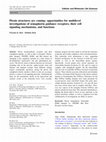
Fig 2 | Conserved tyrosine residues in the cytoplasmic domain of plexin family members. Alignment of protein sequences of human plexin intracellular domains.
In the consensus line (shown under the sequences), amino-acid identity is marked with asterisks and similarity is indicated by dots or columns, as assigned by the
CLUSTALW algorithm. Background colours highlight residues or domains of particular interest. In total, 13 tyrosine residues (Y) are conserved in all plexins (or
in all but one family member) and are shown in red; moreover, three of these residues are included in highly conserved amino-acid stretches (blue background).
The positions of highly conserved tyrosines are indicated on top of the sequences, with reference to the amino-acid coordinates in plexin A1 (accession number
NP-115618.2). Notably, the residues diverging from this consensus in plexin B3 and plexin A4 (underlined) are conserved between the human and mouse genes.
Tyrosine residues that are conserved in at least one entire plexin subfamily are shown in blue. The three conserved arginines found to be functionally required
in the GTPase-activating protein-like motifs are shown in green (Vikis et al, 2002). A grey background highlights the presumptive RhoGTPase-binding domain
(Tong et al, 2008).
©2008 EUROPEAN MOLECULAR BIOLOGY ORGANIZATION
EMBO reports
VOL 9 | NO 9 | 2008 8 6 9
reviews
for semaphorin-dependent myocardial cell motility. Notably, other
transmembrane semaphorins of subclass 6 (such as Sema6B) contain
proline-rich SH3-domain-binding motifs that are potentially implicated in the interaction with cytoplasmic SRC-like tyrosine kinases
and in their functional activation (Comoglio et al, 2004; Eckhardt
et al, 1997).
Potential role of plexin tyrosine phosphorylation
Initial experiments had shown that plexins might become phosphorylated in tyrosine residues when overexpressed (Tamagnone et al,
1999). There is now consistent evidence that plexins might be substrates for associated tyrosine kinases, both receptor type and cytoplasmic (Table 1). Moreover, in the presence of dominant-negative
mutants of ERBB2, MET and FYN that lack kinase activity, the tyrosine
phosphorylation of associated plexins is strongly reduced (Giordano
et al, 2002; Sasaki et al, 2002; Swiercz et al, 2004).
Although the tyrosine phosphorylation of plexins is thought to
exert a regulatory role in semaphorin signalling, this has not yet
been specifically investigated. Tyrosine phosphorylation of plexins could provide docking sites for SH2 or PTB domains (Yaffe,
2002), which are contained in several adaptors and transducer
molecules that are implicated in plexin signalling, such as SRClike tyrosine kinases (SRC, ABL, FAK and PYK2) and the regulatory
subunit of PI(3)K, p85. Moreover, the tyrosine phosphorylation of
plexins could modulate the conformational state of the cytoplasmic domain and allow for the recruitment of specific signal transducers. Recently, oncogenic mutations found in the cytoplasmic
domain of plexin B1—and linked to loss of association with Rac1
and Rnd1—have been found to have a strong impact on plexin
B1 structure and function (Tong et al, 2008; Wong et al, 2007).
Therefore, plexin tyrosine phosphorylation by specific RTKs might
elicit relevant conformational changes in the cytoplasmic tail, differentially regulating the accessibility of specific protein modules.
Along the same lines, as plexin B1 has been previously reported to
differentially regulate Rho activity through the recruitment of either
a RhoGEF or a RhoGAP, one could speculate that plexin B1 tyrosine
phosphorylation mediated by ERBB2 might elicit RhoA activation
by favouring the accessibility of its PDZ-domain-binding sequence.
However, the interaction of plexin B1 with MET could also result in
Rho inhibition by promoting the association of p190 RhoGAP with
the cytoplasmic tail of plexin B1.
The specific tyrosine residues phosphorylated in the cytoplasmic
domain of plexins in response to semaphorin stimulation have not yet
been identified, although this region contains numerous conserved
sites that could be targets of tyrosine kinases. Sequence alignment
reveals the presence of 13 conserved tyrosine residues (Fig 2). Notably,
some of these tyrosines have been found, by using high-throughput
screening experiments, to be phosphorylated in living cells (www.
phosphosite.org). Using the ScanSite algorithm (scansite.mit.edu),
we found that certain conserved tyrosines might be substrates for
known kinases (for example, SRC for Tyr 1833 in plexin A1 and LCK
for Tyr 2053 in plexin B1). Moreover, some of these residues, when
phosphorylated, might become docking sites for SH2- or PTB-domaincontaining proteins. For example, phosphorylated Tyr 1833 in plexin
A1 and the corresponding tyrosine in plexin B1 might bind the SH2
domains found in kinases of the SRC family. Therefore, tyrosine phosphorylation of plexins triggered by semaphorin signals could mediate
the recruitment of distinctive transducers through specific phosphotyrosine docking sites and/or important conformational changes in the
8 7 0 EMBO reports
VOL 9 | NO 9 | 2008
Tyrosine phosphorylation in semaphorin signalling
M. Franco & L. Tamagnone
plexin cytoplasmic tail, thereby allowing for a switch between multiple
intracellular signalling routes. Clearly, these speculative hypotheses
await experimental validation.
In conclusion, tyrosine kinases associated with semaphorins
or semaphorin receptors—and subsequent tyrosine phosphorylation cascades—have an important role in semaphorin functions.
They seem to control the switch between multiple signalling routes
that mediate diverse functional outcomes. Although the tyrosine
phosphorylation of plexins has been observed on semaphorin
stimulation, site-directed mutagenesis experiments are required to
characterize its function. Eventually, the identification of distinctive tyrosine kinases implicated in semaphorin signalling pathways might allow us to test pharmacological inhibitors that target
individual semaphorin functions, and could potentially be used to
modulate processes such as nerve regeneration, immune response
and cancer progression.
ACKNOWLEDGEMENTS
The authors acknowledge all of the members of the Tamagnone laboratory for
helpful discussions. Research activity carried out by M.F. and L.T. is supported
by the Italian Association for Cancer Research (AIRC).
REFERENCES
Artigiani S, Comoglio PM, Tamagnone L (1999) Plexins, semaphorins, and
scatter factor receptors: a common root for cell guidance signals? IUBMB
Life 48: 477–482
Artigiani S, Conrotto P, Fazzari P, Gilestro GF, Barberis D, Giordano S,
Comoglio PM, Tamagnone L (2004) Plexin-B3 is a functional receptor for
semaphorin 5A. EMBO Rep 5: 710–714
Barberis D, Casazza A, Sordella R, Corso S, Artigiani S, Settleman J,
Comoglio PM, Tamagnone L (2005) p190 Rho-GTPase activating protein
associates with plexins and it is required for semaphorin signalling. J Cell Sci
118: 4689–4700
Basile JR, Afkhami T, Gutkind JS (2005) Semaphorin 4D/plexin-B1 induces
endothelial cell migration through the activation of PYK2, Src, and the
phosphatidylinositol 3-kinase–Akt pathway. Mol Cell Biol 25: 6889–6898
Basile JR, Gavard J, Gutkind JS (2007) Plexin-B1 utilizes RhoA and Rho
kinase to promote the integrin-dependent activation of Akt and ERK and
endothelial cell motility. J Biol Chem 282: 34888–34895
Bechara A et al (2008) FAK-MAPK-dependent adhesion disassembly
downstream of L1 contributes to semaphorin3A-induced collapse. EMBO J
27: 1549–1562
Casazza A, Fazzari P, Tamagnone L (2007) Semaphorin signals in cell
adhesion and cell migration: functional role and molecular mechanisms.
Adv Exp Med Biol 600: 90–108
Comoglio PM, Tamagnone L, Giordano S (2004) Invasive growth: a two-way
street for semaphorin signalling. Nat Cell Biol 6: 1155–1157
Conrotto P, Corso S, Gamberini S, Comoglio PM, Giordano S (2004) Interplay
between scatter factor receptors and B plexins controls invasive growth.
Oncogene 23: 5131–5137
Conrotto P, Valdembri D, Corso S, Serini G, Tamagnone L, Comoglio PM,
Bussolino F, Giordano S (2005) Sema4D induces angiogenesis through Met
recruitment by Plexin B1. Blood 105: 4321–4329
Driessens MH, Olivo C, Nagata K, Inagaki M, Collard JG (2002) B plexins
activate Rho through PDZ-RhoGEF. FEBS Lett 529: 168–172
Eckhardt F, Behar O, Calautti E, Yonezawa K, Nishimoto I, Fishman MC (1997)
A novel transmembrane semaphorin can bind c-src. Mol Cell Neurosci 9:
409–419
Falk J et al (2005) Dual functional activity of semaphorin 3B is required for
positioning the anterior commissure. Neuron 48: 63–75
Gherardi E, Love CA, Esnouf RM, Jones EY (2004) The sema domain. Curr
Opin Struct Biol 14: 669–678
Giordano S, Corso S, Conrotto P, Artigiani S, Gilestro G, Barberis D,
Tamagnone L, Comoglio PM (2002) The semaphorin 4D receptor controls
invasive growth by coupling with Met. Nat Cell Biol 4: 720–724
Guo W, Pylayeva Y, Pepe A, Yoshioka T, Muller WJ, Inghirami G, Giancotti FG
(2006) Beta 4 integrin amplifies ErbB2 signaling to promote mammary
tumorigenesis. Cell 126: 489–502
©2008 EUROPEAN MOLECULAR BIOLOGY ORGANIZATION
reviews
Tyrosine phosphorylation in semaphorin signalling
M. Franco & L. Tamagnone
Kikutani H, Kumanogoh A (2003) Semaphorins in interactions between T cells
and antigen-presenting cells. Nat Rev Immunol 3: 159–167
Kruger RP, Aurandt J, Guan KL (2005) Semaphorins command cells to move.
Nat Rev Mol Cell Biol 6: 789–800
Kumanogoh A, Kikutani H (2001) The CD100–CD72 interaction: a novel
mechanism of immune regulation. Trends Immunol 22: 670–676
Maestrini E et al (1996) A family of transmembrane proteins with homology
to the MET–hepatocyte growth factor receptor. Proc Natl Acad Sci USA 93:
674–678
Mitsui N, Inatome R, Takahashi S, Goshima Y, Yamamura H, Yanagi S (2002)
Involvement of Fes/Fps tyrosine kinase in semaphorin3A signaling. EMBO J
21: 3274–3285
Moarefi I, LaFevre-Bernt M, Sicheri F, Huse M, Lee CH, Kuriyan J, Miller WT
(1997) Activation of the Src-family tyrosine kinase Hck by SH3 domain
displacement. Nature 385: 650–653
Morita A et al (2006) Regulation of dendritic branching and spine maturation
by semaphorin3A–Fyn signaling. J Neurosci 26: 2971–2980
Oinuma I, Ishikawa Y, Katoh H, Negishi M (2004) The Semaphorin 4D receptor
Plexin-B1 is a GTPase activating protein for R-Ras. Science 305: 862–865
Olsson AK, Dimberg A, Kreuger J, Claesson-Welsh L (2006) VEGF receptor
signalling: in control of vascular function. Nat Rev Mol Cell Biol 7: 359–371
Pasterkamp RJ, Peschon JJ, Spriggs MK, Kolodkin AL (2003) Semaphorin 7A
promotes axon outgrowth through integrins and MAPKs. Nature 424:
398–405
Perrot V, Vazquez-Prado J, Gutkind JS (2002) Plexin B regulates Rho through the
guanine nucleotide exchange factors leukemia-associated Rho GEF (LARG)
and PDZ-RhoGEF. J Biol Chem 277: 43115–43120
Rohm B, Rahim B, Kleiber B, Hovatta I, Puschel AW (2000) The semaphorin 3A
receptor may directly regulate the activity of small GTPases. FEBS Lett 486:
68–72
Sasaki Y et al (2002) Fyn and Cdk5 mediate semaphorin-3A signaling, which is
involved in regulation of dendrite orientation in cerebral cortex. Neuron 35:
907–920
Shapovalova Z, Tabunshchyk K, Greer PA (2007) The Fer tyrosine kinase
regulates an axon retraction response to Semaphorin 3A in dorsal root
ganglion neurons. BMC Dev Biol 7: 133
Suzuki K et al (2007) Semaphorin 7A initiates T-cell-mediated inflammatory
responses through alpha1beta1 integrin. Nature 446: 680–684
Swiercz JM, Kuner R, Behrens J, Offermanns S (2002) Plexin-B1 directly
interacts with PDZ-RhoGEF/LARG to regulate RhoA and growth cone
morphology. Neuron 35: 51–63
Swiercz JM, Kuner R, Offermanns S (2004) Plexin-B1/RhoGEF-mediated RhoA
activation involves the receptor tyrosine kinase ErbB-2. J Cell Biol 165:
869–880
Swiercz JM, Worzfeld T, Offermanns S (2008) ErbB-2 and Met reciprocally
regulate cellular signaling via plexin-B1. J Biol Chem 283: 1893–1901
Takahashi T, Fournier A, Nakamura F, Wang LH, Murakami Y, Kalb RG,
Fujisawa H, Strittmatter SM (1999) Plexin–neuropilin-1 complexes form
functional semaphorin-3A receptors. Cell 99: 59–69
Takegahara N et al (2006) Plexin-A1 and its interaction with DAP12 in immune
responses and bone homeostasis. Nat Cell Biol 8: 615–622
Tamagnone L, Comoglio PM (2000) Signalling by semaphorin receptors: cell
guidance and beyond. Trends Cell Biol 10: 377–383
Tamagnone L, Giordano S (2006) Semaphorin pathways orchestrate
osteogenesis. Nat Cell Biol 8: 545–547
©2008 EUROPEAN MOLECULAR BIOLOGY ORGANIZATION
Tamagnone L et al (1999) Plexins are a large family of receptors for
transmembrane, secreted, and GPI-anchored semaphorins in vertebrates.
Cell 99: 71–80
Tong Y, Chugha P, Hota PK, Alviani RS, Li M, Tempel W, Shen L, Park HW,
Buck M (2007) Binding of Rac1, Rnd1, and RhoD to a novel Rho GTPase
interaction motif destabilizes dimerization of the plexin-B1 effector domain.
J Biol Chem 282: 37215–37224
Tong Y, Hota PK, Hamaneh MB, Buck M (2008) Insights into oncogenic
mutations of plexin-B1 based on the solution structure of the Rho GTPase
binding domain. Structure 16: 246–258
Toyofuku T et al (2004a) Dual roles of Sema6D in cardiac morphogenesis
through region-specific association of its receptor, Plexin-A1, with off-track
and vascular endothelial growth factor receptor type 2. Genes Dev 18:
435–447
Toyofuku T, Zhang H, Kumanogoh A, Takegahara N, Yabuki M, Harada K,
Hori M, Kikutani H (2004b) Guidance of myocardial patterning in cardiac
development by Sema6D reverse signalling. Nat Cell Biol 6: 1204–1211
Toyofuku T, Yoshida J, Sugimoto T, Zhang H, Kumanogoh A, Hori M, Kikutani H
(2005) FARP2 triggers signals for Sema3A-mediated axonal repulsion. Nat
Neurosci 8: 1712–1719
Trusolino L, Bertotti A, Comoglio PM (2001) A signaling adapter function for
alpha6beta4 integrin in the control of HGF-dependent invasive growth. Cell
107: 643–654
Vikis HG, Li W, Guan KL (2002) The plexin-B1/Rac interaction inhibits PAK
activation and enhances Sema4D ligand binding. Genes Dev 16:
836–845
Winberg ML, Tamagnone L, Bai J, Comoglio PM, Montell D, Goodman CS
(2001) The transmembrane protein Off-track associates with plexins and
functions downstream of semaphorin signaling during axon guidance.
Neuron 32: 53–62
Wong OG et al (2007) Plexin-B1 mutations in prostate cancer. Proc Natl Acad
Sci USA 104: 19040–19045
Yaffe MB (2002) Phosphotyrosine-binding domains in signal transduction. Nat
Rev Mol Cell Biol 3: 177–186
Yamashita N et al (2007) Regulation of spine development by semaphorin3A
through cyclin-dependent kinase 5 phosphorylation of collapsin response
mediator protein 1. J Neurosci 27: 12546–12554
Zhou Y, Gunput RA, Pasterkamp RJ (2008) Semaphorin signaling: progress
made and promises ahead. Trends Biochem Sci 33: 161–170
Luca Tamagnone & Mélanie Franco
EMBO reports
VOL 9 | NO 9 | 2008 8 7 1
Academia.edu no longer supports Internet Explorer.
To browse Academia.edu and the wider internet faster and more securely, please take a few seconds to upgrade your browser.
Tyrosine phosphorylation in semaphorin signalling: shifting into overdrive
Embo Reports, 2008
...Read more
Related Papers
F1000 - Post-publication peer review of the biomedical literature, 2010
Download

The American Journal of Pathology, 2008
Download

Cellular and Molecular Life Sciences, 2012
Download
Developmental Biology, 2011
Download

Cell, 2001
Download

Download


Análisis Plural, 2024
Download


Autores: José Julio García Arranz, Norba-Arte, nº XVII, 1999, pp. 27-40; ISSN: 0213-2214.
Download

Download

