International Journal for Parasitology 36 (2006) 211–217
www.elsevier.com/locate/ijpara
Characterization of LST-R533: Uncovering a novel repetitive
element in Leishmania*
André L. Pedrosa a, Andrea M. Silva b, Jeronimo C. Ruiz b, Angela K. Cruz b,*
b
a
Departamento de Ciências Biológicas, Universidade Federal do Triângulo Mineiro, Uberaba, Minas Gerais, Brazil
Departamento de Biologia Celular e Molecular e Bioagentes Patogênicos, Faculdade Medicina de Ribeirão Preto, Universidade de São Paulo, São Paulo, Brazil
Received 26 June 2005; received in revised form 7 October 2005; accepted 14 October 2005
Abstract
We have previously isolated and sequenced a novel repetitive element, now named LST-R533, which is present in four different regions of one
extremity of Leishmania major chromosome 20. The repeats are polymorphic in size, ranging from 367 to 533 bp and contain an internal 81 bp
sequence with highly conserved segments (14–81 bp long) dispersed throughout the parasite’s genome. These sequences were not found in coding
regions of any predicted gene in L. major Friedlin genome, but are part of untranslated regions of some Leishmania transcripts. Analysis of the
81 bp sequence revealed significant degrees of identity with retrotransposons described in several other organisms. The presence of the sequence
in other species from genus Leishmania was determined by Southern hybridisation and DNA sequencing. This analysis indicated the conservation
of the 81-nucleotide element in all the Leishmania species evaluated. No sequences corresponding to LST-R533 or the 81 bp element were found
on either Trypanosoma brucei or Trypanosoma cruzi databanks.
q 2005 Australian Society for Parasitology Inc. Published by Elsevier Ltd. All rights reserved.
Keywords: Leishmania; Genome analysis; Repetitive sequences; LST-R533; Retrotransposons
1. Introduction
Parasites from the genus Leishmania are the causative
agents of leishmaniasis, a group of diseases that affects an
estimated three million people worldwide. Approximately 30
species of the parasite compose the genus, which is divided in
subgenera Leishmania and Viannia, and 22 of them are
associated with human infections (WHO, 1990). Different
species of the parasite are responsible for causing a wide range
of clinical manifestations, which vary from self-limiting
cutaneous ulcers to the visceral form, a fatal disease if not
treated.
The parasite plays a major role in determining the form and
progression of leishmaniasis (Spath et al., 2003; Naderer et al.,
2004). Although the molecular mechanisms related to parasite
*
Nucleotide sequence data reported in this study are available in the
GenBank under the accession numbers AF339905, AF339906, AF339907,
DQ092335, DQ092336, DQ092337and DQ092338.
* Corresponding author. Address: Avenida Bandeirantes, 3900, Bairro Monte
Alegre, Ribeirão Preto, São Paulo, CEP 14049-900, Brazil. Tel.: C55 163 602
3318; fax: C55 163 633 1786.
E-mail address: akcruz@fmrp.usp.br (A.K. Cruz).
pathogenicity are not well understood, the genetic plasticity of
the organism may be a crucial feature. In fact, this parasite does
not seem to have full ploidy control and several strains of
Leishmania have been shown to be aneuploid or suffer
dramatic alterations in their ploidy (Cruz et al., 1993; Myler
et al., 1999; Ghedin et al., 2004; Martinez-Calvillo et al.,
2005). In addition, Leishmania strains reveal a high degree of
intra- and interspecific chromosomal size polymorphism
probably as a result of recombination processes involving
repetitive elements, frequently observed at chromosomal
extremities. They also show variation of tandemly repeated
sequences (Iovannisci and Beverley, 1989; Ravel et al., 1996;
Sunkin et al., 2000; Ghedin et al., 2004).
In trypanosomatids, repetitive sequences are involved in
central aspects of parasitism such as antigenic variation,
documented in Trypanosoma brucei (Borst and Rudenko,
1994) and gene amplification (reviewed in Beverley, 1991).
Repetitive elements in Leishmania are important components
of circular amplified regions of the genome, which are
frequently associated with development of resistance to drugs
(Singh et al., 2001; Beverley et al., 1984). They may also be of
particular interest for species identification and taxonomy,
0020-7519/$30.00 q 2005 Australian Society for Parasitology Inc. Published by Elsevier Ltd. All rights reserved.
doi:10.1016/j.ijpara.2005.10.002
�212
A.L. Pedrosa et al. / International Journal for Parasitology 36 (2006) 211–217
since several classes of repetitive elements characterised in
protozoan parasites are species-specific (reviewed in FloeterWinter and Shaw, 2001; Wickstead et al., 2003). In
Leishmania, the majority of repetitive sequences described
are associated with chromosomal extremities (Ravel et al.,
1995; Fu and Barker, 1998; Sunkin et al., 2000; Pedrosa et al.,
2001).
We have previously described the isolation and sequencing
of a recombinant cosmid, 117E08 (E8), which represents one
extremity of chromosome 20 of Leishmania major LV39
(Pedrosa et al., 2001; Tosi et al., 1997). The presence of a
repetitive element distributed in three different regions within
the 35 kb of the insert was shown by sequencing, annotation
and hybridisation experiments (Pedrosa et al., 2001). Here
we describe the characterisation of an interspersed element,
LST-R533, which is present as four copies in one of the
extremities of L. major chromosome 20. It bears an internal
81 bp fragment, which is dispersed in the genome of the
different Leishmania species tested and showed significant
sequence identity with sections of retrotransposons characterised in other organisms.
2. Materials and methods
2.1. Leishmania culture and manipulation
Promastigotes of Leishmania (Leishmania) major
LV39 (Rho/SU/59/P), L. (L.) major Friedlin (MHOM/IL/80/Friedlin), L. (L.) major LT252 clonal line CC1 (MHOM/IR/83/IR), Leishmania (Leishmania) amazonensis
(MPRO/BR/1972/M1841-LV-79), Leishmania (Leishmania)
mexicana (MNYC/BZ/1962/M379), Leishmania (Leishmania)
donovani (MHOM/ET/67/HU3), Leishmania (Sauroleishmania) hoogstraali (RHEM/SD/1963/NG26) and Leishmania
(Sauroleishmania) tarentolae (ATCC 30267) were grown at
26 8C in M199 medium (Gibco BRL) supplemented as
described (Kapler et al., 1990). Leishmania (Viannia)
braziliensis 2904 (MHOM/BR/75/M2904) and L. (V.) braziliensis CE3227 (MHOM/BR/94/H-3227) were grown under
similar conditions in a medium further supplemented with 2%
human urine. Cells at density of 1.0–2.0!107 promastigotes/
ml were pelleted at 2000!g for 10 min at 4 8C for genomic
DNA preparation. L. major strains were provided by S.M.
Beverley (Department of Molecular Microbiology, Washington University, St Louis, USA) and P. Bastien (Laboratoire de
Parasitologie-Mycologie, Centre Hospitalier Universitaire de
Montpellier, France). New World and reptile species were
provided by J. Shaw (Departamento de Parasitologia,
Universidade de São Paulo, Brazil).
2.2. Cosmid isolation, sequencing and sequence analysis
Cosmid E8 was isolated from a L. major LV39 genomic
library with the hexameric repeats from the telomeres of
T. brucei as a probe (Tosi et al., 1997) and sequenced using a
transposon-based strategy (Pedrosa et al., 2001). Sequence
searches and multiple sequence alignments were performed by
BLAST (Altschul et al., 1997) and ClustalW (Thompson et al.,
1994), respectively. Sequences described in this work are
available at GenBank under accession numbers AF339905,
AF339906, AF339907, DQ092335, DQ092336, DQ092337
and DQ092338.
2.3. Manipulation of nucleic acids
Episomal DNA was purified using a Plasmid Maxi Kit
(QIAGEN Incorporated, Valencia, CA, USA) and digested
with endonucleases Bam HI, Hind III, Xba I and Xho I as
recommended (New England Biolabs, Beverley, MA). Agarose blocks containing the intact chromosomes of Leishmania
promastigotes were prepared and digested as previously
described (Coburn et al., 1991). Transfer of DNA from agarose
gels to nylon membranes (Gene Screen Plus, NEN Life Science
Products, Boston, MA) was carried out as described (Sambrook
et al., 1989). Promastigote forms of L. major LV39 clonal line
were harvested in the logarithmic and stationary phases of
growth and total RNA was extracted using TRIzol (Gibco
BRL, Grand Island, NY). Amastigotes were obtained from
footpad lesions of infected BALB/c mice 30 days after an
intradermal injection of 1.0!107 stationary phase promastigotes of L. major LV39 and total RNA was extracted using
TRIzol (Antoniazi et al., 2000). Aliquots containing 10 mg of
total RNA from both amastigote and promastigote forms of the
parasite were equilibrated in formamide, formaldehyde and
MOPS buffer (MOPS 0.02 M, sodium acetate 8.0 mM, EDTA
1.0 mM), denatured by heating the sample for 3 min at 95 8C
and fractionated in a 1% agarose formaldehyde/MOPS gel.
2.4. Hybridisation experiments
Gel-purified restriction fragments and PCR products were
radiolabelled by random priming (Feinberg and Vogelstein,
1983) and used as probes in hybridisation experiments.
Southern and northern blotting were conducted as described
(Sambrook et al., 1989). Hybridisations were carried out
overnight at 67 8C, following standard procedures (Sambrook
et al., 1989). Membranes were washed twice in 2!SSPE
(NaCl 0.3 mM, NaH2PO4 20 mM, EDTA 2 mM, pH 7.4), 0.5%
SDS at 67 8C and once in 0.1!SSPE, 0.1% SDS at room
temperature, each time for 15 min (Cruz and Beverley, 1990)
and exposed to a Kodak Diagnostic Film.
2.5. Pulsed field gel electrophoresis
Agarose-embedded chromosomes were separated in 1.0%
agarose gels using a Bio-Rad CHEF-DR II apparatus.
Chromosomes smaller than 1 Mb were resolved under the
following conditions: 4.5 V/cm, ramping from 50 to 120 s for
48 h at 14 8C. Southern blotting, hybridisation and washing
under high stringency conditions were conducted as previously
described (Cruz and Beverley, 1990).
�A.L. Pedrosa et al. / International Journal for Parasitology 36 (2006) 211–217
213
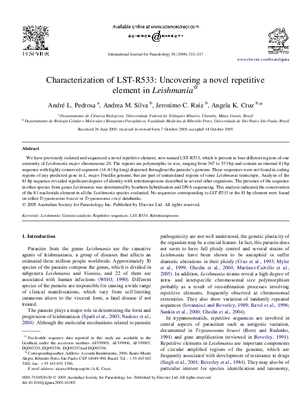
Fig. 1. Isolation of LST-R533. (A) Hind III fragments of cosmid E8 were fractionated in a 0.8% agarose gel and stained with ethidium bromide (1) and the
corresponding blot was probed with the E8-HH1.1 restriction fragment (2). (B) Southern hybridisation of Hind III (1) and Hind III/Bam HI (2) digested Leishmania
major LV39 genomic DNA using the fragment E8-HH1.1 as a probe. The molecular marker used was Hind III-digested lambda DNA. Fragment lengths are shown
on the left side of panels A and B (in kb). Arrowheads indicate the E8-HH1.1 restriction fragment. (C) Northern hybridisation of total RNA from LV39 promastigotes
in the initial and middle logarithmic phases (1 and 2, respectively), in the stationary phase (3) and from lesion amastigotes (4) hybridised with the LST-R533
amplicon (see Fig. 2). (D) Control hybridisation of 0.8 kb Xho I fragment of DHFRTS from L. major. (E) Ethidium bromide stained gel showing ribosomal RNA
bands. The molecular marker is the 0.1–2.0 kb RNA ladder (Invitrogen). (E) Schematic representation of one extremity (50 kb) of chromosome 20 from L. major.
Open boxes represent the four copies of LST-R533. The filled box stands for the chromosomal extremity. The magnified Hind III sites represent restriction fragment
E8-HH1.1. Horizontal lines represent the fragments recognised by the E8-HH1.1 probe in A (numbers indicate the fragment sizes, in kb). H, Hind III; B, Bam HI;
Tel, telomeric repeat.
2.6. PCR amplification
Leishmania DNA embedded in agarose blocks was prepared
for PCR after three successive washes in TE (Tris–HCl
10.0 mM, pH 7.4 and EDTA 1.0 mM), followed by a heating
step in TE (440 mL) at 80 8C for 5 min to melt the agarose.
Aliquots of 5 mL, containing approximately 100 pg of genomic
DNA, were used as templates in a 50 ml-reaction containing
1!PCR buffer (Perkin–Elmer, Foster City, CA), 200 mM
dNTPs, 1.0 mM of each primer and 0.5 units of Taq DNA
polymerase (Perkin–Elmer). Reactions were performed using a
first 3-min step of denaturation at 95 8C, followed by 30 cycles
of 45 s at 95 8C, 40 s at 55 8C, 55 s at 72 8C and a final
extension step of 10 min at 72 8C. The primers used were
LSTR1 (5 0 -GCCCCGCTATCCCTCTGCTGACG-3 0 ) and
LSTR2 (5 0 -GCCTCGCAGACGCTCCCATTGT-3 0 ). PCR
products were visualised in a 1.2% agarose gel with ethidium
bromide staining.
2.7. Cloning
Cosmid E8 was digested with Hind III and the 1.1 kb
fragment was cloned in pUC19. PCR products were purified
using the PEG method (Zhen and Swank, 1993) and cloned in
pGEM using the pGEMR-T Easy Vector kit (Promega
Biosciences Inc.). Ligation products were transformed by
electroporation in Escherichia coli DH10B and selected in
1.5% agar-LB medium containing 50 mg/ml ampicillin.
III-digested cosmid E8 and L. major digested genomic DNA
revealed the expected fragments of 1.1, 4.3 and 5.4 kb (Fig. 1A
and B). Strong hybridisation signals were observed at the high
molecular weight range of Hind III-digested genomic DNA
(Fig. 1B). The shorter Hind III band (w20 kb) originates the
w4.7 kb band seen the Hind III/Bam HI digestion. The higher
molecular weight signal observed in the Hind III digested
genomic DNA is probably due to the presence of several long
restriction fragments with low sequence identity to the probe,
since a corresponding strong signal is not observed in the Hind
III/Bam HI digestion (Fig. 1B, lane 2). Additionally, in the
genomic DNA fainter signals were observed in several
fragments of variable sizes. The available genomic sequence
of L. major Friedlin was searched with the consensus sequence
of the three copies of the element (Ivens et al., 2005). The
fourth copy of the element was identified in the same extremity
of chromosome 20. The restriction map obtained from the
available sequence is consistent with the Southern blot results
(Fig. 1B and E). Sequences obtained from L. major lineages
LV39 and Friedlin were, on average, 99% identical (Fig. 5 and
data not shown). The sequences were previously named LSTR378 (Leishmania Sub-Telomeric Reiterated 378 bp element
(Pedrosa et al., 2001). We have renamed them LST-R533 (the
number corresponds to the size of the longest repeat) and
numbers 1–4 have been added to indicate their position in the
chromosomal end (Fig. 1E).
3.2. Sequence analysis and annotation of LST-R533
3. Results
3.1. Isolation of LST-R533
The hybridisation of the 1.1 kb Hind III restriction fragment
bearing one copy of LST-R533 to blots containing the Hind
Sequence analysis of LST-R533-1, -2, -3 and -4 elements
demonstrated that they are direct repeats in the L. major
genome and polymorphic in size (486, 533, 506 and 367 bp
long, respectively). The core consensus sequence is 322
nucleotides long (Fig. 2) and the mean GC content of
�214
A.L. Pedrosa et al. / International Journal for Parasitology 36 (2006) 211–217
Fig. 2. Multiple sequence alignment of the four copies of LST-R533 found in one extremity of chromosome 20 of Leishmania major Friedlin. Nucleotides used as the
annealing sites for primers LSTR1 (reverse) and LSTR2 (forward) are underlined. Nucleotides in bold represent the 81-nt conserved element.
the repeat is 68.5% (range 68–70%). Annotation of LST-R533
in the complete genomic sequence of L. major Friedlin
confirmed that LST-R533-1 is found juxtaposed to the typical
telomeric and subtelomeric repeats of Leishmania described
previously (Fu and Barker, 1998). LST-R533K2 to -4 are found
at approximate distances of 10.6, 25.6 and 37.4 kb from the
chromosomal extremity (Fig. 1E). Moreover, LST-R533-2, -3
and -4 are located in intergenic regions of 1.4, 1.8 and 2.4 kb,
respectively. All annotated open reading frames (ORFs)
flanking the four copies of LST-R533 are present in L. major
Friedlin genome as single copy sequences.
3.3. Genomic distribution of LST-R533
A BLAST search of LST-R533-2 against the L. major
Friedlin genome revealed an internal region of 81 bp from LSTR533 particularly abundant in the parasite’s genome (Fig. 2,
bold-faced nucleotides). A subsequent BLAST search using the
81 bp element uncovered the presence of sequences ranging
from 14 to 81 bp and with a minimum of 95% identity in 308
different intergenic regions of all chromosomes of the parasite.
Short segments of sequences within the 81-nt element
presented significant identity with retrotransposons described
in several organisms (Table 1) (Jurka and Kapitonov, 1999;
Kapitonov and Jurka, 1999, 2001). These segments coincide
with the regions of the highest sequence conservation within
the L. major genome.
The LSTR-533 PCR-amplified fragment (Fig. 2, region
encompassing the underlined nucleotides) was used as a probe
in a northern blot containing total RNA from promastigotes and
amastigotes of L. major LV39 and the presence of two
transcripts was detected (Fig. 1C). The hybridisation of a probe
representing the dihydrofolate reductase thymidylate synthase
gene (DHFRTS) and the ethidium bromide stained bands of
ribosomal RNA were used as controls for the detection of a
transcript from a single copy gene and for RNA loading and
integrity, respectively (Fig. 1D and E). No portions of LSTR533 were found within annotated coding regions of the L.
major genome. Moreover, we have not found any sequences
with significant identity to both LST-R533 and the 81bp
element in T. brucei or T. cruzi genome databases.
3.4. Distribution of LST-R533 in Leishmania species
A labelled amplicon representing LST-R533 recognised
major chromosomal bands compatible with the approximate
Table 1
Transposable elements presenting sequence identity with the 81 bp-element from Leishmania major
Transposable element
(length in bp)
Organism
Identity (%)
Coordinates (within the
81 bp element)
GenBank accession number/
Reference
Dr000883 (842)
Retrosor1_I (702)
Sz-17 (3386)
L1M2B_5 (3252)
Helitronya1A_CE (3084)
Paltra3_CE (360)
Vandal1 (15,093)
Zebrafish
Sorghum bicolour
Oryza sativa
Homo sapiens
Caenorhabiditis elegans
Caenorhabiditis elegans
Arabidopsis thaliana
100
100
100
100
100
100
100
4–17
8–21
13–26
7–19
67–79
68–80
38–96
AL596027
AF098806
AF111709
Jurka and Kapitonov, 1999
Kapitonov and Jurka, 2001
Kapitonov and Jurka, 1999
Kapitonov and Jurka, 1999
�A.L. Pedrosa et al. / International Journal for Parasitology 36 (2006) 211–217
215
Fig. 3. Distribution of LST-R533 in the genome of Leishmania. (A) PFGE-separated chromosomes of different species of Leishmania: 1, Leishmania major CC1; 2,
L. major Friedlin; 3, Leishmania braziliensis 2904; 4, L. braziliensis CE3227; 5, Leishmania amazonensis; 6, Leishmania mexicana; 7, Leishmania hoogstraali; 8,
Leishmania tarentolae; 9, Leishmania donovani; 10, Molecular marker: lambda DNA concatamers. (B) Hybridisation of the probe LST-R533 to the Southern of the
gel shown in A. C: compression zone.
length of chromosome 20 (760 kb) in all species analysed and a
faint signal for species from the subgenus Sauroleishmania
(Fig. 3A and B). The probe also recognised fainter signals in
other chromosomal bands in Leishmania strains from
subgenera Leishmania, Viannia and Sauroleishmania
(Fig. 3B). The same amplicon was probed to genomic DNA
from L. major digested with Xho I and Xba I. Two to four major
restriction fragments and several other fainter signals were
recognised in all lineages tested were observed (Fig. 4A). In
agreement with pulse field gel electrophoresis (PFGE) results,
only faint signals were detected in lanes corresponding to
species from subgenus Sauroleishmania Fig. 4A and C.
We cloned and sequenced PCR products obtained with
primers LSTR1 and LSTR2 from different species of
Leishmania spp. in order to investigate cross species
conservation of LST-R533. Amplified fragments were polymorphic in size and multiple sequence alignment (ClustalW) of
the Leishmania sequences obtained indicated the conservation
of the region among the different species tested (Fig. 5).
Furthermore, BLAST search revealed that the 81 bp
sequence presents 83% identity with a sequence in the locus
containing a P-type ATPase of L. donovani, which includes
the LdH1A and LdH1B genes. The LdH1A sequence was used
in order to localise the homologous gene in the L. major
Friedlin genome, which bears two copies of the homologous
genes, both annotated as H1A and located at the 53 kb extreme
of chromosome 18. Two copies of internal regions of LSTR533 were found downstream of the second H1A gene in this
chromosome, the first at 2.9 kb (C strand) from the stop codon
and the second at 14.8 kb (K strand).
4. Discussion
Here we describe the characterisation of LST-R533, a
polymorphic repeated element found in inter-ORF regions of
one extremity of L. major chromosome 20. LST-R533-2 is the
longest copy of the element and LST-R533-1 is truncated at the
5 0 -end, whereas LST-R533-3 and K4 are truncated at the 3 0 end. The expression profiles of genes located at this
chromosomal extremity in promastigote forms of the parasite
have been previously investigated and differences in the
expression of genes along the chromosomal extremity studied
were not detected (Pedrosa et al., 2001). Short elements (14–81
nucleotides long) present within the LST-R533 are dispersed
Fig. 4. (A) Hybridisation of the probe LST-R533 to the Xho I (X) and Xba I (Xb) digested genomic DNA from the species shown in Fig. 3. (B) Control hybridisation
with a 0.8 kb Xho I fragment of DHFRTS from Leishmania major showing complete digestion of DNA samples. (C) Agarose gel of the digested genomic DNA
stained by ethidium bromide as a control for DNA loading. Molecular markers used are fragments of Hind III-digested lambda DNA.
�216
A.L. Pedrosa et al. / International Journal for Parasitology 36 (2006) 211–217
Fig. 5. Sequence alignment of the LST-R533 amplified from Leishmania spp. genomic DNA. Multiple sequence alignment of the 81-nucleotide element from
different species of Leishmania: Lm_Fried, Leishmania major Friedlin; Lm_LV39, L. major LV39; Ld_HU3, Leishmania donovani HU3; La_LV79, Leishmania
amazonensis LV79; Lb_2904, Leishmania braziliensis 2904; Lh_NG26, Leishmania hoogstraali. Nucleotides in bold represent the 81-nt conserved element.
and conserved in 308 inter-ORF regions of virtually all L.
major chromosomes. Such sequence conservation within
noncoding regions is compatible with selective pressure and
suggests a functional significance of this short segment for the
parasite. Sequence analysis revealed the presence of two
internal regions of the 81 bp element that presented high
sequence identity with retrotransposons isolated from several
organisms (Jurka and Kapitonov, 1999; Kapitonov and Jurka,
1999, 2001). However, we have not found an intact copy of
such element that could be responsible for its mobilisation in
the Leishmania genome. In fact, the expression of a retroposonlike element in a region spanning the putative promoter from
two variant surface glycoprotein gene expression sites of
T. brucei has already been demonstrated (Lodes et al., 1993).
The detection of L. major transcripts recognised by the LSTR533 probe, associated with the fact that portions of the repeat
were only found in inter-ORF regions, indicates that the
portions of transposable elements described here may be part of
untranslated regions (UTRs) of some transcripts of the parasite.
Sequence data, PFGE hybridisation and restriction fragment
length polymorphism analysis are consistent and allow us to
speculate that different Leishmania species whose complete
genomic sequences are not yet available have a similar
genomic organisation. The observation of strong hybridisation
of the LST-R533 probe to a single chromosomal band in the
PFGE and the fainter signals in other chromosomal bands of all
pathogenic species, associated with L. major genome data,
support the hypothesis that short segments within LST-R533
are transposon relics dispersed throughout the genome of these
parasites. The faint hybridisation signals in PFGE of the
subgenus Sauroleishmania and the conservation of the 81 bp
element sequence are consistent with the lower number of
copies of this small region in this species.
Preliminary comparative analysis between the genomes of
different species of Leishmania revealed a low conservation of
the non-coding regions (Laurentino et al., 2004). Moreover, a
bias of GC content between coding and non-coding regions is a
constant (Ivens et al., 2005; Laurentino et al., 2004). On the
other hand, LST-R533 sequence analysis revealed a GC content
of w68.5%, a considerably high value for an intergenic region
in L. major, whose mean GC content is approximately 57%
(Ivens et al., 2005). Furthermore, the 81 bp conserved element
were found to be distributed throughout the L. major genome
and was present in all the human pathogenic strains of
Leishmania from New and Old World species tested. The
cross-species conservation of the 81 bp element in non-coding
regions is intriguing and allows us to speculate that a formerly
active transposable element became a sequence of functional
relevance for the parasite. This is not the first report indicating
the presence of retrotransposon elements in trypanosomatids
and it adds to previous data published by Ghedin et al. (2004)
demonstrating the occurrence of a retroelement-like sequence
in L. major genome. That element, named LmChr1-DIRE, is
present in one extremity of L. major Friedlin chromosome 1.
Our observation of two conserved blocks of sequence
resembling an inactive retrotransposon dispersed in the
genome of Leishmania is novel and suggests that the dispersion
of this sequence in the genus Leishmania occurred before
separation of Leishmania and Viannia subgenera.
Acknowledgements
We thank Tânia Paula de Aquino Defina and Viviane
Ambrósio Trombela for technical assistance. This work was
supported by Fundação de Amparo à Pesquisa do Estado de
São Paulo (FAPESP) 99/12403-3 (AKC). AMS and JCR are
supported by fellowships from FAPESP.
References
Altschul, S.F., Madden, T.L., Schaffer, A.A., Zhang, J., Zhang, Z., Miller, W.,
Lipman, D.J., 1997. Gapped BLAST and PSI-BLAST: a new generation of
protein database search programs. Nucleic Acids Res. 25, 3389–3402.
Antoniazi, S., Lima, H.C., Cruz, A.K., 2000. Overexpression of miniexon gene
decreases virulence of Leishmania major in BALB/c mice in vivo. Mol.
Biochem. Parasitol. 107, 57–69.
Beverley, S.M., 1991. Gene amplification in Leishmania. Annu. Rev.
Microbiol. 45, 417–444.
�A.L. Pedrosa et al. / International Journal for Parasitology 36 (2006) 211–217
Beverley, S.M., Coderre, J.A., Santi, D.V., Schimke, R.T., 1984. Unstable
DNA amplifications in methotrexate-resistant Leishmania consist of
extrachromosomal circles which relocalize during stabilization. Cell 38,
431–439.
Borst, P., Rudenko, G., 1994. Antigenic variation in African trypanosomes.
Science 264, 1872–1873.
Coburn, C.M., Otteman, K.M., McNeely, T., Turco, S.J., Beverley, S.M., 1991.
Stable DNA transfection of a wide range of trypanosomatids. Mol.
Biochem. Parasitol. 46, 169–179.
Cruz, A., Beverley, S.M., 1990. Gene replacement in parasitic protozoa. Nature
348, 171–173.
Cruz, A.K., Titus, R., Beverley, S.M., 1993. Plasticity in chromosome number
and testing of essential genes in Leishmania by targeting. Proc. Natl Acad.
Sci. USA 90, 1599–1603.
Feinberg, A.P., Vogelstein, B., 1983. A technique for radiolabeling DNA
restriction endonuclease fragments to high specific activity. Anal. Biochem.
132, 6–13.
Floeter-Winter, L.M., Shaw, J.J., 2001. New horizons in the identification and
taxonomy of the Leishmania and the diagnosis of leishmaniasis: the
expansion of molecular techniques. Res. Adv. Microbiol. 4, 63–79.
Fu, G., Barker, D.C., 1998. Characterisation of Leishmania telomeres reveals
unusual telomeric repeats and conserved telomere-associated sequence.
Nucleic Acids Res. 26, 2161–2167.
Ghedin, E., Bringaud, F., Peterson, J., Myler, P., Berriman, M., Ivens, A.,
Andersson, B., Bontempi, E., Eisen, J., Angiuoli, S., Wanless, D., Von Arx,
A., Murphy, L., Lennard, N., Salzberg, S., Adams, M.D., White, O., Hall,
N., Stuart, K., Fraser, C.M., El-Sayed, N.M., 2004. Gene synteny and
evolution of genome architecture in trypanosomatids. Mol. Biochem.
Parasitol. 134, 183–191.
Iovannisci, D.M., Beverley, S.M., 1989. Structural alterations of chromosome 2
in Leishmania major as evidence for diploidy, including spontaneous
amplification of the mini-exon array. Mol. Biochem. Parasitol. 34, 177–
188.
Ivens, A.C., Peacock, C.S., Worthey, E.A., Murphy, L., Aggarwal, G.,
Berriman, M., Sisk, E., Rajandream, M.A., Adlem, E., Aert, R., Anupama,
A., Apostolou, Z., Attipoe, P., Bason, N., Bauser, C., Beck, A., Beverley,
S.M., Bianchettin, G., Borzym, K., Bothe, G., Bruschi, C.V., Collins, M.,
Cadag, E., Ciarloni, L., Clayton, C., Coulson, R.M., Cronin, A., Cruz, A.K.,
Davies, R.M., De Gaudenzi, J., Dobson, D.E., Duesterhoeft, A., Fazelina,
G., Fosker, N., Frasch, A.C., Fraser, A., Fuchs, M., Gabel, C., Goble, A.,
Goffeau, A., Harris, D., Hertz-Fowler, C., Hilbert, H., Horn, D., Huang, Y.,
Klages, S., Knights, A., Kube, M., Larke, N., Litvin, L., Lord, A., Louie, T.,
Marra, M., Masuy, D., Matthews, K., Michaeli, S., Mottram, J.C., MullerAuer, S., Munden, H., Nelson, S., Norbertczak, H., Oliver, K., O’Neil, S.,
Pentony, M., Pohl, T.M., Price, C., Purnelle, B., Quail, M.A.,
Rabbinowitsch, E., Reinhardt, R., Rieger, M., Rinta, J., Robben, J.,
Robertson, L., Ruiz, J.C., Rutter, S., Saunders, D., Schafer, M., Schein, J.,
Schwartz, D.C., Seeger, K., Seyler, A., Sharp, S., Shin, H., Sivam, D.,
Squares, R., Squares, S., Tosato, V., Vogt, C., Volckaert, G., Wambutt, R.,
Warren, T., Wedler, H., Woodward, J., Zhou, S., Zimmermann, W., Smith,
D.F., Blackwell, J.M., Stuart, K.D., Barrell, B., 2005. The genome of the
kinetoplastid parasite, Leishmania major. Science 309, 436–442.
Jurka, J., Kapitonov, V.V., 1999. Sectorial mutagenesis by transposable
elements. Genetica 107, 239–248.
Kapitonov, V.V., Jurka, J., 1999. Molecular paleontology of transposable
elements from Arabidopsis thaliana. Genetica 107, 27–37.
Kapitonov, V.V., Jurka, J., 2001. Rolling-circle transposons in eukaryotes.
Proc. Natl Acad. Sci. USA 98, 8714–8719.
217
Kapler, G.M., Coburn, C.M., Beverley, S.M., 1990. Stable transfection of the
human parasite Leishmania major delineates a 30-kilobase region sufficient
for extrachromosomal replication and expression. Mol. Cell. Biol. 10,
1084–1094.
Laurentino, E.C., Ruiz, J.C., Fazelinia, G., Myler, P.J., Degrave, W., AlvesFerreira, M., Ribeiro, J.M., Cruz, A.K., 2004. A survey of Leishmania
braziliensis genome by shotgun sequencing. Mol. Biochem. Parasitol. 137,
81–86.
Lodes, M.J., Smiley, B.L., Stadnyk, A.W., Bennett, J.L., Myler, P.J., Stuart, K.,
1993. Expression of a retroposon-like sequence upstream of the putative
Trypanosoma brucei variant surface glycoprotein gene expression site
promoter. Mol. Cell. Biol. 13, 7036–7044.
Martinez-Calvillo, S., Stuart, K., Myler, P.J., 2005. Ploidy changes associated
with disruption of two adjacent genes on Leishmania major chromosome 1.
Int. J. Parasitol. 35, 419–429.
Myler, P.J., Audleman, L., deVos, T., Hixson, G., Kiser, P., Lemley, C.,
Magness, C., Rickel, E., Sisk, E., Sunkin, S., Swartzell, S., Westlake, T.,
Bastien, P., Fu, G., Ivens, A., Stuart, K., 1999. Leishmania major friedlin
chromosome 1 has an unusual distribution of protein-coding genes. Proc.
Natl Acad. Sci. USA 96, 2902–2906.
Naderer, T., Vince, J.E., McConville, M.J., 2004. Surface determinants of
Leishmania parasites and their role in infectivity in the mammalian host.
Curr. Mol. Med. 4, 649–665.
Pedrosa, A.L., Ruiz, J.C., Tosi, L.R., Cruz, A.K., 2001. Characterisation of
three chromosomal ends of Leishmania major reveals transcriptional
activity across arrays of reiterated and unique sequences. Mol. Biochem.
Parasitol. 114, 71–80.
Ravel, C., Wincker, P., Bastien, P., Blaineau, C., Pages, M., 1995. A
polymorphic minisatellite sequence in the subtelomeric regions of
chromosomes I and V in Leishmania infantum. Mol. Biochem. Parasitol.
74, 31–41.
Ravel, C., Wincker, P., Blaineau, C., Britto, C., Bastien, P., Pages, M., 1996.
Medium-range restriction maps of five chromosomes of Leishmania
infantum and localization of size-variable regions. Genomics 35, 509–516.
Sambrook, J., Fritsch, E.F., Maniatis, T., 1989. Molecular Cloning: A
Laboratory Manual. Cold Spring Harbor Laboratory Press, Cold Spring
Harbor, NY.
Singh, A.K., Papadopoulou, B., Ouellette, M., 2001. Gene amplification in
amphotericin B-resistant Leishmania tarentolae. Exp. Parasitol. 99, 141–
147.
Spath, G.F., Garraway, L.A., Turco, S.J., Beverley, S.M., 2003. The role(s) of
lipophosphoglycan (LPG) in the establishment of Leishmania major
infections in mammalian hosts. Proc. Natl Acad. Sci. USA 100, 9536–9541.
Sunkin, S.M., Kiser, P., Myler, P.J., Stuart, K., 2000. The size difference
between Leishmania major friedlin chromosome one homologues is
localized to sub-telomeric repeats at one chromosomal end. Mol. Biochem.
Parasitol. 109, 1–15.
Thompson, J.D., Higgins, D.G., Gibson, T.J., 1994. CLUSTAL W: improving
the sensitivity of progressive multiple sequence alignment through
sequence weighting, position-specific gap penalties and weight matrix
choice. Nucleic Acids Res. 22, 4673–4680.
Tosi, L.R., Casagrande, L., Beverley, S.M., Cruz, A.K., 1997. Physical
mapping across the dihydrofolate reductase-thymidylate synthase chromosome of Leishmania major. Parasitology 114, 521–529.
WHO. 1990. World Health Organization Expert Committee: Control of the
Leishmaniasis. WHO Technical Report Series, p. 793.
Wickstead, B., Ersfeld, K., Gull, K., 2003. Repetitive elements in genomes of
parasitic protozoa. Microbiol. Mol. Biol. Rev. 67, 360–375.
Zhen, L., Swank, R.T., 1993. A simple and high yield method for recovering
DNA from agarose gels. Biotechniques 14, 894–898.
�
 Jeronimo Ruiz