CASE REPORT
SURGERY // ONCOLOGY
Skeletal Muscle Metastases and Inferior
Vena Cava Involvement in a Patient
with Clear Cell Renal Cell Carcinoma
and Sarcomatoid Differentiation
Călin Molnar1, Octavian-Sabin Tătaru2, Lucian Mărginean3, Angela Borda4
Surgery Clinic No. I, County Emergency Clinical Hospital, University of Medicine and Pharmacy, Tîrgu Mureș, Romania
Clinic of Urology, County Emergency Clinical Hospital, University of Medicine and Pharmacy, Tîrgu Mureș, Romania
3 Department of Radiology, County Emergency Clinical Hospital, University of Medicine and Pharmacy, Tîrgu Mureș, Romania
4 Department of Pathology, County Emergency Clinical Hospital, University of Medicine and Pharmacy, Tîrgu Mureș, Romania
1
2
CORRESPONDENCE
ABSTRACT
Octavian-Sabin Tătaru
Str. Gheorghe Marinescu nr. 1
540103 Tîrgu Mureș, Romania
Tel +40 758 919 891
E-mail: sabin.tataru@gmail.com
Introduction: Renal cell carcinoma has a propensity to propagate into the renal vein and inferior vena cava. A small percentage has distant metastasis at presentation. Pulmonary, hepatic,
cerebral and bone metastases are common, but skeletal muscle involvement is rare. Case
presentation: We present the case of a 51-year-old patient complaining of right flank pain,
gross hematuria and a painful left laterothoracic mass. Preoperative examination revealed a
tumor in the inferior pole of the right kidney, thrombosis of the right renal vein that extended
into the inferior vena cava and a left laterothoracic tumor. We decided on a preoperative digital
subtraction angiography and selected embolization of the laterothoracic mass. We performed
right radical nephrectomy with vena cava thrombus excision and excision of the left laterothoracic tumor. The pathological examination revealed a clear cell renal carcinoma with sarcomatoid differentiation of the right kidney. Metastases with the above features were noticed in
the right adrenal gland and in the skeletal muscle of the chest wall. Conclusions: The surgical
resection of large renal tumors with associated thrombus within the inferior vena cava is challenging to any surgeon. The preoperative embolization of the metastatic tumor is helpful in the
reduction of pain and intraoperative blood loss.
ARTICLE HISTORY
Received: 1 August, 2016
Accepted: 2 September, 2016
Keywords: inferior vena cava thrombosis, renal vein thrombosis, sarcomatoid clear cell renal
cell carcinoma, skeletal muscle metastasis
INTRODUCTION
Călin Molnar • Str. Gheorghe Marinescu nr. 50,
540136 Tîrgu Mureș, Romania. Tel +40 265 212 111
Lucian Mărginean • Str. Gheorghe Marinescu nr. 50,
540136 Tîrgu Mureș, Romania. Tel +40 265 212 111
Three percent of the solid tumors in adults are found to be renal cell carcinomas
(RCC), with the highest incidence at an age between 50 and 70 years. Pulmonary (50%), lymphatic nodes (35%), hepatic (30%), bone (30%) and adrenal
(5%) metastases are the most frequent in this type of neoplasia.1
Clear cell renal cell carcinoma (CCRCC) metastasizing into muscles is an
atypical discovery. Satake et al. concluded that only 32 cases of skeletal muscle
Angela Borda • Str. Gheorghe Marinescu nr. 38,
540139 Tîrgu Mureș, Romania. Tel +40 265 215 551
Journal of Interdisciplinary Medicine 2016;1(2):197-200
DOI: 10.1515/jim-2016-0039
198
Journal of Interdisciplinary Medicine 2016;1(2):197-200
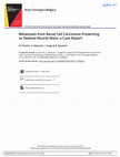
FIGURE 1. Performing a microcatheterization of the intercostal artery and embolization with polyvinyl alcohol, 150–300 µm particles,
to total tumor stasis and devascularization.
metastasis from RCC had been reported until 2009, and
Sountoulides et al. discovered 3 more. Patients with RCC
that present with metastatic involvement of the skeletal
muscles are exceptional, thus making the present case report as set apart from the rest.2
Sarcomatoid RCC (sRCC) has yet to be completely
described. Therefore, it is a very aggressive form of renal neoplasia, due to the incomplete understanding of its
physiopathology and possible form of treatments. Being
an aggresive form of cancer, in which patients present in
an advanced stage of evolution, sRCC is rather uncommon
to medical clinicians that deal with metastatic ailments.3
CASE PRESENTATION
We report the case of a 51-year-old male patient complaining of right flank pain, gross hematuria and a painful
left laterothoracic mass that had evolved over the last 6
months. On examination, the general status was good, and
on local examination a 10 × 10 cm palpable tumor and nonpalpable kidneys were observed. Past personal and family
medical history was not significant.
A thoracic, abdominal and pelvic computerized tomography (CT) scan was performed, that described a right renal
inferior pole tumor, the presence of a right renal vein throm-
bus shortly extending into the inferior vena cava without
lymph nodes involvement, and a left latero-thoracic tumor,
with no other abnormalities identified on CT scan.
Prior to surgery, the patient underwent an embolization
procedure for the laterothoracic tumor. We used the Seldinger technique through a right femoral approach. The tumor vasculature formation of the chest wall was identified,
with blood supply from the VIIIth intercostal artery, which
was embolized to total tumor stasis and devascularization,
followed by pain relief at the tumor site, on the same day
(Figure 1).
Under general anesthesia, within 24 hours from the embolization of the thoracic tumor, we performed right nephrectomy and adrenalectomy, longitudinal resection of
the inferior vena cava, right longitudinal cavoraphy. There
was no drop in the patient’s blood pressure during the inferior vena cava approach.
An incision was made in the center of the laterothoracic
tumor with the complete macroscopical resection of the
tumor situated in the intercostal space between the VIIIth
and IXth rib with minimal blood loss.
The pathological examination revealed a CCRC with
sarcomatoid differentiation of the right kidney (Fuhrman
4). The great majority of the tumor was composed of conventional CCRC, with a Fuhrman 2 nuclear grade. About
5% of the tumor showed a sarcomatoid differentiation,
with rhabdoid appearance of the cells. Rather extensive
necrosis was present in these areas the tumor. Upon immunohistochemical examination, both conventional and
sarcomatoid components expressed CD 10, EMA, AE1/
AE3, CK8, CK18 and were negative for CK 7, CK 20, AMACR, CD117 (Figure 2).
The renal vein contained a tumor thrombus and multiple emboli that were present in the small veins of the
renal sinus. Metastases with conventional CCRC feature
were noticed in the right adrenal gland and in the skeletal
muscle of the chest wall.
The patient was further referred to an oncologist for
chemotherapy.
DISCUSSION
It has been reported that approximately 0.4% of RCC
metastasize to skeletal muscle. Even though the skeletal
muscle presents an abundant blood supply and accounts
for a large surface of the body, metastatic involvement is
very exceptional at this site. The presence of peptidic factors preventing the metastasis and muscular contractions
dislodging anchored tumor cells provide insight into the
rarity of skeletal metastasis.5
Journal of Interdisciplinary Medicine 2016;1(2):197-200
A
B
C
D
199
FIGURE 2. A – Sarcomatoid differentiation with rhabdoid appearance merging from Fuhrman 2 CCRC.
B – Week positivity of CD 10 in the sarcomatoid component of CCRC. C – Strong positivity of AE1/AE3. D –
Positivity of EMA with membrane delineation.
The percentage of sarcomatoid differentiation within
the tumor seems not to influence overall survival in patients with cM1 disease.8 RCC commonly metastasizes to
soft tissues as a single soft tissue deposit developing at any
point ranging from 6 months to 19 years, the maximum
risk being within the initial 5 years after first medical contact. In our reported case, the skeletal muscle metastasis
was synchronous with the primary tumor.6
Renal cell carcinoma metastases present a highly developed vasculature, with increased bleeding described
during the surgery. Major hemorrhage can be prevented
during surgery. Transarterial embolization was found to be
useful in alleviating pain immediately. The procedure can
be safely executed, without including non-target embolization, if a previous angiography is performed for an exact
anatomical evaluation of the vessels, as well as a precise
cathether insertion in the arteries that supply the tumor.
This is why we decided on preoperative embolization of
the left laterothoracic tumor.7
CONCLUSIONS
Cases with sarcomatoid renal cell carcinoma present a
poor prognosis due to its increased rate of recurrence and
high mortality and morbidity, indicating the need for more
effective systemic therapies. Further trials are needed to
discover the reason why skeletal muscle metastases develop in such a small percentage of patients with RCC and
to evaluate the transarterial embolization benefits from an
oncological point of view.
COMPETING INTERESTS
The authors declare that they have no conflict of interests
and that they have no financial interests related to the material in the manuscript.
FUNDING
We state that the authors did not receive any funding for
this manuscript.
CONSENT
Written informed consent was obtained from the patient
for the publication of this report and any accompanying
images. A copy of the written consent is available for review by the Editor-in-chief of this journal.
200
Journal of Interdisciplinary Medicine 2016;1(2):197-200
REFERENCES
1.
2.
3.
Togral G, Arıkan M, Gungor S. Rare skeletal muscle metastasis after radical
nephrectomy for renal cell carcinoma: evaluation of two cases. J Surg
Case Rep. 2014;2014(10):rju101.
Sountoulides P, Metaxa L, Cindolo L. Atypical presentations and rare
metastatic sites of renal cell carcinoma: a review of case reports. J Med
Case Reports. 2011;5:429.
Shuch B1, Bratslavsky G, Linehan WM, Srinivasan R. Sarcomatoid renal cell
carcinoma: a comprehensive review of the biology and current treatment
strategies. Oncologist. 2012;17(1):46-54.
4.
5.
6.
7.
Vipin L, Lohiya S, Windsor K. A large thigh mass: a blood clot or a rare
skeletal muscle metastasis from renal cell carcinoma. SpringerPlus.
2013;2:399.
Ali SH, Chughtai H, Alali F, Diaczok B, Verardi M. Wrist drop: an atypical
presentation of renal cell carcinoma. Am J Med Sci. 2011;342(2):170-173.
Owen RJT. Embolization of musculoskeletal bone tumors. Semin Intervent
Radiol. 2010; 27(2):111-123.
Kim T, Zargar-Shoshtari K, Dhillon J. Using percentage of sarcomatoid
differentiation as a prognostic factor in renal cell carcinoma. Clin
Genitourin Cancer. 2015;13(3):225-230.
Skeletal Muscle Metastases and Inferior Vena Cava Involvement in a Patient with Clear Cell Renal Cell Carcinoma and Sarcomatoid Differentiation
Journal of Interdisciplinary Medicine, 2016
Renal cell carcinoma has a propensity to propagate into the renal vein and inferior vena cava. A small percentage has distant metastasis at presentation. Pulmonary, hepatic, cerebral and bone metastases are common, but skeletal muscle involvement is rare.We present the case of a 51-year-old patient complaining of right flank pain, gross hematuria and a painful left laterothoracic mass. Preoperative examination revealed a tumor in the inferior pole of the right kidney, thrombosis of the right renal vein that extended into the inferior vena cava and a left laterothoracic tumor. We decided on a preoperative digital subtraction angiography and selected embolization of the laterothoracic mass. We performed right radical nephrectomy with vena cava thrombus excision and excision of the left laterothoracic tumor. The pathological examination revealed a clear cell renal carcinoma with sarcomatoid differentiation of the right kidney. Metastases with the above features were noticed in the righ......Read more