Protein Expression and Purification 72 (2010) 254–261
Contents lists available at ScienceDirect
Protein Expression and Purification
journal homepage: www.elsevier.com/locate/yprep
Expression, purification and functional characterization of IjB kinase-2 (IKK-2)
mutants
Sumathy Mathialagan, Gennadiy I. Poda 1, Ravi G. Kurumbail 2, Shaun R. Selness, Troii Hall, Beverly A. Reitz,
Robin A. Weinberg, Nandini Kishore, Gabriel Mbalaviele *
Pfizer Inc., 700 Chesterfield Parkway West, Chesterfield, MO 63017, USA
a r t i c l e
i n f o
Article history:
Received 11 February 2010
Available online 20 February 2010
Keywords:
NEMO
IKK-2
NF-jB
PHA-408
a b s t r a c t
NF-jB signaling plays a pivotal role in a variety of pathological conditions. Because of its central role in
the overall NF-jB regulation, IKK-2 is a viable target for drug discovery. In order to enable structurebased design of IKK-2 inhibitors, we carried out a rational generation of IKK-2 mutants based on
induced-fit docking of a selective IKK-2 inhibitor, PHA-408, into the homology model of IKK-2. One
mutant we have characterized is a catalytically inactive form of IKK-2, D145A IKK-2, wherein the catalytic
aspartic acid, D145 was replaced with alanine. Unlike the WT enzyme, D145A IKK-2 is devoid of kinase
activity despite its ability to bind ATP with high affinity and is not phosphorylated at the T loop. In addition, this mutant binds a diverse collection of inhibitors with comparable binding affinities to WT IKK-2.
Another interesting mutant we have characterized is F26A IKK-2 (F26 is an aromatic residue located at
the very tip of the Gly-rich loop). Pre-incubation of F26A IKK-2 with PHA-408 revealed the role of F26
in the time-dependent binding of this inhibitor. Thus, functional characterization of these mutants provides the first evidence showing the role of a Gly-rich loop residue of a kinase in binding kinetics. These
two mutants along with others that we have identified could be used to validate homology models and
probe the interactions of IKK-2 with a variety of inhibitors.
Ó 2010 Elsevier Inc. All rights reserved.
Introduction
Nuclear factor-jB (NF-jB) is a transcription factor that is ubiquitously expressed and plays a key role in the regulation of a variety of genes involved in autoimmune and inflammatory responses
[1,2]. The NF-jB family consists of five proteins, RelA/p65, NF-jB1
(p50), NF-jB2 (p52), c-Rel and RelB, which form a variety of homodimers and heterodimers that differentially control gene expression [3]. The regulation of NF-jB transcriptional activity is
complex though it is well known that the IKK3 complex, which consists of two IjB kinases, IKK-1 (IKK-a), IKK-2 (IKK-b) and a regulatory subunit, NEMO (NF-jB essential modulator, IKK-c) is critically
* Corresponding author. Fax: +1 636 247 5985.
E-mail address: gabriel.mbalaviele@pfizer.com (G. Mbalaviele).
1
Present address: Ontario Institute for Cancer Research, MaRS Centre, Ont., Canada
M5G 0A3.
2
Present address: Department of Structural Biology, Pfizer, Groton, CT 06340, USA.
3
Abbreviations used: IKK, IjB kinase; NF-jB, nuclear factor kappa B; PKA, protein
kinase A; DAPK1, death-associated protein kinase 1; PAK1, p21-activated kinase 1;
MBP, myelin basic protein; GST, glutathione S-transferase; DTT, dithiothreitol; LPS,
lipopolysaccharide; TNF-a, tumor necrosis factor-a; NEMO, NF-jB essential modulator; IL-1b, interleukin-1b; BAFF, B cell activating factor; RANKL, receptor activator of
NF-jB ligand; LTb, lymphotoxin b; TAK-1, transforming growth factor beta-activated
kinase 1; MEKK-1, extracellular signal-regulated kinase (ERK)/MAPK ERK (MEK)
kinase 1.
1046-5928/$ - see front matter Ó 2010 Elsevier Inc. All rights reserved.
doi:10.1016/j.pep.2010.02.009
involved [4]. Gene knockout studies have clearly demonstrated the
requirement of IKK-2 and NEMO for the overall activation of NFjB. Our current understanding of this pathway is that in resting cells
NF-jB proteins exist in the cytoplasm as latent proteins due to their
association with inhibitory IjB family of proteins (e.g., IjBa, IjBb
and IjBe, which mask the NF-jB nuclear localization signal [5,6].
In response to an appropriate stimulus such as tumor necrosis factor
(TNF)-a, interleukin (IL)-1b, bacterial lipopolysaccharide (LPS), viral
infection or oxidizing agents, the IKK complex is activated and subsequently phosphorylates NF-jB and IjBs, leading to ubiquitination
and degradation of the IjBs and the release of NF-jB [5–7]. The free
NF-jB then translocates into the nucleus to promote gene transcription events [8].
Although IKK-1 and IKK-2 have about 50% overall amino acid
identity (65% identity in their kinase domains) and phosphorylate
IjBs at the same sites, genetic studies have demonstrated that they
have different roles in the overall activation of NF-jB. IKK-1, for
example, is known to function independently of NEMO and play
a key role in the activation of NF-jB via the non-classical pathway,
which is activated by TNF-a family members such as CD40 ligand,
B cell activating factor (BAFF), receptor activator of NF-jB ligand
(RANKL) and lymphotoxin (LT)b [9,10]. This pathway controls the
expression of genes involved in B cell survival and maturation as
well as in peripheral lymphoid organogenesis [11–13]. In contrast,
S. Mathialagan et al. / Protein Expression and Purification 72 (2010) 254–261
IKK-2 appears to be the major player in the activation of NF-jB in
response to pro-inflammatory stimuli, via the NEMO-dependent
classical pathway, causing cellular events, which include inflammatory and immune responses [14]. Additionally, IKK-2 has significantly higher kinase activity (about 20- to 30-fold) compared to
IKK-1 against IjBa and IjBb [15]. Thus, a strong body of evidence
indicates that inhibition of IKK-2 represents a very attractive strategy for modulating NF-jB functions a in a variety of diseases [16].
Whereas it is well established that the activity of IKK-2 requires
its phosphorylation on S177 and S181 in the T loop of the kinase
domain, it is still not clear which kinases phosphorylate this enzyme, though transforming growth factor beta-activated kinase
(TAK)-1, extracellular signal-regulated kinase (ERK)/MAPK ERK
(MEK) kinase (MEKK)-1 and MEKK-3 are the potential candidates
[17,18]. Thus, earlier discovery efforts of pharmacological inhibitors of the NF-jB pathway were indeed aimed at developing ATP
competitive inhibitors of IKK-2 activity [19,20]. These efforts have
led to the identification of several small molecule inhibitors of IKK2 with proven efficacy in relevant cells and animals models of human diseases [21,22]. However, despite over a decade of discovery
efforts, targeting IKK-2 kinase activity has proven to be challenging
as a safe clinical compound has yet to be developed.
When it is feasible, rational structure-based design of inhibitors
is an enabler of drug discovery. However, to date, numerous unsuccessful attempts to solve the crystal structures of IKK-2 have been
undertaken. Recently, the crystal structures of NEMO and IKK
interacting domains have been published, though the IKK peptides
used in this study lack the kinase domain [23]. On the other hand,
the nature of the key amino acid residues that are involved in ATP
binding is unknown. This is despite the fact that the binding patterns of several IKK-2 inhibitors at the ATP site have been characterized [20,22,24]. Therefore, we undertook extensive site-directed
mutagenesis and computational modeling studies in order to generate IKK-2 variants and understand the structure–activity relationship (SAR) of IKK-2 inhibitors and enable structure-based
design of IKK-2 inhibitors. In the process, based on the homology
model of IKK-2 and induced-fit ligand docking, we identified the
roles for F26 and D145 in inhibitor binding, the enzyme catalytic
activity and the binding kinetics of several IKK-2 inhibitors.
Materials and methods
Reagents
Simply Blue SafeStain, Tris–glycine sodium dodecyl sulphate
sample buffer, SEE BLUE molecular weight standards, 4–12% BisTris gels, 10% Tris–glycine gels, nitrocellulose membrane and
MES buffer were purchased from Invitrogen, (Carlsbad, CA), Ndodecylmaltoside from Roche (Pleasanton, CA). Tris PreSet, sodium
fluoride, dithiothreitol (DTT), benzamidine, anti-FLAG M2-agarose
resin and FLAG peptides were purchased from Sigma Chemical
Company (St. Louis, MO). Sodium chloride and glycerol were purchased from J.T. Baker (Phillipsburg, NJ) while the Vivaspin concentrators was obtained from Vivascience (Hanover, Germany). The
MonoQ HR 5/5 column was purchased from Amersham Pharmacia
Biotech (Piscataway, NJ) and the SEC molecular weight standards
came from BioRad (Hercules, CA). SAMTM 96-well biotin capture
plates were purchased from Promega (Madison, WI). [c-33P]- and
[c-35S]- labeled ATP was obtained from Amersham Biosciences
(Piscataway, NJ). Multi-screen plates were obtained from Millipore
Corporation (Billerica, MA), calf intestinal alkaline k phosphatase
from New England Biolabs (Ipwich, MA), and peroxidase-conjugated secondary antibodies from Jackson Immunoresearch Laboratories Inc (West Grove, PA). Phospho-IKK-2 and FLAG IKK-2
antibodies were from Cell Signaling (Danver, MA). cDNA of human
IKK-2 was amplified by reverse transcriptase-polymerase chain
255
reaction from human placental RNA (Clontech, San Jose, CA). High
bind plates, MSD sulfatag streptavidin, MSD antibody diluent and
Read buffer T were purchased from Meso Scale Discovery (Gaithersburg, MD), and phospho-IjBa antibody from Santa Cruz Biotechnology (Santa Cruz, CA).
Inhibitors
PHA-535E (2-amino-6-(2-hydroxy-6-isobutoxyphenyl)-4-(piperidin-3-yl)nicotino nitril2), SC-440 (5-(4-fluorophenyl)-2-ureidothiophene-3-carboxamide), PHA-966 (5-((3-fluorophenyl)ethynyl)-2ureidothiophene-3-carboxamide), SC-514 (5-amino-2,30 -bithiophene-4-carboxamide), SC-108 (1-(4-chlorophenyl)-4-ureido-1H-pyrazole-3-carboxamide), PHA-379 (1-(benzo[d][1,3]dioxol-5-yl)-8-(3chloroisonicotinamido)-4,5-dihydro-1H-benzo[g]indazole-3-carboxamide), PHA-250 (1-(benzo[d][1,3]dioxol-5-yl)-8-(5-chloro-2(4-methylpiperazin-1-yl)isonicotinamido)-4,5-dihydro-1H-benzo[g]indazole-3-carboxamide), PHA-068E (N1-(1,8-dimethylimidazo[1,2-a]quinoxalin-4-yl)ethane-1,2-diamine) were synthesized at
Pfizer, Inc. (St. Louis, MO). The inhibitors were dissolved in DMSO
and stored at 20 °C as 10 mM aliquots.
Cloning and expression
The IKK-2 cDNA was amplified using reverse oligonucleotide
primer that incorporated the peptide sequence for a FLAG epitope
tag at the C-terminus of the IKK-2 coding region (DYKDDDDKD).
The IKK-2-FLAG cDNA was subcloned into the baculovirus vector
pFastBac. This construct was then used as a template to introduce
the F26A, F26W, M96L, K106N, K106Q, D145A and Y169F mutations. The mutagenesis was done using the Stratagene Quikchange
Mutagenesis Kit. The presence of the mutation was confirmed by
DNA sequencing.
Isolation and purification of IKK-2 wild type and mutants
Cells expressing C-terminal FLAG-tagged IKK-2 WT and mutants including F26A, F26W, M96L, K106N, K106Q, D145A and
Y169F proteins were suspended at 1 l fermentation per 100 ml buffer A (20 mM Tris PreSet, pH 8.10, 150 mM NaCl, 0.30 mM n-dodecylmaltoside, 20 mM NaF, 10% glycerol, 0.50 mM DTT, 5 mM
benzamidine). Microfluidization was used to lyse the cells, the
pH adjusted to 8.1 using 50% NaOH and the suspension centrifuged
at 38,000g for 30 min. Anti-FLAG antibody (8 ml) was added to the
supernatant and the protein allowed to batch bind for 2 h with
rocking. The antibody resin slurry was poured into an XK 16/10
column, washed with 15 column volumes of buffer A followed by
5 column volumes of buffer B (buffer A containing 500 mM NaCl).
IKK-2 variants were eluted using 5 column volumes of buffer A
containing FLAG peptide (0.1 mg/ml).
IKK-2 kinase assay
Initially, IKK-2 kinase activity was measured using 10 lM biotinylated IjBa peptide (Biotin-Gly-Leu-Lys-Lys-Glu-Arg-Leu-Leu-AspAsp-Arg-His-Asp-Ser32-Gly-Leu-Asp-Ser36-Met-Lys-Asp-Glu-Glu),
10 lM [c-33P]ATP (3000 Ci/mmol specific activity) and 45 nM of
D145A IKK-2 or WT IKK-2 in a 50 ll reaction consisting of 25 mM
HEPES, pH 7.6, 2 mM MnCl2, 2 mM MgCl2,10 mM NaF, 5 mM DTT
and 0.1% BSA. After incubation at 25 °C for 30 min, 25 ll was transferred to SAMTM 96-well biotin capture plate. Each well was then
washed successively with 800 ll of 2 M NaCl, 1.2 ml of 2 M NaCl containing 1% H3PO4, 400 ll of H2O and 200 ll of 95% ethanol. The plates
were air-dried, 25 ll of scintillation fluid was added to each well and
the phosphorylated IjBa peptide was determined using Top Count
NXT (Packard Instrument Co, Waltham, MA).
256
S. Mathialagan et al. / Protein Expression and Purification 72 (2010) 254–261
The kinase assay for WT and other mutants (F26A, F26W, M96L,
K106N, K106Q and Y169F) were done using Meso Scale Technology
high bind 96-well plates coated with anti p-IjBa antibody at 5 ng/
well, after validation experiments and demonstration of data correlation with the above radioactive assay. The high bind plates
were air-dried over night, and the plates were blocked for 1 h with
slow rocking at RT with MSD blocking buffer A (Meso Scale Discovery, Gaithersburg, MD). Kinase activity was measured in the presence of 1 lM ATP, 5 lM biotinylated IjBa peptide in 25 mM
HEPES, 2 mM MnCl2, 2 mM MgCl2, 10 mM NaF and 200 pM of
IKK-2 WT or mutants in a volume of 30 ll. The plates were incubated with slow rocking for 30 min, washed twice with TBST,
and MSD sulfatag streptavidin was added at 2 lg/ml in a volume
of 25 ll. After 90 min incubation, the plates were washed twice
with TBST, followed by 150 ll MSD read buffer T at 2 dilution
containing DDH2O, and the plates were read using an MSD sector
imager 6000. To test the effects of the inhibitors on kinase activity,
the enzymes were pre-incubated or not with the inhibitors for 1 h
before adding the solution containing IjBa peptide as described
above.
or D145A IKK-2) or (20 lg for HUVEC-derived samples) were separated by SDS–PAGE and transferred to nitrocellulose membranes
(at 26 V for 1 h). The membranes were then blocked with 5% dry
powdered milk, and reconstituted in Tris-buffered saline
(100 mM Tris, pH 8.0, 150 mM NaCl) containing 0.05% Tween 20
(TBST) for 30 min at room temperature for IKK-2 variants. To detect phosphorylated IKK-2, the blots were incubated overnight
with primary antibody (1:500, anti-phospho-IKK-2) in 1% milk in
TBST and then washed four times in TBST and incubated with peroxidase-conjugated goat anti-rabbit (1:5000) for 1 h. Enhanced
chemiluminescence (ECL plus) was used for detection. To detect
total IKK-2, the membranes were stripped with Restore™ Western
Blot Stripping buffer, and probed overnight with anti-FLAG IKK-2
antibody at a final concentration of 0.1 lg/ml final in TBST containing 1% milk. The blots were washed as above and probed with peroxidase-conjugated goat anti-mouse secondary antibodies
(1:5000) for 1 h, and washed as described above. Antibody labeling
of protein bands were detected using fluorescent secondary antibodies. Detection was done using Li-COR Odyssey Scanner.
Molecular modeling
ATP binding assay
The binding of ATP and inhibitors to WT IKK-2 and mutants was
analyzed using anti-FLAG antibody immobilized on M2-agarose.
The binding assay was conducted using 96-well Millipore multiscreen filter plates. The binding reaction consists of 56 nM WT
IKK-2 or D145A IKK-2 and 0–0.6 lM concentrations of cold and
[c-35S]ATP (specific activity > 1000 Ci/mmol) in 50 ll of reaction
volume in kinase buffer (25 mM HEPES, pH 7.6, 2 mM MnCl2,
2 mM MgCl2, 10 mM NaF, 5 mM DTT and 0.1% BSA). Nonspecific
binding was defined by the addition of 1000 unlabelled ATP. Following 2 h incubation at 4 °C, the reaction mixtures were filtered,
and the filter was subjected to a rapid wash with 200 ll cold PBS
(without CaCl2 and MgCl2). The filter plates were air-dried, 30 ll
of scintillation fluid were added to each well, and the bound
[c-35S]ATP was counted using a Top Count NXT as described previously [24]. Grafit program was used to fit the data. To test the effects of the inhibitors on [c-35S]ATP binding, they were preincubated or not with the enzyme for 1 h before incubating the
mixtures with [c-35S]ATP as described above.
Phosphatase treatment
FLAG-tagged WT and D145A IKK-2 (4–8 lg) were immunoprecipitated with anti-FLAG M2-agarose beads (16 ll of anti-FLAG
agarose/lg of proteins) in 5 ml of ELISA buffer (20 mM Tris–HCl,
pH 7.2, 150 mM NaCl, 0.1% bovine serum albumin and 0.05%
Tween 20) for 2 h at 4 °C. The immobilized proteins were pelleted
by centrifugation, washed once successively with ELISA buffer
(20 mM Tris–HCl, pH 7.2, 150 mM NaCl, 0.1% BSA and 0.05% Tween
20), and then with kinase buffer (25 mM HEPES, pH 7.6, 2 mM
MnCl2, 2 mM MgCl2, 10 mM NaF, 5 mM DTT and 0.1% BSA). Samples were re-suspended in a buffer containing 50 mM Tris–HCl,
pH 7.6, 0.1 mM EDTA and 2 mM MnCl2, then were incubated for
30 min at room temperature in the presence of 500 U k phosphatase/lg IKK-2. The phosphatase was removed from the immobilized kinases by washing 3 times with ELISA buffer at room
temperature, and re-suspended samples in kinase buffer were then
subjected to [c-35S]ATP binding as described above.
Western analysis
Tris–glycine SDS sample buffer (Invitrogen, Carlsbad, CA) was
added to samples, which then were heated for 5 min at 90 °C. Equal
amounts of proteins (25 ng for buffer- or phosphatase-treated WT
The homology model of the IKK-2 kinase domain (300 amino
acids) was built using a protein kinase A (PKA) in-house crystal
structure in complex with a low-molecular-weight pyrrolopyrimidine inhibitor as a template. The protein sequence of human IKK-2
was retrieved from SwissProt (Accession No. O14920, October
2001). Although the full length IKK-2 contains 756 amino acids,
we performed a BLAST search using IKK-2 1–300, and identified
three proteins with available crystal structures: DAPK1 (32% sequence identity in the kinase domain with IKK-2), PAK1 (30%)
and PKA (30%). Although death-associated protein kinase 1
(DAPK1) and p21-activated kinase 1 (PAK1) exhibit higher or similar sequence identity with IKK-2 in the kinase domain (32% and
30%, correspondingly) we selected PKA as a template to build a
homology model of IKK-2 for a number of reasons. PKA has 7 amino acids in the hinge region as IKK-2 does. However, both DAPK1
and PAK1 also have seven amino acids in the hinge (B. Lunney, personal communication). While this fact justifies selection of PKA as
a template it alone does not provide advantages over DAPK1 and
PAK1. An important point is that PKA has the same gate-keeper
residue (Met, M96 in IKK-2) as IKK-2 does while DAPK1 has Leu
(L93). This provides an advantage of using PKA as a template for
building a homology model over DAPK1. While PAK1 also has
Met (M344) as gate-keeper, it has Arg residue (R299) in place of
the unique solvent-exposed lysine located across the hinge (K106
in IKK-2). We used the induced-fit docking (IFD) workflow from
Schrödinger, Inc. for flexible ligand docking of a variety of chemical
series of IKK-2 inhibitors to afford structure-based analog design
and thus speed up lead optimization programs of IKK-2 inhibitors
as small molecule therapeutics in the treatment of rheumatoid
arthritis. Coordinates from the structurally-conserved regions were
assigned from PKA due to high sequence identity. The activation
loop was built and refined using multi-step energy minimization
procedure while the rest of the protein was kept fixed and gradually released tethering on the backbone of the A-loop. PHA-408 in
its putative protein-bound conformation optimized at Becke3LYP/
6-31G* level with Jaguar (Jaguar, version 6.5, 2005, Schrödinger,
LLC, New York, NY) was hand-docked initially and the whole protein–ligand complex was refined via multi-step energy minimization cycles with first the ligand and protein backbone fixed.
Positional constraints on the ligand and protein backbone was
gradually eliminated to afford a starting geometry of the IKK-2/
PHA-408 complex for further ligand docking with the IFD workflow
from Schrödinger, Inc. [25]. At the first stage of IFD, the protein and
ligand van der Waals radii have been scaled down to 0.5 and 20
257
S. Mathialagan et al. / Protein Expression and Purification 72 (2010) 254–261
poses was generated and stored. Then, a 6 Å shell of residues adjacent to the ligand was optimized with the Prime Induced Fit procedure and the final docking was done with extra precision (XP)
Glide scoring with post-docking minimization. During the IFD procedure it was observed that some ligands flip at the binding site
allowing the carboxamide hinge region recognition element to
bind either to both the backbone NH and the carbonyl of C99 or
the backbone NH of C99 and backbone carbonyl of Q97. It is worth
mentioning that the conformation of the A-loop is the weakest part
of the model but with the exception of Y169 (next to the conserved
in protein kinases DFG motif, which is DLG in IKK-2) no other residues were in the close contact with PHA-408.
Results
Induced-fit docking of PHA-408 into IKK-2 homology model
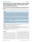
PHA-408 (Fig. 1A) is a potent and selective inhibitor of IKK-2 [26].
Based on the induced-fit docking procedure described in Methods,
we sought to understand the most likely binding mode of PHA408. The carboxamide moiety is binding to both the backbone NH
and carbonyl of residue C99 of IKK-2 (Fig. 1B). This hydrogen bond
in the hinge region is known as a key interaction in most protein
kinases with the exception of the Pim kinase family (PIM1, PIM2
and PIM3) that is devoid of this key proton donor NH as a hinge binder. The 4-fluoro-N-phenyl group is directed towards the solvent in
the sugar region, while the pyridine is engaged with the Y169 side
chain via a hydrogen bond and is also engaged with F26 at the very
tip of the Gly-rich loop with a p–p parallel-displaced type of interaction. The carboxamide linker that connects the tricyclic core and the
pyridine moiety forms a hydrogen bond with the catalytic aspartic
acid, D166, from the conserved DFG kinase motif (DLG in IKK-2).
The appended piperazine group seems to form a mono-dental
hydrogen bond with the conserved aC-Glu residue from the C-helix
that is known to be important in activation. The LUDI binding free
energy [27] for PHA-408 was 17.35, while the HT binding score
(estimated binding free energy in kCal/mol) [28] was 15.0. Based
A
on these elaborate docking experiments that treat both the ligand
and the protein binding site as flexible, we speculated on the IKK-2
amino acid residues that may form key interactions with PHA-408.
Rational design of IKK-2 mutants
Based on the binding mode of PHA-408 at the ATP site of IKK-2
(Fig. 1B), we selected four points for site-directed mutagenesis
studies: F26, K106, D145 and Y169. F26 is located at the very
end of the Gly-rich loop of IKK-2, and as it was shown from the
docking experiments, forms a p–p parallel-displaced interaction
with the pyridine group of PHA-408. It is known that at this position a large number of protein kinases have a bulky Tyr or Phe residue that adopts a conformation either under the Gly-rich loop (as
in our homology model) or could be pointing out towards the Aloop. Substitution of F26 with Ala (F26A) or with Trp (F26W) would
probe the role of this residue in ligand binding and duration of action. IKK-2 has a bulky Met, M96, as a gate-keeper. Thus, we
thought that replacing M96 with a smaller amino acid, for example, Leu (M96L) could potentially modulate ligand binding affinities. K106 is located in the solvent-exposed region across the
hinge of IKK-2 and forms an ion-pair with D103. This K106 residue
is unique for IKK-2, thus allowing us to probe the substitution of
this residue with a neutral Asn (K106N) and negatively charged
Glu (K106Q). We also pursued mutagenesis of Asp 145, which is
likely to play the role of the catalytic base in the overall kinase
reaction based on sequence alignment with cyclic AMP dependent
protein kinase A (PKA). Mutation of the corresponding Asp in the
yeast kinase renders the enzyme inactive [29]. D166 in PKA
(D145 in IKK-2) is believed to engage the hydroxyl group from
the Ser/Thr residue of the substrate, which eventually leads to
the formation of phosphorylated Ser or Thr on the protein substrate. Y169 was chosen because it forms a Hydrogen bond with
the pyridine nitrogen of PHA-408 in our model. It is also an adjacent bulky residue in the conserved protein kinase DFG motif.
Thus, we decided to mutate this Y169 to Phe (Y169F). The various
IKK-2 mutants generated for this study are shown in Table 1.
B
F26
M96
N
NH
O
H2N
N
N
O
K44
N
Cl
Q61
N
C99
K106
F
D166
Y169
D103
K147 D145
Fig. 1. Induced-fit docking of PHA-408 into IKK-2 homology model. (A) Chemical structure of PHA-408. (B) Putative dominant conformation of PHA-408 bound to the IKK-2
homology model. The binding mode was obtained using the induced-fit docking (IFD) workflow in Schrödinger, Inc. [25]. Only key binding site residues as well as residues
involved in our site-directed mutagenesis studies are shown for simplicity.
258
S. Mathialagan et al. / Protein Expression and Purification 72 (2010) 254–261
Table 1
WT and IKK-2 mutants.
WT IKK-2
Mutant IKK-2
F26
F26A
F26W
M96
M96L
K106
K106N
K106Q
D145
D145A
Y169
Y169F
D145A mutation causes loss of IKK-2 activity
IKK-2 variants are expressed at high levels in insect cells
F26A, F26W, M96L, K106N, K106Q, D145A, Y169F and WT IKK-2
were all constructed as C-terminal FLAG-tagged proteins. Each
construct was expressed in SF9 insect cells, and anti-FLAG antibodies were used for protein purification. Coomassie blue staining of
the gels shows that purification using the anti-FLAG resin resulted
in purity of greater than 85% for each protein (Fig. 2). These proteins were expressed at high levels in SF9 cells (2–15 mg/l range).
Western blot analysis using anti-FLAG antibodies confirmed the
expression of WT and mutants of IKK-2 (data not shown). In order
to address concerns of potential concentration induced aggregation, both the diluted and concentrated forms of the proteins were
WT
F26A
1 2 3 4 5 6
evaluated by size exclusion chromatography. Samples with a 10fold difference in concentration (1 vs. 10 mg/ml) showed no difference in peak shape or purity and their retention times were similar,
suggesting increased concentration did not induce aggregation
(data not shown). Thus, the availability of these variants of IKK-2
enabled us to assess the functional relevance of the affected residues outside and within the ATP binding cavity in IKK-2.
WT and IKK-2 mutants, including F26A, F26W, M96L, K106N,
K106Q, D145A and Y169F were evaluated for IKK-2 kinase activity
using the S32 and S36 containing IjBa peptide as substrate. The
data shows that all mutants exhibited kinase activity comparable
to WT IKK-2 with the exception of D145A IKK-2, which showed
0% of WT IKK-2 activity (Fig. 3). D145A IKK-2 lacked kinase activity
despite being tested with saturating concentrations of the substrate (Fig. 3B). Thus, these findings revealed that D145 is an
important residue for the ability of IKK-2 to phosphorylate IjBa
peptide in vitro.
D145A IKK-2 is unphosphorylated, but still binds ATP
We sought to understand the mechanisms of D145A IKK-2 failure to phosphorylate IjBa peptide. Since ATP binding is the first
K106Q
1 2 3 4 5 6
1 2 3 4 5 6
D145A
Y169F
1 2 3 4 5 6 7 8 9
1 2 3 4 5 6
Fig. 2. IKK-2 mutants are expressed at high levels in insect Sf9 cells. The supernatants from cell lysates were incubated with anti-FLAG antibody for 2 h. The antibody resin
slurry was poured into an XK 16/10 column, washed, and the proteins were eluted using buffer A containing FLAG peptide. The gels were stained with Coomassie blue. For
WT, F26A, K106Q and Y169F: lane 1, MW standards; lane 2, flow through; lane 3, buffer wash; lane 4, 0.50 M NaCl wash; lane 5, continued 0.50 M NaCl wash; lane 6, eluate.
For D145A: Lane 1, MW standards; lane 2, flow through; lane 3, buffer wash #1; lane 4, buffer wash #2; lane 5, buffer wash #3; lane 6, buffer wash #4; lane 7, 0.50 M NaCl;
lane 8, eluate; lane 9, continued eluate. 25 ll out of 15 ml eluate were loaded, thus indicating that the proteins were expressed at high levels.
B
A 160
100
% WT IKK-2 control
% WT IKK-2 control
140
120
100
80
60
40
80
60
40
20
20
0
0
WT
F26A
F26W
M96L
K106N K106Q Y169F No enzyme
WT
D145A No enzyme
Fig. 3. D145A mutation causes loss of IKK-2 activity. (A) Vehicle (no enzyme), WT or IKK-2 mutants (0.2 nM each) were incubated with 1 lM ATP, 5 lM IjBa peptide.
Phosphorylated IjBa peptide was captured by the anti p-IjBa antibody coated on the MSD high bind plates. (B) Vehicle (no enzyme), WT or IKK-2 mutants (45 nM each) were
incubated with 1 lM ATP, 10 lM IjBa peptide using the [c-33P]ATP/SAM plate assay. Kinase activity was expressed as % of WT IKK-2. The data shows loss of kinase activity
due to the D145A mutation. The data represent the average of triplicate determinations from one representative experiment with error bars indicating S.D.
259
S. Mathialagan et al. / Protein Expression and Purification 72 (2010) 254–261
step in substrate phosphorylation by kinases, we investigated the
ability of D145A IKK-2 to bind ATP. We found that D145A IKK-2
binds ATP with Kd (50 ± 9 nM) and Bmax (2.59 ± 0.78 pmol) comparable to WT IKK-2 (70 ± 5 nM, and 2.38 ± 0.04 pmol, respectively
(Fig. 4A). The inset shows that D145A IKK-2 ran similarly to WT
IKK-2 on SDS–PAGE, and that equal quantities of proteins were
used for the binding assay. Consistent with these findings, WT
and D145A IKK-2 bound various ATP competitive inhibitors of
IKK-2, including PHA-408 [26], with equal affinity (Fig. 4B).
Since IKK-2 activity is regulated by phosphorylation at S177 and
S181 in the T loop, to gain further insights into the mechanisms of
the D145A-induced IKK-2 loss of activity, we determined the phosphorylation status of WT and D145A IKK-2. We found that WT, but
not D145A IKK-2, reacted with anti-p-IKK-2 (S181) antibody, an effect that was abolished upon treatment of WT IKK-2 with k phosphatase (Fig. 4C). Interestingly, whereas de-phosphorylated WT
IKK-2 failed to bind ATP (Fig. 4D), D145A IKK-2 bound ATP with
same affinity before or after treatment with k phosphatase
(Fig. 4E). Collectively, the data indicate that the failure of D145A
IKK-2 to phosphorylate IjBa peptide was due to its inability to
be phosphorylated at S181 rather than its ability to bind ATP.
F26 is required for time-dependent binding of PHA-408
Using the [c-35S]ATP binding assay, we found that PHA-408 and
PHA-250, but not ADP, SC-514, PHA-379, PHA-440, PHA-108, PHA535E, PHA-966 and PHA-068E, bind to WT IKK-2 in a time-dependent manner (Fig. 5A and data not shown). Since (i) PHA-408 is so
far the only IKK-2 inhibitor that is known to bind tightly to IKK-2
[26]; (ii) D145A is catalytically inactive; and (iii) our docking model predicted that F26, K106 and Y169 interact with PHA-408, we
therefore determined the role of these residues in the binding
kinetics of PHA-408. PHA-408 was pre-incubated or not with
IKK-2 variants for 1 h, and kinase activity (Fig. 5A–D) or [c-35S]ATP
binding (Fig. 5E) was measured. We found that the F26A, but not
the K106Q, D145A and Y169F mutations, resulted in loss of the
time-dependent binding (P10-fold) of PHA-408 compared to WT
IKK-2 (Fig. 5, and Table 2).
S
B
SC-514
SC-514
S
10
Cl
Cl
WT IKK-2
D145A IKK-2
IKK
3
2
2
Inp Supts IP
WT
1
D145A
0
0
0.2
0.4
[γ-35S]ATP(μM)
0.6
H 2N
N
H
Cl
N N
1
O
0.5
0.4
0.3
H2N
N
N
H
O H
2
0.02
N
PHA-966
PHA-408
H 2N
O
.1
.02 .03 .05
F
SC-440
N
N
N
H
N N
35
[γ-- S] ATP bound (pmol)
Inp
uts
Su
pts
Bu
ffe
λp r
pa
se
Inp
uts
Su
pts
Bu
ffe
λp r
pa
se
S NH
NH22
N
N
H
O H O
O
.2 .3 .4 .5
1
2
D145A IKK-2 IC 50 (μM)
3 4 5
10
S NH2
N
O H O
D
IKK-2
H2N
N
Cl
O
H2N
C
N
NH2
F
0.2
N NH2
OH
D145A
PHA-068E
N N
O
N N
H
O
O
NH2
NH2
ADP
0.1
PHA-535E
PHA-535E NH
IKK-2 Flag
N
O
O
0.05
0.04
0.03
p-IKK-2
NN
O
O
H2N
SC-108
SC-108
E
1.4
Buffer
λ phosphatase
1.2
1
0.8
0.6
0.4
0.2
0
0
0.2
0.4
[γ-35S]ATP(μM)
0.6
[γ--35S] ATP bound (pmol)
WT IKK - 2 IC 50 (μM)
[γ-35S]ATP bound (pmol)
A
5
4
buffer
λ phosphatase
1.4
1.2
1
0.8
0.6
0.4
0.2
0
0
0.2
0.4
[γ-35S]ATP(μM)
0.6
Fig. 4. Unphosphorylated D145A IKK-2, but not de-phosphorylated WT IKK-2, binds ATP. (A) Binding of [c-35S]ATP to WT or D145A IKK-2 is specific and saturable.
Immunoprecipitated WT (s) and D145A IKK-2 (d) were incubated with increasing concentrations of [c-35S]ATP, followed by a rapid separation of bound from free ligand.
Inset, Western blot analysis of IKK-2 from inputs, supernatants or immunoprecpitated samples. (B) Spot fire representation of IC50 values of various IKK-2 inhibitors against
WT IKK-2 activity and D145A IKK-2 ([c-35S]ATP binding). (C) Western blot analysis of p-IKK-2 and total IKK-2 from inputs, supernatants, buffer and k phosphatase-treated
samples. (D and E) Binding of [c-35S]ATP to buffer (s)- and k phosphatase (d)-treated WT and D145A IKK-2. De-phosphorylated IKK-2 variants were generated by treating
immunoprecipitated proteins with k phosphatase. Inp, inputs; supts, supernatants; IP, immunoprecipitates; k ppase, k phosphatase. The data represent the average of
triplicate determinations from one representative experiment with error bars indicating S.D.
260
S. Mathialagan et al. / Protein Expression and Purification 72 (2010) 254–261
A
120
WT
Pre-incubation
No pre-incubation
40
60
40
20
0
0
D
1
Y169F
Pre-incubation
No pre-incubation
40
80
60
40
0
E
120
10
0.00001 0.001
0.1
PHA-408 (μM)
10
D145A
Pre-incubation
No pre-Incubation
100
% control
60
K106Q
Pre-incubation
No pre-incubation
20
0.00001 0.001
0.1
PHA-408 (μM)
80
% control
80
20
0.01
0.0001
PHA-408 (μM)
% control
% control
% control
60
120
100
100
80
100
With Pre-incubation
No Pre-Incubation
120
100
C
F26A
B
80
60
40
20
20
0
0.00001 0.001
0.1
PHA-408 (μM)
10
0
0.00001 0.001
0.1
PHA-408 (μM)
10
Fig. 5. F26 is required for the duration of action of PHA-408. Time-dependent effect of PHA-408 on WT (A), F26A (B), K106Q (C), Y169F (D) or D145A (E). PHA-408 was preincubated (s) or not (d) for 1 h with 0.2 nM WT or IKK-2 mutants (A–D) or with 56 nM WT IKK-2 or D145A IKK-2 (E). The phosphorylation of IjBa peptide (A–D) or
[c-35S]ATP binding (E) in the presence of PHA-408 was measured 30 or 120 min after incubation. The data shows that F26A mutation resulted in the loss of time-dependent
binding of PHA-408 to IKK-2. The data represent the average of triplicate determinations from one representative experiment with error bars indicating S.D.
Table 2
Quantitative data on the effects of PHA-408 on the function of IKK-2 variants. PHA408 was pre-incubated or not with IKK-2 variants for 1 h, and kinase activity
(phosphorylation of IjBa peptide) or [c-35S]ATP binding () was measured. The data
shows that the F26A, but not the K106Q, D145A and Y169F mutations resulted in loss
of the time-dependent binding (P10-fold) of PHA-408. WT, wild type.
F26A IKK-2
K106Q IKK-2
Y169F IKK-2
D145A IKK-2*
WT IKK-2
Pre-incubation with
PHA-408 (IC50: lM)
No pre-incubation with
PHA-408 (IC50: lM)
0.024 ± 0.004
0.001 ± 0.0001
0.001 ± 0.0003
0.0019 ± 0.003
0.0011 ± 0.0002
0.030 ± 0.007
0.013 ± 0.001
0.026 ± 0.012
0.052 ± 0.007
0.022 ± 0.002
Discussion
We expressed several IKK-2 variants in SF9 insect cells, which
were purified to greater than 85% using anti-FLAG antibodies. Protein expression was confirmed by Western blot analysis using antiFLAG antibodies. We found that IKK-2 variants were expressed at
high levels in SF9 cells. Potential concerns of concentration induced aggregation were ruled out as both the diluted and concentrated forms of the proteins analyzed by size exclusion
chromatography showed no difference in peak shape or purity,
and their retention times were similar. Thus, the availability of
these variants of IKK-2 enabled us to assess the functional relevance of the affected residues outside and within the ATP binding
cavity in IKK-2.
Several kinases, including TAK-1 and MEKK-1 are known to
activate IKK-2 whose phosphorylation at Ser residues (S177 and
S181) in the activation loop is required for its catalytic activity
[18,30,31]. We made an unexpected observation that in contrast
to the WT enzyme, D145A IKK-2 expressed in Sf9 cells was not
phosphorylated at S181. We also observed that unphosphorylated
D145A IKK-2 was still capable of binding not only ATP, but also
IKK-2 inhibitors at the adenine binding site. Furthermore, despite
binding ATP, D145A IKK-2 was unable to phosphorylate IjBa peptide. Although we did not analyze S177 phosphorylation in this
study, the D145A IKK-2 findings were unexpected since the WT enzyme lost its ability to bind nucleotides upon de-phosphorylation
by k phosphatase, which removes phosphate indiscriminately.
The reason why this IKK-2 variant was not phosphorylated is not
clear, as it was as soluble as the WT enzyme. It may be that
D145A IKK-2 was not phosphorylated efficiently in Sf9 cells due
to the absence of the trimeric holocomplex containing NEMO and
IKK-1, which is found in mammalian cells. This possibility is unlikely since WT IKK-2 was consistently phosphorylated under the
same experimental conditions. Instead, we speculate that it may
be the result of the D145A mutation inducing changes in protein
conformation. Although beyond the scope of this manuscript, future studies should examine the structure of D145A IKK-2 more
closely and determine whether its features are restricted to its
expression in Sf9 cells or translate to mammalian biological systems. Specifically, it would be important to determine the activity
of D145A IKK-2 expressed in mammalian cells as well as the functional consequence of D145A mutation on NF-jB signaling in these
cells in response to inflammatory stimuli.
It should be mentioned that the homology modeling of the kinase domain of IKK-2 shows that D145 is outside of the nucleotide
binding site. Thus, mutation of this amino acid to alanine should
not cause significant perturbation of the nucleotide binding site
and this would explain why D145A IKK-2 was still competent of
binding ATP. Mechanistically, the side chain carboxylate of D145
in the homology model forms hydrogen bonds with N150 and
T185. It also forms an ion-pair with K147. Comparison of the
homology model of IKK-2 with other protein kinases suggests that
N150 is likely to stabilize the binding of Mg2+ATP at the inter-domain cleft of IKK-2 by directly interacting with Mg2+ and the terminal phosphates of ATP. T185 is part of the IKK-2 activation loop and
is likely to be involved in the overall kinase catalytic reaction. Similarly, K147 is also presumed to have a catalytic role. Thus, D145, in
S. Mathialagan et al. / Protein Expression and Purification 72 (2010) 254–261
addition to its primary role as the catalytic base in the kinase reaction, also facilitates relaying the phosphorylation state of the activation loop to the nucleotide binding site. This role arises from its
unique location linking the conformation of the activation loop to
the ATP site and because of the multitude of direct interactions
formed by the side chain carboxylate of D145 with residues from
ATP site and the activation loop. It is very likely that de-phosphorylation of the activation loop in the WT enzyme causes a significant
perturbation in the conformation of D145 side chain such that
N150 can no longer provide appropriate stabilization for the binding of Mg2+ATP at the ATP site of IKK-2. This would explain why the
WT enzyme is incompetent of binding ATP upon de-phosphorylation of the activation loop. In contrast, the D145A IKK-2 lacks the
critical D145 side chain and is able to accommodate a suitable conformation of N150 that can form appropriate interactions with
Mg2+ and the terminal phosphates of ATP. Consistent with these
findings, WT and D145A IKK-2 bound various ATP competitive
inhibitors of IKK-2, including PHA-408 [26], with equal affinity.
Finally, based on the homology model of IKK-2 and the putative
binding mode of PHA-408 identified by the induced-fit docking
(Fig. 1), we selected F26, M96, K106 and Y169 to explore their role
in ATP and ligand binding as well as their mechanism of action. We
found that while F26W, M96L, K106N, K106Q and Y169F mutations did not affect neither IKK-2 catalytic activity nor duration
of action of PHA-408 for unknown reasons, nevertheless, our studies demonstrated a unique role of F26 in time-dependent inhibition of IKK-2. Indeed, pre-incubation of F26A IKK-2 with PHA408 showed that F26, an aromatic residue located at the very tip
of the Gly-rich loop, is required for the time-dependent inhibition
of IKK-2 activity by this inhibitor. Our studies for the first time reveal the pivotal role of the Gly-rich loop residue, F26, in the binding kinetics of PHA-408 to IKK-2, which may explain the extended
duration of action of this inhibitor as reported recently [26]. Thus,
these findings could be used in design of novel potent IKK-2 inhibitors with extended duration of action both in vitro and in vivo.
In conclusion, we have demonstrated that the D145A mutation
prevented phosphorylation of IKK-2 in Sf9 cells, and as a result,
rendered the enzyme inactive though it was unexpectedly still
capable of binding ATP. In addition, we demonstrated that the
F26A mutant abolished the time-dependent component in the
inhibition of IKK-2 by PHA-408. Our findings shed light into the
catalytic mechanisms of IKK-2, identify residues at the ATP site
that interact with PHA-408 and provide better insights for rational
design of potent IKK-2 inhibitors with slow off kinetics.
References
[1] M. Karin, F.R. Greten, NF-[kappa]B: linking inflammation and immunity to
cancer development and progression, Nat. Rev. Immunol. 5 (2005) 749–759.
[2] Q. Li, I.M. Verma, NF-[kappa]B regulation in the immune system, Nat. Rev.
Immunol. 2 (2002) 725–734.
[3] G. Bonizzi, M. Karin, The two NF-[kappa]B activation pathways and their role
in innate and adaptive immunity, Trends Immunol. 25 (2004) 280–288.
[4] M.S. Hayden, S. Ghosh, Shared Principles in NF-[kappa]B Signaling, Cell 132
(2008) 344–362.
[5] Z. Chen, J. Hagler, V.J. Palombella, F. Melandri, D. Scherer, D. Ballard, T.
Maniatis, Signal-induced site-specific phosphorylation targets I kappa B alpha
to the ubiquitin-proteasome pathway, Genes Dev. 9 (1995) 1586–1597.
[6] J.A. DiDonato, F. Mercurio, M. Karin, Phosphorylation of I kappa B alpha
precedes but is not sufficient for its dissociation from NF-kappa B, Mol. Cell.
Biol. 15 (1995) 1302–1311.
[7] K. Brown, S. Gerstberger, L. Carlson, G. Franzoso, U. Siebenlist, Control of I
kappa B-alpha proteolysis by site-specific signal-induced phosphorylation,
Science 267 (1995) 1485–1488.
[8] S. Ghosh, M.S. Hayden, New regulators of NF-[kappa]B in inflammation, Nat.
Rev. Immunol. 8 (2008) 837–848.
[9] E. Niederberger, G. Geisslinger, The IKK-NF-{kappa}B pathway: a source for
novel molecular drug targets in pain therapy?, FASEB J 22 (2008) 3432–3442.
[10] M. Neumann, M. Naumann, Beyond I{kappa}Bs: alternative regulation of NF{kappa}B activity, FASEB J. 21 (2007) 2642–2654.
261
[11] L. Yin, L. Wu, H. Wesche, C.D. Arthur, J.M. White, D.V. Goeddel, R.D. Schreiber,
Defective lymphotoxin-beta receptor-induced NF-kappa B transcriptional
activity in NIK-deficient mice, Science 291 (2001) 2162–2165.
[12] S. Fagarasan, R. Shinkura, T. Kamata, F. Nogaki, K. Ikuta, K. Tashiro, T. Honjo,
Alymphoplasia (aly)-type nuclear factor {kappa}B-inducing kinase (NIK)
causes defects in secondary lymphoid tissue chemokine receptor signaling
and homing of peritoneal cells to the Gut-associated lymphatic tissue system,
J. Exp. Med. 191 (2000) 1477–1486.
[13] A. Fütterer, K. Mink, A. Luz, M.H. Kosco-Vilbois, K. Pfeffer, The lymphotoxin
[beta] receptor controls organogenesis and affinity maturation in peripheral
lymphoid tissues, Immunity 9 (1998) 59–70.
[14] Z.-W. Li, W. Chu, Y. Hu, M. Delhase, T. Deerinck, M. Ellisman, R. Johnson, M.
Karin, The IKKbeta subunit of Ikappa B kinase (IKK) is essential for nuclear
factor kappa B activation and prevention of apoptosis, J. Exp. Med. 189 (1999)
1839–1845.
[15] N. Kishore, Q.K. Huynh, S. Mathialagan, T. Hall, S. Rouw, D. Creely, G. Lange, J.
Caroll, B. Reitz, A. Donnelly, H. Boddupalli, R.G. Combs, K. Kretzmer, C.S. Tripp,
IKK-i and TBK-1 are enzymatically distinct from the homologous enzyme IKK2. comparative analysis of recombinant human IKK-i, TBK-1, and IKK-2, J. Biol.
Chem. 277 (2002) 13840–13847.
[16] J. Strnad, J.R. Burke, I[kappa]B kinase inhibitors for treating autoimmune and
inflammatory disorders: potential and challenges, Trends Pharmacol. Sci. 28
(2007) 142–148.
[17] G. Takaesu, R.M. Surabhi, K.-J. Park, J. Ninomiya-Tsuji, K. Matsumoto, R.B.
Gaynor, TAK1 is critical for I[kappa]B kinase-mediated activation of the nF[kappa]B pathway, J. Mol. Biol. 326 (2003) 105–115.
[18] S. Ghosh, M. Karin, Missing pieces in the NF-[kappa]B puzzle, Cell 109 (2002)
S81–S96.
[19] J.R. Burke, M.A. Pattoli, K.R. Gregor, P.J. Brassil, J.F. MacMaster, K.W. McIntyre,
X. Yang, V.S. Iotzova, W. Clarke, J. Strnad, Y. Qiu, F.C. Zusi, BMS-345541 is a
highly selective inhibitor of Ikappa B Kinase that binds at an allosteric site of
the enzyme and blocks NF-kappa B-dependent transcription in mice, J. Biol.
Chem. 278 (2003) 1450–1456.
[20] D. Wen, Y. Nong, J.G. Morgan, P. Gangurde, A. Bielecki, J. DaSilva, M. Keaveney,
H. Cheng, C. Fraser, L. Schopf, M. Hepperle, G. Harriman, B.D. Jaffee, T.D. Ocain,
Y. Xu, A selective small molecule I{kappa}B Kinase beta inhibitor blocks
nuclear factor {kappa}B-mediated inflammatory responses in human
fibroblast-like synoviocytes, chondrocytes, and mast cells, J. Pharmacol. Exp.
Ther. 317 (2006) 989–1001.
[21] P.L. Podolin, J.F. Callahan, B.J. Bolognese, Y.H. Li, K. Carlson, T.G. Davis, G.W.
Mellor, C. Evans, A.K. Roshak, Attenuation of murine collagen-induced arthritis
by a novel, potent, selective small molecule inhibitor of I{kappa}B kinase 2,
TPCA-1 (2-[(aminocarbonyl)amino]-5-(4-fluorophenyl)-3-thiophenecarboxamide), occurs via reduction of proinflammatory cytokines and antigeninduced T cell proliferation, J. Pharmacol. Exp. Ther. 312 (2005) 373–381.
[22] K. Ziegelbauer, F. Gantner, N.W. Lukacs, A. Berlin, K. Fuchikami, T. Niki, K.
Sakai, H. Inbe, K. Takeshita, M. Ishimori, H. Komura, T. Murata, T. Lowinger, K.B.
Bacon, A selective novel low-molecular-weight inhibitor of I[kappa]B kinase[beta] (IKK-[beta]) prevents pulmonary inflammation and shows broad antiinflammatory activity, Br. J. Pharmacol. 145 (2005) 178–192.
[23] M. Rushe, L. Silvian, S. Bixler, L.L. Chen, A. Cheung, S. Bowes, H. Cuervo, S.
Berkowitz, T. Zheng, K. Guckian, M. Pellegrini, A. Lugovskoy, Structure of a
NEMO/IKK-associating domain reveals architecture of the interaction site,
Structure 16 (2008) 798–808.
[24] N. Kishore, C. Sommers, S. Mathialagan, J. Guzova, M. Yao, S. Hauser, K. Huynh,
S. Bonar, C. Mielke, L. Albee, R. Weier, M. Graneto, C. Hanau, T. Perry, C.S. Tripp,
A selective IKK-2 inhibitor blocks NF-{kappa}B-dependent gene expression in
interleukin-1{beta}-stimulated synovial fibroblasts, J. Biol. Chem. 278 (2003)
32861–32871.
[25] W. Sherman, T. Day, M.P. Jacobson, R.A. Friesner, R. Farid, Novel procedure for
modeling ligand/receptor induced fit effects, J. Med. Chem. 49 (2006) 534–553.
[26] G. Mbalaviele, C.D. Sommers, S.L. Bonar, S. Mathialagan, J.F. Schindler, J.A.
Guzova, A.F. Shaffer, M.A. Melton, L.J. Christine, C.S. Tripp, P.-C. Chiang, D.C.
Thompson, Y. Hu, N. Kishore, A novel, highly selective, tight binding I{kappa}B
kinase-2 (IKK-2) inhibitor: a tool to correlate IKK-2 activity to the fate and
functions of the components of the NF-{kappa}B pathway in arthritis relevant
cells and animal models, J. Pharmacol. Exp. Ther. (2009). jpet.108.143800.
[27] J.J. Buccafusco, J.W. Beach, A.V. Terry Jr., Desensitization of nicotinic
acetylcholine receptors as a strategy for drug development, J. Pharmacol.
Exp. Ther. 328 (2009) 364–370.
[28] E.R. Butelman, T.E. Prisinzano, H. Deng, S. Rus, M.J. Kreek, Unconditioned
behavioral effects of the powerful {kappa}-opioid hallucinogen salvinorin A in
nonhuman primates: fast onset and entry into cerebrospinal fluid, J.
Pharmacol. Exp. Ther. 328 (2009) 588–597.
[29] C.S. Gibbs, M.J. Zoller, Rational scanning mutagenesis of a protein kinase
identifies functional regions involved in catalysis and substrate interactions, J.
Biol. Chem. 266 (1991) 8923–8931.
[30] M. Delhase, M. Hayakawa, Y. Chen, M. Karin, Positive and negative regulation
of IB kinase activity through IKK subunit phosphorylation, Science 284 (1999)
309–313.
[31] F. Mercurio, H. Zhu, B.W. Murray, A. Shevchenko, B.L. Bennett, J.w. Li, D.B.
Young, M. Barbosa, M. Mann, A. Manning, A. Rao, IKK-1 and IKK-2: cytokineactivated I{kappa}B kinases essential for NF-B activation, Science 278 (1997)
860–866.
Academia.edu no longer supports Internet Explorer.
To browse Academia.edu and the wider internet faster and more securely, please take a few seconds to upgrade your browser.
Expression, purification and functional characterization of I?B kinase-2 (IKK-2) mutants
Protein Express Purif, 2010
...Read more
Related Papers
Protein Expression and Purification, 2010
Download

PLOS One, 2011
Download
Bioorganic & Medicinal Chemistry Letters, 2004
Download

Journal of Molecular Graphics & Modelling, 2010
Download

Journal of Biological Chemistry, 2002
Download

Bioorg Medicinal Chem Letter, 2011
Download

Bioorg Medicinal Chem Letter, 2007
Download

Bioorganic & Medicinal Chemistry Letters, 2007
Download