Cerebral Cortex December 2011;21:2838--2849
doi:10.1093/cercor/bhr084
Advance Access publication April 28, 2011
Functional and Dysfunctional Brain Circuits Underlying Emotional Processing of Music in
Autism Spectrum Disorders
Andrea Caria1,2, Paola Venuti2 and Simona de Falco2
1
Institute of Medical Psychology and Behavioral Neurobiology, Eberhard-Karls-University of Tübingen, Tübingen D-72074, Germany
and 2Department of Cognitive Science and Education, University of Trento, Rovereto 38068, Italy
Address correspondence to Andrea Caria, Institute of Medical Psychology and Behavioural Neurobiology, Eberhard-Karls-University of Tübingen,
Gartenstr. 29, D-72074 Tuebingen, Germany. Email: andrea.caria@uni-tuebingen.de.
Keywords: Asperger, autism spectrum disorders, emotion, fMRI, music
Introduction
Autism spectrum disorders (ASDs)—including autism, Asperger
syndrome (AS), and pervasive developmental disorders not
otherwise specified (ICD-10, WHO 1993; Diagnostic and
Statistical Manual of Mental Disorders (DSM-IV), APA
1994)—are neurodevelopment disorders with an estimated
incidence of 6:1000 (Chakrabarti and Fombonne 2001; CDC
2007; Levy et al. 2009).
Despite intersubject variability, dramatic impairments of
interpersonal behavior, communication, and empathy are core
features of ASD (APA 1994; Pelphrey et al. 2002). A deficit in
the ability to express and understand emotions has often been
hypothesized to be an important correlate of such social
impairments (Hobson 1986; Trevarthen 1998; Pelphrey et al.
2002). Behavioral studies on ASD have documented difficulties
in the recognition of facial expressions of emotions (Weeks and
Hobson 1987; Celani et al. 1999; Adolphs et al. 2001; Dawson
et al. 2004; Boraston et al. 2008) and of emotional prosody
(Rutherford et al. 2002; McCann and Peppé 2003; Golan et al.
2006, 2007). Deficits in the processing of nonverbal emotional
cues have been found to relate with the level of social
dysfunction of individuals with ASD (Braverman et al. 1989).
Moreover, poor or unusual emotional prosody often characterizes speech productions of individuals with ASD (Paul et al.
2005), and the atypical expression of emotions is included as
Ó The Author 2011. Published by Oxford University Press. All rights reserved.
For permissions, please e-mail: journals.permissions@oup.com
criteria in the most used diagnostic observation tool for ASD
(Autism Diagnostic Observation Schedule-Generic [ADOS-G],
Lord et al. 2000).
Neuroimaging studies enable the exploration of the neurobiological correlates of such a core deficit in emotion
processing. The majority of studies have concentrated on
responses to facial expressions, highlighting in most cases
hypoactivation of fusiform gyrus and amygdala (Critchley et al.
2000; Pierce et al. 2001; Hall et al. 2003, 2010; Hubl et al. 2003;
Schultz et al. 2003; Piggot et al. 2004; Wang et al. 2004; Deeley
et al. 2007; Hadjikhani et al. 2007). However, the observation of
atypical brain responses in ASD during visual exposure to facial
expressions of emotions could be explained by their abnormal
visual inspection of faces (Boucher and Lewis 1992; Klin et al.
2002; Corden et al. 2007) or by the disruptive value that such
a social-salient stimulus exerts on individuals whose diagnosis
specifically impairs the social domain (Klin 2008).Furthermore,
although studying brain responses to affective facial expressions helps to elucidate emotional reactions induced by others
in an interpersonal context, it does not capture the whole
features of emotional processing that could be elicited by
nonsocial affective stimuli.
To date, little is known about individuals with ASD’s ability to
perceive emotions conveyed by nonsocial stimuli. Only recently Silani et al. (2008) investigated brain response to
emotionally arousing stimuli in AS using pictures from the
International Affective Picture System (IAPS; Lang et al. 2008)
that includes images of various kinds of emotional scenes. In
response to unpleasant compared with neutral pictures, the
authors found greater activity in the inferior orbitofrontal
cortex, but not amygdala, in control participants, suggesting
a stronger basic response to emotions in this group.
During the past years, neuroscience research has demonstrated that music is a valuable tool to study emotion (Koelsch
2005a, 2005b). Music has been found to be capable of inducing
strong and consistent positive and negative emotions in
neurotypical (NT) individuals (Koelsch et al. 2006; Mitterschiffthaler et al. 2007). Moreover, the emotional responses
evoked by music are quite comparable across different musical
categories and subjects (Peretz and Hebert 2000; Trehub 2003;
Fritz et al. 2009).
Neuroimaging studies on NT adults have brought to light the
neural correlates of music processing that include a network of
limbic and paralimbic structures implicated in reward and
emotion. Specifically, activations of amygdala, hippocampus,
parahippocampal gyrus, insula, temporal poles, ventral striatum,
orbitofrontal cortex, and cingulate cortex were observed in
response to music (Blood and Zatorre 2001; Salimpoor et al. 2009).
Downloaded from https://academic.oup.com/cercor/article/21/12/2838/299898 by guest on 07 March 2022
Despite intersubject variability, dramatic impairments of sociocommunicative skills are core features of autistic spectrum
disorder (ASD). A deficit in the ability to express and understand
emotions has often been hypothesized to be an important correlate
of such impairments. Little is known about individuals with ASD’s
ability to sense emotions conveyed by nonsocial stimuli such as
music. Music has been found to be capable of evoking and
conveying strong and consistent positive and negative emotions in
healthy subjects. The ability to process perceptual and emotional
aspects of music seems to be maintained in ASD. Individuals with
ASD and neurotypical (NT) controls underwent a single functional
magnetic resonance imaging (fMRI) session while processing
happy and sad music excerpts. Overall, fMRI results indicated that
while listening to both happy and sad music, individuals with ASD
activated cortical and subcortical brain regions known to be
involved in emotion processing and reward. A comparison of ASD
participants with NT individuals demonstrated decreased brain
activity in the premotor area and in the left anterior insula,
especially in response to happy music excerpts. Our findings shed
new light on the neurobiological correlates of preserved and altered
emotional processing in ASD.
�with ASD. However, in line with recent neuroimaging studies
focusing on the brain response to emotional scenes with social
as well as nonsocial valence (Silani et al. 2008; Bird et al. 2010),
we expected to find decreased activity in individuals with ASD
compared with NT controls in areas involved with high-level
awareness of own emotional states.
Two methodological consideration regarding this study
should be noted. First, considering the possible variability in
music preferences across individuals, participants were asked to
select their preferred happy and sad music pieces, which were
then employed as stimuli in our experiment together with
validated musical pieces used in previous fMRI investigations
(Mitterschiffthaler et al. 2007). We expected that self-selected
stimuli would enhance the emotional response compared with
the ‘‘standard’’ musical excerpts. Second, to increase sample
homogeneity, we included in the study only participants with
AS, enabling us to conduct our investigation on individuals with
a clearer diagnosis compared with pervasive developmental
disorder not otherwise specified, and a less severe symptomatology compared to classic autism.
Results from this inquiry promised to enhance our knowledge of emotional skills and deficit in ASD and provide the
neurobiological bases for the interventions based on music
therapy which seem to facilitate communication in these
patients (de Falco and Venuti 2006; Kern and Aldrige 2006;
Kern et al. 2007).
Materials and Methods
Participants
Altogether 22 individuals voluntarily participated in this study. Eight
were individuals with AS (6 men; age range 19--37 years; mean age
23.40 years, standard deviation [SD] 7.03) and 14 were NT participants
(6 men; age range 19--32 years; mean age 24.30 years, SD 3.02).
Participants were recruited through Internet advertisement. They had
no history of major psychiatric disorders (other than AS) or medical
illness affecting brain function (e.g., psychosis or epilepsy) and did not
have intellectual delay. They were nonmusicians and received no
specific music education. All participants with AS received a clinical
diagnosis from an independent clinician based on the DSM-IV and ICD10 criteria. To confirm the diagnosis, all participants were also
administered with the ADOS-G (Lord et al. 2000) and, when age
appropriate (n = 6), with the Asperger Gilliam Asperger’s Disorder
Scale (GADS, Gilliam 2001) and the Krug Asperger’s Disorder Index
(KADI, Krug and Arick 2003). Six participants met the diagnostic
criteria for ASD at the ADOS and reached a high probability of AS at
GADS and/or KADI scales; the other 2 participants reached a high
probability of AS at both GADS and KADI scales.
Intelligence was measured through the Wechsler Adult Intelligence
Scale-Revised (WAIS-R) (Wechsler 1981). All participants gave written
informed consent for their participation in the study. The experimental
procedures were approved by the ethical committee for experiments
involving humans at the University of Trento.
Stimuli
Stimuli consisted of 10 happy musical excerpts, 10 sad musical
excerpts, and 10 control stimuli. Half of the emotional stimuli were
the happy and sad musical pieces used in a previous study
(Mitterschiffthaler et al. 2007) and consisting of famous classical
musical pieces from 18th, 19th, and 20th century Western, herein
named ‘‘standard.’’ The second half of the stimuli were preferred happy
and sad musical pieces selected by the participants, herein named
‘‘favorite.’’ Participants were asked to select their favorite happy and sad
instrumental pieces. As for the control stimuli, we did not employ noise
stimuli in order to minimize experimental group’s discomfort. Stimuli
consisting of random sequences of tones with no rhythmic structure
Cerebral Cortex December 2011, V 21 N 12 2839
Downloaded from https://academic.oup.com/cercor/article/21/12/2838/299898 by guest on 07 March 2022
Following the clinical insights of a particular interest and
disposition for music in individuals with ASD, researchers have
empirically documented that music does represent a domain of
preserved or even enhanced abilities in ASD (Sloboda et al.
1985; Treffert 1989; Young and Nettlebeck 1995; Mottron et al.
1999, 2000; Heaton et al. 2001). As an example, individuals with
ASD show intact or superior musical pitch processing (Mottron
et al. 2000; Bonnel et al. 2003; Heaton 2003, 2005). Although
such musical abilities have been mostly observed in autistic
musical savants, there are also indications about spared musical
skills and potential in autistic individuals who are not savants
(Heaton 2009). Moreover, behavioral studies have reported on
the ability of individuals with ASD to properly identify the
positive and negative emotional valence of sad and happy music
stimuli (Heaton et al. 1999; Allen et al. 2009) and, recently,
Quintin et al. (forthcoming) demonstrated the preserved ability
of adolescents with AS to recognize musical emotion as
belonging to one of 4 categories: happy, sad, scared, or peaceful.
Although emotional reactions to music seem to be essentially
preserved in ASD, the access to a full range of emotion words
to describe them is impaired (Allen et al. 2009, 2010).
Consistently, previous studies on alexithymia in ASD reported
that high-functioning ASD individuals have difficulties in highlevel analysis of their own emotional states and reactions
(Hill et al. 2004; Berthoz and Hill 2005; Bird et al. 2010).
Possible relations between ASD preserved ability of basic
emotion recognition in music and their unusual pattern of
emotional responsiveness and behavior within the interpersonal
domain have been discussed according to 2 of the main theories
about ASD (Heaton et al. 1999; Quintin et al. forthcoming).
Hobson’s theory (1993), hypothesizing a basic deficit in interpersonal relatedness—early spontaneous ability to read others
emotional expressions—to be the core dysfunction of ASD,
would explain a preserved ability to experience emotions
through music that does not imply direct interpersonal interaction (Heaton et al. 1999). In a similar fashion, difficulties in
meta-representations of others mind posited to be the ASD core
deficit according to the theory of mind hypothesis (Baron-Cohen
et al. 1985; Baron-Cohen 1995) would not apply to music as no
mental representations are needed to appreciate the affective
aspects of music (Heaton et al. 1999). More generally, the
emotion processing deficits in ASD arising in response to social
situations as specified in the DSM-IV (APA 1994) but not in
response to non primarily social stimuli, such as music, may be
thus specific to the social domain (Quintin 2010).
Hence, emerging evidence emphasizes the role of musical
stimuli in studying emotion processing in ASD at a neurobiological
level. As music is not primarily social in its nature, investigation of
the emotional response to music may deepen our knowledge
about the neurobiological bases of emotion processing in ASD,
going beyond the impaired interpersonal domain.
In an attempt to identify the neural correlates of emotion
processing in ASD, we used functional magnetic resonance
imaging (fMRI) during the processing of happy and sad music
excerpts. Our goal was to investigate emotion processing in
individuals with ASD within the music domain, which
represents an area of interest and preserved abilities. Based
on previous behavioral data (Heaton et al. 1999; Allen et al.
2009; Allen and Heaton 2010; Quintin at al. 2010), we
hypothesize that music induces activity in some of limbic and
paralimbic structures usually connected to reward and emotion
(Blood and Zatorre 2001; Salimpoor et al. 2009) in individuals
�and no melodic contour (Deutsch 1999; Janata, Birk et al. 2002;
Levitin and Menon 2003) were instead used. All the auditory stimuli
were digitized sound files (sampling rate = 22 050 kHz, 16 bit
resolution, stereo) normalized to the same root mean square level
and presented at a comfortable loudness level. Participants were asked
to assess their individual emotional state induced by the selected
stimuli prior to the functional MRI data acquisition so that we also
reduced novelty effects of auditory material. During stimuli assessment
and fMRI data acquisition, participants passively attended to the musical
excerpts.
Behavioral Data Analysis
Our purpose was to verify how participants with AS, compared with the
control group, explicitly rated the emotional valence and arousal of the
stimuli for both happy and sad music. Moreover, we wanted to
investigate if preference influenced the scores attribution. Separate
analyses of variance (ANOVAs) were carried out on the valence and
arousal scores to reach these objectives, with either Group (AS vs. NT)
and Preference (favorite vs. standard) or group and connotation (happy
vs. sad) as factors. Statistical analysis of the behavioral data was
performed with the statistical package SPSS 14.0 (SPSS Inc.).
fMRI Analysis
Functional data were first preprocessed using standard routines
(Supplementary Material). For each participant, an analytic design
matrix was constructed modeling onsets and duration of each trial as
epochs convolved with a canonical hemodynamic response function. At
the first level, for each single subject, the different types of music
corresponding to the 5 experimental conditions (favorite happy and
sad music—FH and FS, standard happy and sad music—SH and SS and
control stimuli CS) were modeled as separate regressors and interrogated to derive contrast images for second-level group analysis. All
regressors were then incorporated into a general linear model. Motion
correction parameters created during the realignment stage were
2840 Functional and Dysfunctional Brain Circuits in Austism Spectrum Disorders
d
Results
Behavioral Data
Valence
Mean valence scores are reported in Figure 1. No main effect
for group emerged in any of the ANOVAs carried out on the
valence scores with either Group and Preference or Group and
Connotation as factors. Specifically, no group differences
emerged in the ability to accurately rate more positive the
happy excerpts compared with the sad ones both within
favorite (F1,19 = 130.57, P < 0.01) and within standard music
(F1,19 = 645.61, P < 0.01). However, a Group 3 Connotation
interaction effect was found for standard music (F1,19 = 8.50,
P < 0.05): Although a main effect of connotation was found at
an univariate level in both groups, this effect was stronger in
NT group compared with AS group.
Arousal
Mean valence scores are reported in Figure 1. No main or
interaction effect for group emerged in any of the ANOVAs
carried out on the arousal scores except for a Group 3
Connotation interaction for favorite music (F1,19 = 4.57, P <
0.05). In particular, within favorite music, although no
significant differences in the arousal scores between sad and
happy excerpts were found in either group, a trend of
increased arousal for happy excerpt was found in AS group
only. Also, within standard music, participants in both groups
Caria et al.
Downloaded from https://academic.oup.com/cercor/article/21/12/2838/299898 by guest on 07 March 2022
Experimental Protocol
Two different pseudorandom sequences of stimuli, one starting with
happy music and one starting with sad, were administered to the
participants to exclude that the order of presentation had an effect on
the emotional response. The selected stimuli were randomly presented
in a block design consisting of 30-s epochs of musical excerpts (happy
and sad alternated) and 30 s of control stimuli interspersed with 16 s of
rest. To minimize interference effects due to rapid switching between
one affective state to another, 2 stimuli with the same emotional
characteristics were presented consecutively. The same order of the
stimuli presentation was used for each participant during both the
stimuli assessment and fMRI data acquisition (Supplementary Material).
The former, performed out of the scanner room, was based on the
subjective emotional valence and arousal measured with the SelfAssessment Manikin (SAM) (Bradley and Lang 1994). SAM is a nonverbal
pictorial assessment for measuring pleasure, aversion, and arousal
associated with a person’s affective reaction; valence and arousal
dimensions vary along a 9-point scale (valence: from 1 = extremely
negative to 9 = extremely positive; arousal: from 1 = calm to 9 =
exciting). Several studies have found that individuals with highfunctioning autism (Baron-Cohen et al. 1997; Neumann et al. 2006)
can recognize facial expressions of basic emotions. Moreover, previous
studies have successfully administered SAM to adults with highfunctioning ASD (Wilbarger et al. 2009); schematic pictorial representations of facial expressions have also been used as self-report tool in
studies on children with ASD (Heaton et al. 1999).
Before the experiment, participants were briefed about the
experimental tasks and SAM ratings and were trained to execute the
ratings. After listening to each music piece, subjects were presented
with the 2 SAM valence (6 s) and arousal (6 s) scales in close
succession. The final selection of subjective rating was performed by
positioning a red outline on the chosen level of the scale. Participants
were provided with 2 buttons allowing movements of the cursor in the
left and right directions.
included in the analysis as a covariate of no interest to model residual
effects due to head motion. Contrast images of each class of stimuli
compared with control stimuli were created. The main contrasts of
interest were happy music (favorite + standard) > control stimuli, sad
music (favorite + standard) > control stimuli, favorite > standard within
happy and favorite > standard within sad. Second-level analysis in the
AS group was obtained using a fixed-effect analysis following guidelines
provided in Friston et al. (1999). The reason for performing a fixedeffect analysis was based on several aspects: the reduced number of
participants being too small to perform a random-effect analysis, the
good reproducibility between participants of the activation patterns,
the minimal intersessions variability as a single fMRI session was
acquired, and the homogeneity of the selected group in terms of age
and diagnosis.
As for the NT group, a second-level random-effects analysis was
performed to allow inferences across participants that generalize to
the population. The resulting contrast t-maps obtained from the
first-level (intrasubject) analysis were entered into a full factorial
design in SPM5 with preference (favorite and standard) and valence
(happy and sad) as within-subject factors. The same contrasts of
interest considered for the AS group were assessed in the NT
group analysis. SPM {t}maps of the AS and NT groups were corrected
for multiple comparisons across the whole brain. Significance
levels were set at P < 0.05, corrected using cluster-wise false
discovery rate (FDR) correction (Genovese et al. 2002; Chumbley
and Friston 2009); only clusters with a size of k > 10 voxels
were considered. The surviving activated voxels were superimposed
on high-resolution magnetic resonance scans of a standard
brain (MNI); brain regions were labeled anatomically according to
Tzourio-Mazoyer et al. (2002).
Group differences between AS and NT were assessed performing ttests for independent samples on the first-level contrast images
generated for each group. Threshold significance for functional imaging
data was P < 0.01, corrected for multiple comparisons at the cluster
level (k = 10). For helping clarity and readability of the manuscript, NT
results are considered only for comparisons with AS group.
�equally rated arousing the happy excerpts compared with the
sad ones (F1,19 = 0.89, ns). Moreover, arousal scores attributed
to favorite happy music were higher compared with the
standard ones in both groups (F1,19 = 12.50, P < 0.005), and
both groups rated standard sad music as less arousing
compared with favorite sad music (F1,19 = 27.56, P < 0.001).
fMRI Results of the AS Group
fMRI analysis revealed significant blood oxygenation level-dependent (BOLD) responses (P < 0.05, FDR) in cortical and
subcortical brain regions (Tables 1 and 2, Fig. 2) underlying
music perception and emotional processing.
Happy Music
Happy music, with respect to control stimuli activated the left
supramarginal gyrus (Brodmann’s area, BA40), the primary
auditory cortex (BA42) bilaterally, and the right auditory
association area (BA21). Enhanced BOLD response was also
observed in the inferior frontal gyrus (BA44, 45) and
cerebellum bilaterally, the right insula (BA47), the putamen,
and caudate nucleus (Table 1, Fig. 2a). When favorite musical
pieces were contrasted to standard, enhanced activity in
several bilateral brain regions was observed (Table 1, Fig. 2b).
Specifically, active regions were the medial prefrontal cortex
(mPFC) (BA8, 9), the posterior cingulate cortex (BA31, 26) and
precunes (BA30) the left posterior insula, the ventromedial
(BA11) and frontopolar (BA10) cortices, and the lingual gyrus
(BA17). In addition, activity in the primary (BA41) and
secondary auditory cortex (BA21) was also measured.
Sad Music
While AS participants listened to sad music in comparison with
control stimuli, a significant activation was observed only in
the right cerebellum using the cluster-wise FDR corrected P
value (Table 2, Fig. 2c). On the contrary, sad favorite excerpts
activated bilaterally temporal regions (BA22, 38), the inferior
frontal gyrus (BA44, 45) and the cerebellum, the right supramarginal gyrus (BA40), the right ventral tegmental area
(VTA)/substantia nigra, the right hippocampus, the left insula,
the left precuneus, the right medial and frontopolar prefrontal
cortex (BA8, 10), and the right premotor regions (BA6)
(Table 2, Fig. 2d).
Results on NT of the same contrasts of interest are shown in
Tables 3 and 4 and Figure 2.
Table 1
Asperger group—happy music
Location
Standard and favorite [ control stimuli
Supramarginal gyrus
Superior temporal gyrus
Superior temporal pole
Cerebellum
Middle temporal gyrus
Cerebellum
Superior temporal gyrus
Inferior frontal gyrus/pars opercularis
Anterior insula
Inferior frontal gyrus/pars triangularis
Inferior frontal gyrus/pars triangularis
Putamen
Caudate nucleus
Favorite [ standard
Superior frontal gyrus
Middle frontal gyrus
Cerebellum
Middle temporal gyrus
Medial superior frontal gyrus
Middle cingulate gyrus
Posterior cingulate gyrus
Precuneus
Posterior insula
Supplementary motor area
Rectus gyrus
Frontal superior medial gyrus
Lingual gyrus
Thalamus
Inferior frontal gyrus/pars triangularis
Superior temporal gyrus
Side
Coordinates (MNI)
BA
t value
L
R
R
L
R
R
L
R
R
R
L
R
R
�51, �39, 27
69, �30, 18
54, 3, �9
�27, �72, �39
69, �33, 3
45, �54, �45
�69, �36, 21
63, 12, 21
57, 30, �6
36, 18, 24
�39, 15, 21
24, 9, 12
18, 6, 15
40
42
38
7.79
6.81
6.28
6.25
5.91
5.83
5.76
5.76
5.22
4.42
4.18
3.67
3.51
R
R
L
R
L
R
R
L
L
R
L
L
R
R
L
L
15, 57, 30
33, 33, 51
�3, �81, �27
69, �18, �21
�3, 48, 45
6, �27, 36
6, �48, 24
�9, �51, 18
�33, �33, 21
3, �18, 48
�3, 48, �15
0 57, �6
6, �84, �6
3, �15, 9
�3, 63, 63
�42, �42, 12
21
42
44
47
45
45
8/9
8
21
8
31
26
30
48
6
11
10
17
45
41
6.16
6.46
5.58
5.27
5.21
4.94
4.89
4.68
4.34
4.31
4.29
4.23
3.57
3.38
3.25
3.21
Note: Significant enhanced activations (SPM t-maps) during processing of happy music in
participants with AS.
Between Group fMRI Analysis
While listening to happy music with respect to control stimuli,
NT individuals compared with AS participants activated the
right premotor cortex, the supplementary motor area and the
cerebellum bilaterally (Fig. 3a, Table 5). Favorite happy musical
excerpts compared with standard activated in NT group more
than AS group the supplementary motor area and the left
anterior insula/frontal operculum (BA47, 48) (Fig. 3b, Table 5).
While listening to sad music with respect to control stimuli, NT
individuals compared with AS participants activated the right
supramarginal gyrus, the right superior temporal gyrus, the
supplementary motor area, the left frontal operculum, and the
left cerebellum (Fig. 3a, Table 5). Favorite sad musical excerpts
compared with standard activated in NS group more than AS
Cerebral Cortex December 2011, V 21 N 12 2841
Downloaded from https://academic.oup.com/cercor/article/21/12/2838/299898 by guest on 07 March 2022
Figure 1. Behavioral data. FH 5 favorite happy. SH 5 standard happy. FS 5 favorite sad. SS 5 standard sad. CS 5 control stimuli.
�Table 2
Asperger group—sad music
Location
Coordinates
(MNI)
R
51, �54, �39
R
R
L
R
R
R
R
R
R
R
R
L
L
R
L
R
R
R
L
R
69, �30, 3
57, 6, �9
�63, �42, 15
63, �42, 15
63, �42, 15
69, �27, 21
54, 30 15
60, 15, 9
36, �39, �33
45, 12, 54
69, �33, 30
�51, 15, �3
�18, �93, �21
12, �21, �9
�24, �81, 51
6, 21, 70
21, �27, �12
36, 60, 12
�45, �18, 0
0, 15, 69
Brodmann
area (BA)
t value
5.55
22
38
22
22
22
18
45
44
6
40
45
7
8
10
13
6
9.16
9.12
8.0
7.68
7.68
6.0
5.89
5.78
5.69
5.17
5.14
5.14
5.06
5.06
5.05
4.74
4.53
4.45
4.0
3.52
Note: Significant enhanced activations (SPM t-maps) during processing of sad music in
participants with AS.
group the left and right premotor cortex only (Fig. 3b, Table 5).
No significant activations were observed when AS group was
compared with healthy controls.
Discussion
The present fMRI study investigated the neural correlates of
emotional processing in AS individuals during listening to
happy and sad music. A comparison with NT individuals aimed
to describe functional and dysfunctional brain circuits underlying music-evoked emotions in ASD. The experimental
design entailed the presentation of a set of validated happy and
sad musical pieces (Mitterschiffthaler et al. 2007) and a set of
self-selected favorite happy and sad musical excerpts. By using
favorite pieces, we hypothesized an enhanced emotional
response to music in AS and reduced potential confounds
due to variability of musical preference. Behavioral ratings of
the music pieces, collected before the scanning procedure,
overall indicated no differences between the 2 groups in the
ability to correctly identify the valence of the stimuli, although
the distinction of happy and sad music was more extreme in
NT individuals. Explicit arousal in response to the music
excerpts was also similar in the 2 groups. Moreover, an effect of
preference was found in both groups, that is, self-selected
excerpts were generally rated more arousing and with stronger
emotional valence than standards ones. Our behavioral results
are in line with the literature depicting music as a domain of
preserved ability and interest in ASD (Heaton et al. 1999, 2008;
Bonnel et al. 2003; Heaton 2003, 2009). Our findings also
support those of a recent investigation by Quintin and
colleagues (2010) which revealed no differences between
adolescents with AS and NT controls in their ability to correctly
identify the emotional valence of music excerpts. It appears
that although ASD individuals have prominent deficits in
processing complex emotional cues within the social context,
their ability to appropriately identify the emotional content of
2842 Functional and Dysfunctional Brain Circuits in Austism Spectrum Disorders
d
Caria et al.
Downloaded from https://academic.oup.com/cercor/article/21/12/2838/299898 by guest on 07 March 2022
Standard and favorite [ control stimuli
Cerebellum
Favorite [ standard
Middle temporal gyrus
Superior temporal pole
Superior temporal gyrus
Superior temporal gyrus
Superior temporal gyrus
Lingual gyrus
Inferior frontal gyrus/pars triangularis
Inferior frontal gyrus/pars opercularis
Cerebellum
Middle frontal gyrus
Supramarginal gyrus
Inferior frontal gyrus/pars triangularis
Cerebellum
VTA/substantia nigra
Precuneus
Medial superior frontal
Hippocampus
Superior frontal gyrus
Posterior insula
Supplementary motor area
Side
music—a complex nonsocial affective stimulus—is largely
preserved.
fMRI results indicated that while listening to music individuals with AS-activated brain regions known to be involved
in the processing of syntactic, temporal, rhythmic and pitch
information such as the left supramarginal gyrus, the superior
temporal gyrus and pole bilaterally, the supplementary motor
area, and the cerebellum (Riecker et al. 2000; Maess et al. 2001;
Janata et al. 2002; Koelsch et al. 2002, 2005; Tillmann et al.
2003; Callan et al. 2006; Peck et al. 2009). This is line with the
literature reporting preserved ability in AS individuals to
perceive musical structure and increased sensitivity to musical
pitch and timbre (Heaton et al. 1999, 2008; Bonnel et al. 2003;
Heaton 2003, 2009). More interestingly, several emotion- and
reward-related brain areas were also observed in the main
contrasts of interest as discussed below.
During happy excerpts presentation, enhanced brain activity
was observed in the right anterior insula, anterior part of
superior temporal pole, putamen, and caudate nucleus. The
right anterior insula has been associated with subjective perception of emotional states (Craig 2002, 2003) and awareness
of emotionally salient stimuli (Critchley et al. 2004, Craig
2009). It has been posited that anterior insula, along with
anterior cingulate cortex, is a key substrate for conscious
emotion experience and for central representation of autonomic arousal as it seems to integrate visceral, attentional, and
emotional information (Dagleish 2004). The right insular
cortex is also involved in sound detection, nonverbal processing, and auditory temporal processing (Bamiou et al. 2003;
Levitin and Menon 2003). Consistently, activity in the right
anterior insula was already observed in previous studies when
healthy individuals listened to pleasant music (Koelsch et al.
2006). Moreover, activity within the anterior part of superior
temporal pole (BA38) seems to play a role in emotional
processes, in particular in high-level processing of perceptual
inputs to visceral emotional responses (Olson et al. 2007).
Activity in the dorsal striatum, caudate nucleus and putamen,
has been shown to be involved in reward-related responses
(Balleine et al. 2007). The observed activations in the anterior
insula as well as in the dorsal striatum indicate that happy
music represents a strong emotional and rewarding stimulus
for AS individuals as also confirmed by behavioral data.
Within happy music, favorite with respect to standard
excerpts, besides activating brain areas involved in musical
structure processing, also elicited activity in the dorsal regions
of the mPFC (BA8, 9), the left precuneus, the right posterior
cingulum, the medial orbitofrontal cortex (BA10, 11), the left
posterior insula and the right thalamus. The mPFC is involved in
the perception of pleasant stimuli and judgments regarding
self-relevance and affect (Aharon et al. 2001; Bartels and Zeki
2004; Ochsner et al. 2004). Recent evidence (Janata 2009)
indicates that the mPFC represents a neural substrate for
associating music, emotions, and memory. Activity within the
retrosplenial cortex, the precuneus and the posterior cingulate
cortex, has been also associated with memory retrieval (Cabeza
and Nyberg 2000; Janata et al. 2007), autobiographical memory
(Fink et al. 1996; Maguire 2001; Piefke et al. 2003), and episodic
memory processes (Krause et al. 1999) in healthy individuals.
Whether these processes are related to recall music descriptors, such as the tonality or melodic passages or to recall of
autobiographical memories, or both, cannot be here discerned.
Studies on memory on high-functioning ASDs, although not
�univocally, report an impairment in certain aspects of episodic
memory and memory recall (Bowler et al. 2000; Gardiner et al.
2003; Williams et al. 2005, 2006). However, no studies have
thus far specifically investigated the music-evoked memory
processing in these individuals.
Favorite happy music seems to be perceived as more intense
and able to induce a stronger emotional response with respect
to standard stimuli as indicated by activity in the orbitofrontal
cortex and in the mPFC, which are known to be modulated by
emotional responses to music and the perceived pleasantness
of music (Blood et al. 1999; Blood and Zatorre 2001; Brown
et al. 2004). Activity in the orbitofrontal cortex (BA10, 11) has
been associated with the perception of pleasant emotional
stimuli (Aharon et al. 2001; Karama et al. 2002; O’Doherty et al.
2003; Bartels and Zeki 2004; Hamann et al. 2004; Aron et al.
2005; David et al. 2005; Ferretti et al. 2005; Fisher et al. 2005;
Sabatinelli et al. 2007). Orbitofrontal cortex critically contributes to emotional processing in the human brain (Kringelbach
2005) and it is supposed to be involved in monitoring the
reward value of many different reinforcers (Kringelbach and
Rolls 2004). Activity within the orbitofrontal cortex as well as
in the right thalamus was previously observed during pleasant
emotional responses to music and associated with reward and
emotional arousal respectively (Blood and Zatorre 2001).
In individuals with AS, sad musical pieces, favorite and
standard together, compared with control stimuli, elicited
activity in the cerebellum only, whereas the right VTA/
substantia nigra and the right hippocampus in addition to
auditory brain areas such as BA22, 38 and musical structure
analysis--related areas were activated when favorite were
compared with standard pieces. Activity within a network of
mesolimbic structures involved in reward/motivation and
emotional processing including the VTA/substantia nigra was
previously reported during listening to standard pieces in the
classical repertoire (Blood and Zatorre 2001; Menon and
Levitin 2005). The VTA and substantia nigra are crucial for
reward processing as dopamine neuron cell bodies projecting
to the nucleus accumbens are located in this mesolimbic
region (Nicola et al. 2000; Berridge and Robinson 2003). As
previously observed in healthy subjects (Mitterschiffthaler et al.
2007), sad stimuli, although elicited a differential pattern of
activity with respect to happy music—involving the hippocampus and VTA/substantia nigra—, do represent a pleasant
and rewarding stimuli for AS individuals. This result also
supports the observed preserved ability in AS to recognize
happy and sad music (Heaton 1999; Quintin 2010).
Altogether our functional data of AS group reveal the
involvement of brain regions implicated in emotion and reward
and corresponding to those reported in studies on emotional
processing of pleasant music in healthy individuals (Blood et al.
1999; Blood and Zatorre 2001; Menon and Levitin 2005;
Koelsch 2005a; Koelsch et al. 2005; Mitterschiffthaler et al.
Cerebral Cortex December 2011, V 21 N 12 2843
Downloaded from https://academic.oup.com/cercor/article/21/12/2838/299898 by guest on 07 March 2022
Figure 2. Highest activated brain regions that respond to happy and sad music in participants with AS (red) and NTs (green). (a) Favorite þ standard [ control stimuli within
happy. (b) Favorite [ standard within happy. (c) Favorite þ standard [ control stimuli within sad. (d) Favorite [ standard within sad.
�Table 3
Control group—happy music
Location
Coordinates
(MNI)
Brodmann
area (BA)
R
L
R
L
R
L
R
L
R
R
R
R
L
L
R
L
54, 0, 48
�6, 3, 75
6, 9, 72
�27, �60, �27
45, 30, 0
�51, �36, 21
30, �57, �30
�24, 6, 9
48, �33, 0
24, 9, 12
69, �33, 24
48, �42, 6
�36, 12, 3
�36, 33, 6
51, 15, 0
�12, �12, 6
R
R
L
R
L
R
L
L
L
L
L
L
L
R
R
L
R
R
R
L
27, 48, 42
33, 33, 51
39, �27, 12
33, 33, 0
�9, �24, �42
3, 27, 39
�30, 18, �9
�36, �30, 15
�15, �54, 39
�63, �45, 18
�9, 42, 21
�15, �9, 6
�18, 12, 12
6, �30, �18
0, �24, 42
�27, 3, 9
6, 9, 9
33, �39, �39
12, 66, 3
12, �6, 0
6
6
6
45
42
21
40
42
13
45
44
9
8
41
32
6
13
48
22
10
31
10
t value
6.44
6.15
5.66
5.37
5.27
5.26
4.63
4.51
4.33
4.25
4.22
4.20
4.13
4.05
3.98
3.10
7.11
6.46
6.05
5.75
5.59
5.57
5.55
5.53
5.41
5.38
5.34
5.34
5.25
5.24
5.08
5.03
4.91
4.86
4.82
4.81
Note: Significant enhanced activations (SPM t-maps) during processing of happy music in healthy
controls (NT).
Table 4
Control group—sad music
Location
Side
Standard and favorite [ control stimuli
Precentral gyrus
R
Insula
L
Insula
R
Inferior frontal gyrus
L
Favorite [ standard
No active areas
Coordinates
(MNI)
Brodmann
area (BA)
t value
54, 3, 48
�39, 21, 0
42, 30, 0
�54, 21, �3
6
47
47
47
5.14
4.97
4.95
4.70
Note: Significant enhanced activations (SPM t-maps) during processing of sad music in healthy
controls (NT).
2007; Salimpoor et al. 2009). Furthermore, fMRI results confirm
behavioral studies on emotion recognition in the musical
domain in ASD that, more consistently than those in the visual
domain, indicate that emotion perception in music is not out of
norms. Impairment of emotion processing seems to be face
specific (Schultz et al. 2000; Pelphrey et al. 2002; Gross 2004;
Spezio et al. 2007) and does not appear across other domains
such as music. This may be due to the strength of music in
inducing emotional states and to the fact that emotional
processing of music does take place out of interpersonal and
social context. This pattern of results is consistent with the
2844 Functional and Dysfunctional Brain Circuits in Austism Spectrum Disorders
d
Caria et al.
Downloaded from https://academic.oup.com/cercor/article/21/12/2838/299898 by guest on 07 March 2022
Standard and favorite[ control stimuli
Precentral gyrus
Supplementary motor area
Supplementary motor area
Cerebellum
Inferior frontal gyrus/pars triangularis
Superior temporal gyrus
Cerebellum
Putamen
Middle temporal gyrus
Putamen
Supramarginal gyrus
Superior temporal gyrus
Insula
Inferior frontal gyrus/pars triangularis
Inferior frontal gyrus/pars opercularis
Thalamus
Favorite [ standard
Superior frontal gyrus
Middle frontal gyrus
Heschl gyrus
Anterior cingulate gyrus
Pons
Supplementary motor area
Anterior insula
Posterior insula
Precunes
Superior temporal gyrus
Frontal superior medial gyrus
Thalamus
Caudate nucleus
Midbrain
Middle cingulate gyrus
Putamen
Caudate nucleus
Cerebellum
Frontal superior medial gyrus
Thalamus
Side
theories of autism outlined in the introduction, hypothesizing
either a basic deficit in interpersonal relatedness (Hobson
1993) or difficulties in meta-representations of others’ mind
(Baron-Cohen et al. 1985; Baron-Cohen 1995). In fact, it is not
necessary any knowledge about the states of mind or the
emotional intentions of the music composer to appreciate the
emotional connotations embedded within the musical compositions (Heaton et al. 1999).
AS and NT individuals showed activity in many common
brain areas during processing of happy music. Direct comparisons of NT group with respect to AS group revealed higher
activation in premotor regions and cerebellum. Premotor
regions together with cerebellum are implicated in processing
of rhythmic and temporal components of music (Grahn and
Brett 2007; Zatorre et al. 1994). Decreased activity in these
areas in AS might reflect an altered rhythm perception and
tracking or a diminished cortical motor preparation for
vocalization/covert singing (Riecker et al. 2000; Callan et al.
2006; Peck et al. 2009). Functional and anatomical alterations of
the cerebellum have been observed in studies of individuals
with ASD (Courchesne et al. 1994; Courchesne 1995; Hashimoto
et al. 1995). Moreover, motor deficit has been frequently
described in AS (Green et al. 2002; Weimer et al. 2001) and
motor skill impairment was also correlated with the severity of
AS (Hilton 2007). The comparison between NT and AS
individuals on the favorite > standard happy music contrast
revealed a diminished left insula/frontal operculum activity in
participants with AS. Previous studies on healthy participants
reported enhanced left insula activity in response to pleasant
music (Blood and Zatorre 2001; Menon and Levitin 2005).
Similarly, when listening to sad music, the NT group showed
increased activation in premotor areas as well as in the left
frontal operculum. Therefore, hypoactivation of the left insula/
frontal operculum emerged as the only atypicality in AS
individuals’ emotional processing of music.
Our findings indicate that in contrast to individuals with AS’s
low performance within social and interpersonal domains, they
seem to have a preserved ability in processing affect in musical
stimuli (Heaton et al. 1999; Boso et al. 2009). Indeed, the
observed activity within brain regions such as the mPFC, the
orbitofrontal cortex, the dorsal striatum, the thalamus as well as
the VTA/substantia nigra and the hippocampus suggests that
affective components of music were processed at different levels.
Specifically, a physiological level of emotional processing (firstorder emotional experience, Damasio 1999; Critchley et al. 2001;
LeDoux 2003) seems to be preserved as brain response to music
was observed in the mesolimbic and limbic regions known to be
involved in reward and emotion. Moreover, activity in the
prefrontal cortex suggests a higher level of emotional processing
which could be linked either to reward (Rolls 1990, 1996;
Damasio 1996) or to top-down regulation of intense emotional
responses (Davidson et al. 1990; Davidson and Irwin 1999).
On the other hand, AS showed reduced activity in the left
anterior insula with respect to the healthy controls during
affective music perception. Robust evidence exists about the
crucial role of the anterior insular cortex in the representation of
internal bodily states of arousal as well as emotional awareness or
second-order (interoceptive) awareness (Critchley et al. 2001;
LeDoux 2003) andalexithymia (Bird et al. 2010).
The deficits in social and empathic skills and in particular the
difficulties in the cognitive processing of emotions in ASD have
been connected to alexithymia (literarely ‘‘being without
�words for emotions’’), a subclinical condition characterized by
difficulties in perceiving, identifying, and describing feelings
and emotions. Bermond (1997) drawn a distinction between
‘‘type I alexithymia’’ in which affective responses are reduced
or absent and ‘‘type II alexithymia’’ in which affective arousal is
present, but the individual is unable to gain cognitive
awareness of the nature of the emotions. Several studies
indicated a compromised emotional awareness—type II alexithymia—in ASD individuals (Hurlburt et al. 1994; Hill et al.
2004; Ben Shalom et al. 2006; Rieffe et al. 2006; Silani et al.
2008; Allen and Heaton 2010). In particular, Berthoz and Hill
(2005) found a high incidence of type II alexithymia in ASD
group compared with controls, but no significant difference in
type I alexithymia between controls and autism group. Silani
et al. (2008) reported an association between a reduced
response in the anterior insula and self-reported poor
awareness of own and others feelings in high-functioning
autism/Asperger individuals. In a subsequent study, the same
authors (Bird et al. 2010) observed a reduced activation of the
left anterior insula in individuals with ASD compared with
control participants when exposed to empathic pain stimuli.
They reported that alexithymia, measured with the Toronto
Alexithymia Scale—a standard questionnaire sensitive to type II
alexithymia—mediated the empathy deficits in ASD. Hypoactivation of the left anterior insula in response to music in our AS
group compared with the NT group provides further confirmation of the importance of this region as the site of
differences in sensitivity to emotion-inducing stimuli in autism.
According to a recent hypothesis (Shalom 2009), a higher
level of emotional processing might be used to compensate
type II alexithymia in ASD. This compensatory mechanism
would be mediated by the medial prefrontal regions. In this
study, activity in these regions (BA10, 11) was observed during
both ‘‘favorite’’ happy and sad musical excerpts, although no
Table 5
Between group comparison NT [ AS
Location
Happy
Standard and favorite [ baseline
Precentral gyrus
Supplementary motor area
Cerebellum
Cerebellum
Favorite [ standard
Supplementary motor area
Insula
Inferior frontal gyrus (frontal operculum)
Sad
Standard and favorite [ baseline
Supramarginal gyrus
Inferior temporal gyrus
Precentral gyrus
Supplementary motor area
Inferior frontal gyrus (frontal operculum)
Cerebellum
Favorite [ standard
Precentral gyrus
Precentral gyrus
Side
Coordinates
(MNI)
Brodmann
area (BA)
t value
R
R
R
L
27, �15, 75
6, �15, 60
36, �84, �27
0, �87, �33
6
6
5.34
3.73
3.23
3.13
L
L
L
�12, 12, 70
�39, 6, 15
�39, 24, �6
6
48
47
5.07
3.29
3.10
R
R
R
L
L
L
39, �60, 31
48, �60, �9
27, �15, 75
�15, �3, 63
�54, 18, 15
33, �87, �30
39
37
6
6
47
3.91
3.84
3.65
3.61
3.65
3.38
L
R
�21, 3, 60
21, �6, 57
6
6
3.99
3.61
Note: Significant enhanced activations (SPM t-maps) during processing of happy and sad music
in healthy controls (NT) compared with individuals with AS.
emotional assessment of our participants was specifically
required.
Finally, our findings may help to explain the reported efficacy
of music therapies in ASD (de Falco and Venuti 2006; Kern and
Aldridge 2006). Music constitutes a domain of preserved skills
and interest and a powerful and intelligible affective stimulus
that emotionally captures and rewards ASD individuals as well as
NTs. Although music does not have a primary social connotation,
it can be regarded as a nonverbal form of communication able to
consistently convey affective meaning, which can be therefore
Cerebral Cortex December 2011, V 21 N 12 2845
Downloaded from https://academic.oup.com/cercor/article/21/12/2838/299898 by guest on 07 March 2022
Figure 3. Highest activated brain regions in healthy controls compared with participants with AS. (a) Favorite þ standard [ control stimuli within happy (red) and sad (blue). (b)
Favorite [ standard within happy (red) and sad (blue). PMC 5 premotor cortex. SMA 5 supplementary motor area. R 5 right.
�used to facilitate emotion comprehension and to increase
communicative skills in ASD patients.
Conclusions
Supplementary Material
Supplementary material
oxfordjournals.org/.
can
be
found
at:
http://www.cercor.
Funding
EU Marie Curie Reintegration grant and the Provincia
Autonoma di Trento.
Notes
We are very grateful to all of our volunteers and also to their families
where assistance was given. We would like to thank Asperger Onlus for
their support in recruitment. Also, we thank Gianpaolo Basso, Claudio
Boninsegna, and Alessia Giovenzana for their sensitive and professional
assistance to patients during the experiment, and the Laboratorio di
Osservazione e Diagnostica Funzionale of the University of Trento for
the diagnostic assessments. The fMRI experiment was performed at the
Center for Mind/Brain Sciences (CiMeC) of the University of Trento.
Conflict of Interest: None declared.
References
Adolphs R, Sears L, Piven J. 2001. Abnormal processing of social
information from faces in autism. J Cogn Neurosci. 13:232--240.
Aharon I, Etcoff N, Ariely D, Chabris CF, O’Connor E, Breiter HC. 2001.
Beautiful faces have variable reward value: fMRI and behavioral
evidence. Neuron. 32:537--551.
Allen R, Heaton P. 2010. Autism, music, and the therapeutic potential of
music in alexithymia. Music Percept. 27(4):251--261.
Allen R, Hill E, Heaton P. 2009. ’Hath charms to soothe.’ An exploratory
study of how high-functioning adults with ASD experience music.
Autism. 13(1):21--41.
Aron A, Fisher H, Mashek DJ, Strong G, Li H, Brown LL. 2005. Reward,
motivation, and emotion systems associated with early-stage intense
romantic love. J Neurophysiol. 94:327--337.
Balleine BW, Delgado MR, Hikosaka O. 2007. The role of the dorsal
striatum in reward and decision-making. J Neurosci. 27:8161--8165.
Bamiou D, Musiek FE, Luxon LM. 2003. The insula (Island of Reil) and
its role in auditory processing. Literature review. Brain Res Rev.
42:143--154.
2846 Functional and Dysfunctional Brain Circuits in Austism Spectrum Disorders
d
Caria et al.
Downloaded from https://academic.oup.com/cercor/article/21/12/2838/299898 by guest on 07 March 2022
This study substantially enhances our knowledge about the
neurobiological correlates of emotion processing in ASD.
Previous studies concentrated on emotions perceived in social
stimuli, such as faces, leaving the neural correlates of emotions
conveyed by nonsocial stimuli largely unexplored. By analyzing
brain response to affective music, we highlighted for the first
time several preserved cortical and subcortical circuits underlying affect and reward in individuals with ASD. Despite the
generally impaired perception of emotions of ASD individuals
in social situations, we demonstrated that they do possess
relatively intact perception of emotions when listening to
music. Moreover, patients with respect to control individuals
showed a specific hypoactivation of the left anterior insula,
which is considered pivotal for awareness of emotional states
and second-level emotional process—a well-documented deficit in ASD. Our results also provide a neurobiological
justification for the use of music therapies in ASD, which seem
to enhance emotional skills and facilitate communication in
these patients.
Baron-Cohen S. 1995. Mindblindness: an essay on autism and theory of
mind. Cambridge (MA): MIT Press.
Baron-Cohen S, Leslie AM, Frith U. 1985. Does the autistic child have
a ‘‘theory of mind?’’. Cognition. 21:37--46.
Baron-Cohen S, Wheelwright S, Joliffe T. 1997. Is there a ‘‘language of
the eyes’’? Evidence from normal adults and adults with autism or
Asperger syndrome. Vis Cogn. 4:311--331.
Bartels A, Zeki S. 2004. The neural correlates of maternal and romantic
love. Neuroimage. 21:1155--1166.
Ben Shalom D, Mostofsky SH, Hazlett RL, Goldberg MC, Landa RJ,
Faran Y, McLeod DR, Hoehn-Saric R. 2006. Normal physiological
emotions but differences in expression of conscious feelings in
children with high-functioning autism. J Autism Dev Disord.
36:395--400.
Bermond B. 1997. Brain and alexithymia. In: Vingerhoets A, Bussel F,
Boelhouwer J, editors. The (non)expression of emotions in health
and disease. Tilburg (The Netherlands): Tilburg University Press.
p. 115--130.
Berridge KC, Robinson TE. 2003. Parsing reward. Trends Neurosci.
26(9):507--513.
Berthoz S, Hill EL. 2005. The validity of using self-reports to assess
emotion regulation abilities in adults with autism spectrum
disorder. Eur Psychiatry. 20(3):291--298.
Bird G, Silani G, Brindley R, White S, Frith U, Singer T. 2010. Empathic
brain responses in insula are modulated by levels of alexithymia but
not autism. Brain. 133:1515--1525.
Blood AJ, Zatorre RJ. 2001. Intensely pleasurable responses to music
correlate with activity in brain regions implicated in reward and
emotion. Proc Natl Acad Sci U S A. 98:11818--11823.
Blood AJ, Zatorre RJ, Bermudez P, Evans AC. 1999. Emotional responses
to pleasant and unpleasant music correlate with activity in
paralimbic brain regions. Nat Neurosci. 2:382--387.
Bonnel AC, Mottron L, Peretz I, Trudel M, Gallun E, Bonnel AM. 2003.
Enhanced pitch sensitivity in individuals with autism: a signal
detection analysis. J Cogn Neurosci. 15:226--235.
Boraston ZL, Corden B, Miles LK, Skuse DH, Blakemore SJ. 2008. Brief
report: perception of genuine and posed smiles by individuals with
autism. J Autism Dev Disord. 38(3):574--580.
Boso M, Comelli M, Vecchi T, Barale F, Politi P. 2009. Exploring musical
taste in severely autistic subjects: preliminary data. Ann N Y Acad
Sci. 1169:332--335.
Boucher J, Lewis V. 1992. Unfamiliar face recognition in relatively able
autistic children. J Child Psychol Psychiatry. 3:843--859.
Bowler DM, Gardiner JM, Grice SJ. 2000. Episodic memory and
remembering in adults with Asperger syndrome. J Autism Dev
Disord. 30:295--304.
Bradley MM, Lang PJ. 1994. Measuring emotion: the self-assessment
manikin and the semantic differential. J Behav Ther Exp Psychiatry.
25:49--59.
Braverman M, Fein D, Lucci D, Waterhouse L. 1989. Affect comprehension in children with pervasive developmental disorders.
J Autism Dev Dis. 19:301--316.
Brown S, Martinez MJ, Parsons LM. 2004. Passive music listening
spontaneously engages limbic and paralimbic systems. Neuroreport.
15:2033--2037.
Cabeza R, Nyberg L. 2000. Imaging cognition. II. An empirical review of
275 PET and fMRI studies. J Cogn Neurosci. 12:1--47.
Callan DE, Tsytsarev V, Hanakawa T, Callan AM, Katsuhara M,
Fukuyama H, Turner R. 2006. Song and speech: brain regions
involved with perception and covert production. Neuroimage.
31:1327--1342.
CDC 2007. Surveillance summaries. MMWR Morbid Mortal Wkly Rep.
56:1--28.
Celani G, Battacchi MW, Arcidiacono L. 1999. The understanding of the
emotional meaning of facial expressions in people with autism. J
Autism Dev Dis. 29:57--66.
Chakrabarti S, Fombonne E. 2001. Pervasive developmental disorders in
preschool children. JAMA. 285:3093--3099.
Chumbley JR, Friston KJ. 2009. False discovery rate revisited: FDR and
topological inference using Gaussian random fields. Neuroimage.
44:62--70.
�Genovese CR, Lazar NA, Nichols T. 2002. Thresholding of statistical
maps in functional neuroimaging using the false discovery rate.
Neuroimage. 15:870--878.
Gilliam JE. 2001. Gilliam Asperger’s disorder scale: examiner’s manual.
Austin (TX): Pro-Ed.
Golan O, Baron-Cohen S, Hill J. 2006. The Cambridge Mindreading
(CAM) Face-Voice Battery: testing complex emotion recognition in
adults with and without Asperger syndrome. J Autism Dev Dis.
36:169--183.
Golan O, Baron-Cohen S, Hill J, Rutherford MD. 2007. The ‘Reading the
Mind in the Voice’ test-revised: a study of complex emotion
recognition in adults with and without autism spectrum conditions.
J Autism Dev Dis. 37:1096--1106.
Grahn JA, Brett M. 2007. Rhythm and beat perception in motor areas of
the brain. J Cogn Neurosci. 19:893--906.
Green D, Baird G, Barnett AL, Henderson L, Huber J, Henderson SE.
2002. The severity and nature of motor impairment in Asperger’s
syndrome: a comparison with specific developmental disorder
motor function. J Child Psychol Psychiatry. 43:655--668.
Gross TF. 2004. The perception of four basic emotions in human and
non human faces by children with autism and other developmental
disabilities. J Abnorm Child Psychol. 32(5):469--480.
Hadjikhani N, Joseph RM, Snyder J, Tager-Flusberg H. 2007. Abnormal
activation of the social brain during face perception in autism. Hum
Brain Mapp. 28:441--449.
Hall GB, Doyle KA, Goldberg J, West D, Szatmar P. 2010. Amygdala
engagement in response to subthreshold presentations of anxious
face stimuli in adults with autism spectrum disorders: preliminary
insights. PLoS One. 5:e10804.
Hall GB, Szechtman H, Nahmias C. 2003. Enhanced salience and emotion
recognition in Autism: a PET study. Am J Psychiatry. 160:1439--1441.
Hamann S, Herman RA, Nolan CL, Wallen K. 2004. Men and women differ
in amygdala response to sexual stimuli. Nat Neurosci. 7:411--416.
Hashimoto H, Tayama M, Murakawa K, Yoshimoto T, Miyazaki M,
Harada M, Kuroda Y. 1995. Development of the brainstem and
cerebellum in autistic patients. J Autism Dev Disord. 25:1--18.
Heaton P. 2003. Pitch memory, labeling and disembedding in autism. J
Child Psychol Psychiatry. 44:543--551.
Heaton P. 2009. Assessing musical skills in autistic children who are not
savants. Philos Trans R Soc B Biol Sci. 364:443--447.
Heaton P, Pring L, Hermelin B. 2001. Musical processing in high
functioning children with autism. The biological foundations of
music. Ann N Y Acad Sci. 930:443--444.
Heaton P, Williams K, Cummins O, Happè F. 2008. Autism and pitch
processing splinter skills: a group and sub-group analysis. Autism.
12:21--37.
Heaton P. 2005. Interval and contour processing in autism. J Autism Dev
Dis. 8:1--7.
Heaton P, Pring L, Hermelin B. 1999. A pseudo-savant: a case of
exceptional musical splinter skills. Neurocase. 5:503--509.
Hill E, Berthoz S, Frith U. 2004. Cognitive processing of own emotions
in individuals with autistic spectrum disorders and in their relatives.
J Autism Dev Disord. 34:229--235.
Hilton C. 2007. Relationship between motor skill impairment and
severity in children with Asperger syndrome disorder. Res Autism
Spectrum Disord. 1:339--349.
Hobson RP. 1986. The autistic child’s appraisal of expressions of
emotion. J Child Psychol Psychiatry Allied Discip. 27:321--342.
Hubl D, Bolte S, Feineis-Matthews S, Lanfermann H, Federspiel A,
Strik W, Poustka F, Dierks T. 2003. Functional imbalance of visual
pathways indicates alternative face processing strategies in autism.
Neurology. 61:1232--1237.
Hurlburt RT, Happè F, Frith U. 1994. Sampling the form of inner
experience in three adults with Asperger syndrome. Psychol Med.
24:385--395.
Janata P, Birk JL, Horn JDV, Leman M, Tillmann B, Bharucha JJ. 2002.
The cortical topography of tonal structures underlying Western
music. Science. 298:2167--2170.
Janata P, Tillmann B, Bharucha J. 2002. Listening to polyphonic music
recruits domain-general attention and working memory circuits.
Cogn Affect Behav Neurosci. 2:121--140.
Cerebral Cortex December 2011, V 21 N 12 2847
Downloaded from https://academic.oup.com/cercor/article/21/12/2838/299898 by guest on 07 March 2022
Corden B, Chilvers R, Skuse D. 2007. Avoidance of emotionally arousing
stimuli predicts social-perceptual impairment in Asperger’s syndrome. Neuropsychologia. 46:137--147.
Courchesne, E., Townsend, J. P., & Saitoh, O. 1994. The brain in infantile
autism: Posterior fossa structures are abnormal. Neurology. 44:214--223.
Courchesne E. 1995. New evidence of cerebellar and brainstem hypoplasia
in autistic infants, children, and adolescents: the MRI imaging study by
Hashimoto and colleagues. J Autism Dev Dis. 25:19--22.
Craig AD. 2002. How do you feel? Interoception: the sense of the
physiological condition of the body. Nat Rev Neurosci. 3(8):
655--666.
Craig AD. 2003. Interoception: the sense of the physiological condition
of the body. Curr Opin Neurobiol. 13(4):500--505.
Craig AD. 2009. How do you feel—now? The anterior insula and human
awareness. Nat Rev Neurosci. 10:59--70.
Critchley HD, Daly EM, Bullmore ET, Williams SC, Van Amelsvoort T,
Robertson DM, Rowe A, Phillips M, McAlonan G, Howlin P, et al.
2000. The functional neuroanatomy of social behavior: changes in
cerebral blood flow when people with autistic disorder process
facial expressions. Brain. 123:2203--2212.
Critchley HD, Mathias CJ, Dolan RJ. 2001. Neuroanatomical basis for
first and second-order representations of bodily states. Nat Neurosci. 4:207--212.
Critchley HD, Wiens S, Rotshtein P, Ohman A, Dolan RJ. 2004. Neural
systems supporting interoceptive awareness. Nat Neurosci. 7(2):
189--195.
Dagleish T. 2004. The emotional brain. Nat Rev Neurosci. 5:582--589.
Damasio AR. 1996. The somatic marker hypothesis and the possible
functions of the prefrontal cortex. Philos Trans R Soc Lond B Bio Sci.
351:1413--1420.
Damasio AR. 1999. The feeling of what happens: body and emotion in
the making of consciousness. New York: Harcourt Brace.
David SP, Munafo MR, Johansen-Berg H, Smith SM, Rogers RD,
Matthews PM, Walton RT. 2005. Ventral striatum/nucleus accumbens activation to smoking-related pictorial cues in smokers and
nonsmokers: a functional magnetic resonance imaging study. Biol
Psychiatry. 58:488--494.
Davidson RJ, Ekman P, Saron C, Senulis J, Friesen WV. 1990. Approachwithdrawal and cerebral asymmetry: emotional expression and
brain physiology I. J Pers Soc Psychol. 58:330--341.
Davidson RJ, Irwin W. 1999. The functional neuroanatomy of affective
style. Trends Cogn Sci. 3:11--21.
Dawson G, Toth K, Abbott R, Osterling J, Munson J, Estes A, Liaw J.
2004. Early social attention impairments in autism: social orienting,
joint attention, and attention to distress. Dev Psychol. 40:271--283.
de Falco S, Venuti P. 2006. E’ possibile aumentare l’attenzione condivisa
in soggetti con disturbo dello spettro autistico? G Ital Disabl. 6:14--27.
Deeley Q, Daly EM, Surguladze S, Page L, Toal F, Robertson D, Curran S,
Giampietro V, Seal M, Brammer MJ, et al. 2007. An event related
functional magnetic resonance imaging study of facial emotion
processing in Asperger syndrome. Biol Psychiatry. 62:207--217.
Deutsch D. 1999. Grouping mechanisms in music. In: Deutsch D, editor.
Psychology of music. 2nd ed.. San Diego (CA): Academic Press.
p. 299--348.
Ferretti A, Caulo M, Del Gratta C, Matteo RD, Merla A, Montorsi F,
Pizzela V, Pompa P, Rigatti P, Rossini PM, et al. 2005. Dynamics of
male sexual arousal: distinct components of brain activation
revealed by fMRI. Neuroimage. 26:1086--1096.
Fink GR, Markowitsch HJ, Reinkemeier M, Bruckbauer T, Kessler J, Heiss WD. 1996. Cerebral representation of one’s own past: neural networks
involved in autobiographical memory. J Neurosci. 16:4275--4282.
Fisher H, Aron A, Brown LL. 2005. Romantic love: an fMRI study of
a neural mechanism for mate choice. J Comp Neurol. 493:58--62.
Friston KJ, Holmes AP, Worsley KJ. 1999. How many subjects constitute
a study. Neuroimage. 10:1--5.
Fritz T, Jentschke S, Gosselin N, Sammler D, Peretz I, Turner R,
Friederici AD, Koelsch S. 2009. Universal recognition of three basic
emotions in music. Curr Biol. 19:573--576.
Gardiner JM, Bowler DM, Grice SJ. 2003. Further evidence of preserved
priming and impaired recall in adults with Asperger’s syndrome. J
Autism Dev Disord. 33:259--269.
�2848 Functional and Dysfunctional Brain Circuits in Austism Spectrum Disorders
d
Mottron L, Peretz I, Belleville S, Rouleau N. 1999. Absolute pitch in
autism: a case study. Neurocase. 5:485--501.
Mottron L, Peretz I, Menard E. 2000. Local and global processing of
music in high-functioning persons with autism: beyond central
coherence? J Child Psychol Psychiatry. 41:1057--1065.
Neumann D, Spezio ML, Piven J, Adolphs R. 2006. Looking you in the
mouth: abnormal gaze in autism resulting from impaired top-down
modulation of visual attention. Soc Cogn Affect Neurosci.
1:194--202.
Nicola SM, Surmeier J, Malenka RC. 2000. Dopaminergic modulation of
neuronal excitability in the striatum and nucleus accumbens. Annu
Rev Neurosci. 23:185--215.
O’Doherty J, Winston J, Critchley H, Perrett D, Burt DM, Dolan RJ. 2003.
Beauty in a smile: the role of medial orbitofrontal cortex in facial
attractiveness. Neuropsychologia. 41:147--155.
Ochsner KN, Knierim K, Ludlow DH, Hanelin J, Ramachandran T,
Glover G, Mackey SC. 2004. Reflecting upon feelings: an fMRI study
of neural systems supporting the attribution of emotion to self and
other. J Cogn Neurosci. 16:1746--1772.
Olson IR, Plotzker A, Ezzyat Y. 2007. The enigmatic temporal pole:
a review of findings on social and emotional processing. Brain.
130:1718--1731.
Paul R, Augustyn A, Klin A, Volkmar F. 2005. Perception and production
of prosody by speakers with autistic spectrum disorders. J Autism
Dev Dis. 35:205--220.
Peck K, Galgano JF, Branski RC, Bogomolny D, Ho M, Holodny AI,
Kraus DH. 2009. Event-related functional MRI investigation of vocal
pitch variation. Neuroimage. 44:175--181.
Pelphrey KA, Sasson NJ, Reznick JS, Paul G, Goldman BD, Piven J. 2002.
Visual scanning of faces in autism. J Autism Dev Dis. 32:249--261.
Peretz I, Hebert S. 2000. Toward a biological account of music
experience. Brain Cogn. 42:131--134.
Piefke M, Weiss PH, Zilles K, Markowitsch HJ, Fink GR. 2003.
Differential remoteness and emotional tone modulate the neural
correlates of autobiographical memory. Brain. 126:650--668.
Pierce K, Muller RA, Ambrose J, Allen G, Courchesne E. 2001. Face
processing occurs outside the fusiform ‘face area’ in autism:
evidence from functional MRI. Brain. 124:2059--2073.
Piggot J, Kwon H, Mobbs D, Blasey C, Lotspeich L, Menon V,
Bookheimer S, Reiss AL. 2004. Emotional attribution in highfunctioning individuals with autistic spectrum disorder: a
functional imaging study. J Am Acad Child Adolesc Psychiatry.
43:473--480.
Quintin E-M,
Bhatara A, Poissant H, Fombonne E, Levitin DJ.
Forthcoming. Emotion Perception in Music in High-Functioning
Adolescents With Autism Spectrum Disorders. J Autism Dev Disord.
Riecker A, Ackermann H, Wildgruber D, Dogil G, Grodd W. 2000.
Opposite hemispheric lateralization effects during speaking and
singing at motor cortex, insula and cerebellum. Neuroreport.
11:1997--2000.
Rieffe C, Meerum Terwogt M, Kotronopoulou K. 2006. Awareness of
single and multiple emotions in high-functioning children with
autism. J Autism Dev Disord. 37:455--465.
Rolls ET. 1990. A theory of emotion, and its application to understanding the neural basis of emotion. Cognit Emotion. 4:161--190.
Rolls ET. 1996. The orbitofrontal cortex. Philos Trans R Soc Lond B Biol
Sci. 351:1433--1443.
Rutherford MD, Baron-Cohen S, Wheelwright S. 2002. Reading the mind
in the voice: a study with normal adults and adults with Asperger
syndrome and high functioning autism. J Autism Dev Dis. 32:189--194.
Sabatinelli D, Bradley MM, Lang PJ, Costa VD, Versace F. 2007. Pleasure
rather than salience activates human nucleus accumbens and medial
prefrontal cortex. J Neurophysiol. 98:1374--1379.
Salimpoor VN, Benovoy M, Longo G, Cooperstock JR, Zatorre RJ. 2009.
The rewarding aspects of music listening are related to degree of
emotional arousal. PLoS One. 4:14.
Schultz RT, Gauthier I, Klin A, Fulbright RK, Anderson AW, Volkmar F,
Skudlarski P, Lacadie C, Cohen DJ, Gore JC. 2000. Abnormal ventral
temporal cortical activity during face discrimination among
individuals with autism and Asperger syndrome. Arch Gen
Psychiatry. 57(4):331--340.
Caria et al.
Downloaded from https://academic.oup.com/cercor/article/21/12/2838/299898 by guest on 07 March 2022
Janata P, Tomic ST, Rakowski SK. 2007. Characterisation of musicevoked autobiographical memories. Memory. 15:845--860.
Janata P. 2009. The neural architecture of music-evoked autobiographical memories. Cereb Cortex. 19:2579--2594.
Karama S, Lecours AR, Leroux JM, Bourgouin P, Beaudoin G, Joubert S,
Beauregard M. 2002. Areas of brain activation in males and females
during viewing of erotic film excerpts. Hum Brain Mapp. 16:1--13.
Kern P, Aldridge D. 2006. Using embedded music therapy interventions
to support outdoor play young children with autism in an inclusive
community-based child care program. J Music Ther. 43:270--294.
Kern P, Wolery M, Aldridge D. 2007. Use of songs to promote
independence in morning greeting routines for young children. J
Autism Dev Dis. 37:1264--1271.
Klin A 2008. Three things to remember if you are an fMRI researcher of
face processing in autism spectrum disorders. Biol Psychiatry.
64:549--551.
Klin A, Jones W, Schultz R, Volkmar F, Cohen D. 2002. Visual fixation
patterns during viewing of naturalistic social situations as predictors
of social competence in individuals with autism. Arch Gen
Psychiatry. 59:809--816.
Koelsch S. 2005a. Investigating emotion with music: neuroscientific
approaches. Ann N Y Acad Sci. 1060:457--461.
Koelsch S. 2005b. Neural substrates of processing syntax and semantics
in music. Curr Opin Neurobiol. 15:1--6.
Koelsch S, Fritz T, Schluze K, Alsop D, Schlaug G. 2005. Adults and
children processing music: an fMRI study. Neuroimage.
25:1068--1076.
Koelsch S, Gunter TC, von Cramon DY, Zysset S, Lohmann G,
Friederici AD. 2002. Bach speaks: a cortical ‘language-network’
serves the processing of music. Neuroimage. 17:956--966.
Koelsch S, Fritz T, Cramon DY, Muller K, Friederici AD. 2006.
Investigating emotion with music: an fMRI study. Hum Brain Mapp.
27:239--250.
Krause BJ, Schmidt D, Mottaghy FM, Taylor J, Halsband U, Herzog H,
Tellmann L, Muller-Gartner HW. 1999. Episodic retrieval activates
the precuneus irrespective of the imagery content of word pair
associates. A PET study. Brain. 122:255--263.
Kringelbach ML. 2005. The human orbitofrontal cortex: linking reward
to hedonic experience. Nat Rev Neurosci. 6:691--702.
Kringelbach ML, Rolls ET. 2004. The functional neuroanatomy of the
human orbitofrontal cortex: evidence from neuroimaging and
neuropsychology. Prog Neurobiol. 72:341--372.
Krug DA, Arick JR. 2003. Krug Asperger’s disorder index. Austin (TX):
PRO-ED.
Lang PJ, Bradley MM, Cuthbert BN. 2008. International affective picture
system (IAPS): affective ratings of pictures and instruction manual.
Technical report A-7. Gainesville (FL): University of Florida.
LeDoux J. 2003. The emotional brain, fear, and the amygdala. Cell Mol
Neurobiol. 23:727--738.
Levitin DJ, Menon V. 2003. Musical structure is processed in ‘‘language’’
areas of the brain: a possible role for Brodmann Area 47 in temporal
coherence. Neuroimage. 20(4):2142--2152.
Levy SE, Mandel DS, Schultz RT. 2009. Autism. Lancet.
374(9701):1627--1638.
Lord C, Risi S, Lambrecht L, Cook EH, Leventhal BL, DiLavore PC, Pickles A,
Rutter M. 2000. The autism diagnostic observation schedule-generic:
a standard measure of social and communication deficits associated
with the spectrum of autism. J Autism Dev Dis. 30:205--223.
Maess B, Koelsch S, Gunter TC, Friederici AD. 2001. ‘Musical syntax’ is
processed in the area of Broca: an MEG-study. Nat Neurosci.
4:540--545.
Maguire EA. 2001. Neuroimaging studies of autobiographical event
memory. Philos Trans R Soc Lond B Biol Sci. 356:1441--1451.
McCann J, Peppé S. 2003. Prosody in autistic spectrum disorders:
a critical review. Int J Lang Comm Dis. 38:325--350.
Menon V, Levitin DJ. 2005. The rewards of music listening: response
and physiological connectivity of the mesolimbic system. Neuroimage. 28:175--184.
Mitterschiffthaler MT, Fu CHJ, Dalton JA, Andrew CM, Williams SCR.
2007. A functional MRI study of happy and sad affective states
evoked by classical music. Hum Brain Mapp. 28:1150--1162.
�Wang AT, Dapretto M, Hariri AR, Sigman M, Bookheimer SY. 2004.
Neural correlates of facial affect processing in children and
adolescents with autism spectrum disorder. J Am Acad Child
Adolesc Psychiatry. 43:481--490.
Wechsler D. 1981. Manual for the Wechsler Adult Intelligence ScaleRevised. New York: Psychological Corporation.
Weeks SJ, Hobson RP. 1987. The salience of facial expression for
autistic children. J Child Psychol Psychiatry Allied Discip.
28:137--151.
Weimer AK, Schatz AM, Lincoln A, Ballantyne AO, Trauner DA. 2001.
‘‘Motor’’ impairment in Asperger syndrome: evidence for a deficit in
proprioception. J Dev Behav Pediatr. 22:92--101.
Wilbarger LJ, McIntosh DN, Winkielman P. 2009. Startle modulation in
autism: positive affective stimuli enhance startle response. Neuropsychologia. 47:1323--1331.
Williams DL, Goldstein G, Minshew NJ. 2005. Impaired memory for
faces and social scenes in autism: clinical implications of memory
dysfunction. Arch Clin Neuropsychol. 20:1--15.
Williams DL, Goldstein G, Minshew NJ. 2006. The profile of memory
function in children with autism. Neuropsychology. 20:21--29.
Young RL, Nettlebeck T. 1995. The abilities of a musical savant and his
family. J Autism Dev Dis. 25:231--324.
Zatorre RJ, Evans A, Meyer E. 1994. Neural mechanisms underlying
melodic perception and memory for pitch. J Neurosci. 14:
1908--1919.
Cerebral Cortex December 2011, V 21 N 12 2849
Downloaded from https://academic.oup.com/cercor/article/21/12/2838/299898 by guest on 07 March 2022
Schultz RT, Grelotti D, Klin A, Kleinman J, van der Gaag C, Marois R,
Skudlarski P. 2003. The role of the fusiform face area in social
cognition: implications for the pathobiology of autism. Philos Trans
R Soc Lond B Biol Sci. 358:415--427.
Shalom DB. 2009. The medial prefrontal cortex and integration in
autism. Neuroscientist. 15:589--598.
Silani G, Bird G, Brindley R, Singer T, Frith C, Frith U. 2008. Levels of
emotional awareness and autism: an fMRI study. Soc Neurosci. 3:97--112.
Sloboda JA, Hermelin B, O’Connor N. 1985. An exceptional musical
memory. Music Percept. 3(2):155--169.
Spezio ML, Adolphs R, Hurley RSE, Piven J. 2007. Analysis of face gaze in
autism using ‘‘Bubbles’’. Neuropsychologia. 45(1):144--151.
Tillmann B, Janata P, Bharucha JJ. 2003. Activation of the inferior frontal
cortex in musical priming. Cogn Brain Res. 16:145--161.
Treffert DA. 1989. Extraordinary people: understanding ‘idiot savants’.
New York: Harper and Row.
Trehub SE. 2003. The developmental origins of musicality. Nat
Neurosci. 6:669--673.
Trevarthen C. 1998. Children with autism: diagnosis and intervention to
meet their needs. London: Jessica Kingsley Publishers.
Tzourio-Mazoyer N, Landeau B, Papathanassiou D, Crivello F, Etard O,
Delcroix N, Mazoyer B, Joliot M. 2002. Automated anatomical
labeling of activations in SPM using a macroscopic anatomical
parcellation of the MNI MRI single-subject brain. Neuroimage.
15:273--289.
�
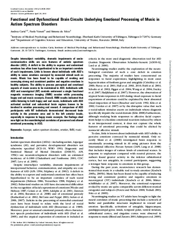
 S. Falco