INSTRUMENTATION & METHODOLOGY
Autonomic Arousal Index: an Automated Detection Based on Peripheral Arterial
Tonometry
Giora Pillar,1 Amir Bar,2 Arie Shlitner,1 Robert Schnall,2 Jacob Shefy,2 and Peretz Lavie1
1Sleep
Laboratory, Bruce Rappaport Faculty of Medicine, Technion - Israel Institute of Technology, Haifa, Israel; 2Itamar Medical Ltd., Caesarea,
Israel
46.2±14.4 years, BMI 28.5±5.4 kg/m2). All underwent a whole night PSG
with simultaneous PAT recording. The PSG recordings were blindly manually analyzed for arousals based on American Academy of Sleep
Medicine (AASM) criteria, while PAT was scored automatically.
There was a significant correlation between PSG and PAT arousals
(R=0.82, p<0.0001) with a good agreement across a wide range of values,
with a ROC curve having an area under the curve (AUC) of 0.88.
We conclude that automated analysis of the peripheral arterial tonometry
signal can detect EEG arousals from sleep, in a relatively quick and reproducible fashion.
Key words: Sleep; autonomic nervous system; arousals; peripheral arterial tonometry; sympathetic activation.
INTRODUCTION
nomic parameters such as heart rate and blood pressure, which
are not necessarily associated with any visible EEG changes.
This has led several authors to suggest that markers of autonomic activation (e.g., continuous blood pressure monitoring or pulse
transit time) should be included in routine polysomnographic
(PSG) recordings to assess the true extent of sleep fragmentation
in the form of autonomic arousal index (AAI).13-17 Piston et al.
reported a good correlation between PTT and EEG frequency
shifts in response to external stimuli in normal subjects.14 The
PTT could also, to some extent, detect sleep-disordered breathing
events.13 Argod et al. found a reasonable agreement between
standard scoring to PTT in detecting nonapneic obstructive respiratory events but reported a very high interobserver variability
in the scoring of both (30%-37%).17
The recently developed technique of peripheral arterial
tonometry (PAT) has been shown to be a sensitive marker of both
tonic18 and transient sympathetic arousal during sleep.19 Here,
we sought to examine and validate the efficacy of an automatic
analysis of this signal along with the PAT-derived pulse rate in
the detection of arousals from sleep.
SLEEP FRAGMENTATION IN THE FORM OF FREQUENT
“BRIEF AROUSALS” HAS BEEN IMPLICATED IN DAYTIME IMPAIRMENT OF COGNITIVE AND PSYCHOMOTOR PERFORMANCE, PARTICULARLY IN SLEEP RELATED BREATHING DISORDERS.1,2 Thus, the number of arousals
is a useful marker of sleep quality, which is independent of traditional sleep-quality markers such as sleep efficiency and sleep
latency. Based on the AASM criteria, an arousal from sleep is
scored when there is a notable EEG shift toward a higher frequency for at least 3 seconds but no more than 15 seconds during
all non-REM (NREM) stages of sleep, assuming sleep is recorded prior to and following the event for at least 10 seconds. Since
EEG alpha waves or mixed frequency waves are common during
rapid-eye movement (REM) sleep, the definition of arousal during REM sleep relies on a combination of EEG arousal and
increased EMG or body movements.3 Since these criteria are not
always clear-cut and are commonly difficult to determine, large
inter-scorer variability in scoring arousals from sleep has been
reported.4,5 Thus, an automatic and reliable method to detect
arousals has been sought.
It is well documented that arousals from sleep are associated
with transient sympathetic activation.6-12 The term “autonomic
arousal” is used to denote transient changes during sleep in auto-
METHODS
Subjects
The study group consisted of 96 adult subjects, with 11 being
healthy volunteers and 85 being patients referred with suspected
obstructive sleep apnea (OSA) (Table 1). The average age was
46 years, with the patients older than the healthy volunteers, and
the average BMI was 28.5kg/m2 with the patients heavier than
the healthy volunteers. Twenty-three percent of the patients were
hypertensive, and 5% were known to have ischemic heart disease. None of the patients had congestive heart failure. All the
patients were drawn from the Technion Sleep Disorders Center
Disclosure Statement
Consultants or Employees of Itamar-Medical Ltd.
Submitted for publication October 2001
Accepted for publication March 2002
Address correspondence to: Peretz Lavie, PhD, Sleep Laboratory, Faculty of
Medicine, Gutwirth Building, Technion-Israel Institute of Technology, Haifa
32000, ISRAEL
SLEEP, Vol. 25, No. 5, 2002
541
AAI: An Automated Detection—Pillar et al
Downloaded from https://academic.oup.com/sleep/article-abstract/25/5/541/2750145 by guest on 11 June 2020
Summary: Arousals from sleep are associated with increased sympathetic activation and are therefore associated with peripheral vasoconstriction. We hypothesized that digital vasoconstrictions as measured by
peripheral arterial tonometery (PAT), combined with an increase in pulse
rate, would accurately reflect arousals from sleep, and can provide an
autonomic arousal index (AAI).
Based on a previously studied group of 40 sleep apnea patients simultaneously recorded by both polysomnography (PSG) and PAT systems, an
automated algorithm using the PAT signal (and pulse rate derived from it)
was developed for detection of arousals from sleep. This was further validated in a separate group of 96 subjects (85 patients referred with suspected obstructive sleep apnea and 11 healthy volunteers mean age
�sleep was scored as an arousal, while during REM sleep, an
increase in EMG was required as well. In both cases at least 10
seconds of sleep period was required prior to and following the
event in order to be scored as an arousal. Arousal index (ARI)
was calculated by dividing the total number of arousals by the
number of hours of sleep. Respiratory events were scored
according to the AASM guidelines for measurement in clinical
research.22 The respiratory disturbance index (RDI) was calculated as the number of apneas plus hypopneas divided by the
number of hours of sleep. Periodic limb movements (PLM) were
also scored based on AASM criteria, and the PLM index (PLMI)
was calculated similarly to the other indices.
Table 2—Distribution of the study population according to RDI
The Peripheral Arterial Tonometry System
All participants underwent a whole night PSG; (EEG 4214;
Nihon Kohden, Kogyo Co., Tokyo, Japan) with simultaneous
recordings of PAT signal (Site-PAT 200; Itamar- Medical Ltd.
Caesarea, Israel). Prior to bedtime, patients completed a sleep
questionnaire including physical data (e.g., weight and height),
general health condition and medical history, medication usage,
sleep habits, and the Epworth sleepiness scale (ESS).20 Lights off
were no later than midnight, and lights were on at 06:00.
The PAT signal was measured using the Sleep PAT-200TM
device (Itamar-Medical LTD, Caesarea, Israel), which is a novel
specifically designed noninvasive finger plethysmograph.18,19
The PAT probe consists of an optical sensor housed in a self pressurized finger-mounted shell, which generates a uniform pressure
field surrounding the distal two phalanges of the finger
(~50mmHg). The optical devices serve to measure pulsatile volume changes while the uniform pressure field unloads the arterial wall tension, improves the dynamic range of the measurement,
and helps to reduce artifacts. The signal is further filtered (0.2
to 20Hz) and transferred as an analog input to an auxiliary direct
current (DC) input channel of the PSG, where it is recorded
simultaneously with the other standard channels. Upon completion of the recordings, the PSG data file, which includes the PAT
data alongside the rest of the PSG data, is retrieved for sleep scoring and PAT automatic analysis, using specific software. This
software employs algorithms based on weighting two features of
the PAT signal, each of which indicates sympathetic surge:
amplitude attenuation (reflecting vasoconstriction) and pulserate increase (comparable to heart-rate increase). The automatic
algorithm was developed using a previous cohort of 40 patients
(training set). The PAT arousal event is scored when one of the
following has occurred:
1.
The PAT signal amplitude attenuation, relative to
baseline, of at least 50% or
2.
The PAT signal amplitude attenuation of at least 30%,
relative to baseline, with concurrent increase of more
than 10% in pulse rate.
Pulse-rate changes were derived from the PAT signal itself and
the parallelism between a PAT-amplitude decrease and pulse-rate
increase was defined using an overlapping window of 30 seconds.
Polysomnography
Scoring
Overnight PSG was performed according to standard laboratory protocol, using computerized PSG with the following channels: two EEG (C3-A2 and O2-A1), EOG, submental EMG, arterial oxygen saturation, nasal-oral airflow (thermistors), electrocardiogram (ECG), chest and abdominal wall motion (piezo electrodes), bilateral anterior tibialis EMG, snoring (microphone
placed one meter above the bed, and body position. Sleep was
staged according to standard criteria.21 Arousals were defined
according to the AASM guidelines.3 Any EEG frequency shift
for at least 3 seconds but no more than 15 seconds during NREM
The PSG recordings were manually analyzed for sleep stages,
respiratory disturbances, movements, and arousals based on
ASDA criteria, with the scorer being completely blinded to the
PAT signal. To determine scoring reliability, a second scorer
scored 20 records independently (randomly selected). The correlation between the results of the two scorers with respect to the
total number of AASM-defined arousals was 0.96. This correlation is substantially higher than the previously reported ones,
possibly as the two scorers have worked together for a long period of time (>15years). The scored data file, also including the
Breathing Disorder
Severity
Normal
(RDI: 0-10)
Mild
(RDI:11-20)
Moderate
(RDI: 21-40)
Severe
(RDI: 40+)
Control
Patients
Overall
N
%
N
%
N
%
7
63.6
21
24.7
28
29.2
4
36.4
18
21.1
22
22.9
0
0
19
22.4
19
19.8
0
0
27
31.8
27
28.1
population and were referred for evaluation because of presumed
OSAS. Normal volunteers were without any complaints of sleep
disruption, daytime sleepiness, or snoring and were free of any
disease or use of any medication. They were recruited via advertisements in the Technion’s Faculty of Medicine. The suspected
OSA patients were excluded if they were on alpha-blocking medications or in unstable clinical condition. The study was
approved by the Rambam Medical Center IRB, and patients
signed an informed consent form prior to being studied.
Protocol
SLEEP, Vol. 25, No. 5, 2002
542
AAI: An Automated Detection—Pillar et al
Downloaded from https://academic.oup.com/sleep/article-abstract/25/5/541/2750145 by guest on 11 June 2020
Table 1—Age and body habitus characteristics of the study
group.
Control
Patients
Overall
Age
N
11
85
96
(years)
Mean
22.3
49.3
46.2
STD
6.6
12.0
14.4
Height
Mean
172.8
179.9
172.0
(cm)
STD
6.8
10.3
9.9
Weight
Mean
65.9
87.4
85.0
(kg)
STD
10.5
18.9
19.4
BMI
Mean
22.0
29.3
28.5
(Kg/m2)
STD
2.9
5.1
5.4
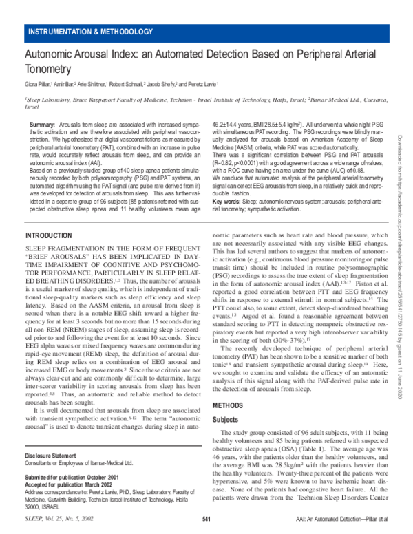
�ASDA-ARI vs. PAT-ARI
140
120
80
60
40
r=0.82
20
0
0
20
40
60
80
100
120
140
PAT-ARI
Figure 1—As can be seen, a very high and statistically significant correlation (R=0.82, p<0.0001) was found between the PAT-ARI (PAT-based arousal index) and the
ASDA-ARI (arousal index based on ASDA criteria). The red dots represent patients with PLMs.
recorded raw PAT signal, was then retrieved for automatic identification of PAT-based arousal events. This analysis takes into
consideration the sleep staging as interpreted by the PSG scorer
and processes the PAT signal within the sleep periods only.
The average ESS score for the whole group was 9 ± 6.4, with
18% of participants reporting severe sleepiness (ESS>16), and
48% reporting virtually no sleepiness (ESS<8). The distribution
of the study population according to RDI is presented in Table 2.
The average RDI for the whole group was 30 ± 28 events per
hour. As can be seen, the group had a wide range of sleep-disordered breathing, distributed from normal breathing (primarily 7
of the 11 normal volunteers, but also 21 of the referred patients)
to severe OSAS. The average ASDA-ARI (EEG arousals) for the
whole group was 27 ± 24 arousals per hour while the average
PAT-AAI for the group was 29 ± 19 arousals per hour.
Ten of the patients had also PLM, with an average PLM index
of 32 ± 23 movements per hour.
Figure 1 displays a scatter graph of the ASDA-ARI (gold standard) versus the PAT-AAI values for the whole study population
with the calculated correlation coefficient. There was a good and
statistically significant correlation between the two parameters
(R=0.82, p<0.0001). There was also a significant correlation
between RDI and both measures of arousals (0.92 for ASDA-ARI
and 0.80 for PAT-AAI, p<0.05 for both). Analyzing the correlations between ASDA-ARI and PAT-AAI separately for the subjects with no OSA (RDI of 10 or less) and OSA patients revealed
correlation coefficients of 0.58 and 0.84 respectively (p<0.05 for
both). Figure 2 displays the Bland-Altman plot for ASDA-ARI
and the PAT-AAI for the study population. There was a good
agreement between the two parameters across a wide range of
arousal indexes. Figure 3 shows the ROC curve for identifying
patients with pathologic AAI with the PAT (defined by a threshold of 20 arousals per hour of sleep23) when taking the ASDAbased scoring as “gold standard”, with an AUC of 0.88.
Since PLM may be associated with subcortical autonomic
arousals, occasionally without meeting criteria to be scored as
Statistical Analysis
Analysis was performed to assess the correlation between the
PAT-based AAI (PAT-AAI) and the ASDA-based arousal index
derived from the PSG scoring (ASDA-ARI) using Pearson correlation coefficient and a Bland-Altman plot. In order to evaluate
the efficacy of the PAT automatic analysis as a potential tool for
diagnosing a disorder of arousals from sleep, a receiver operating
characteristic (ROC) curve based on a threshold of AAI=2023 for
abnormality was plotted and its area under the curve (AUC) was
calculated. This curve joins points that represent the sensitivity
and specificity for all of possible PAT-AAI thresholds on an X-Y
plane (x=1-specificity, y=sensitivity) for all possible values of
the PAT-AAI thresholds. The area under the ROC curve (i.e.,
AUC) is considered a measure of the overall efficacy of the
score–an AUC value of 0.5 indicates a nonsignificant score for
separating normals from patients with pathologic AAI, and a
value close to 1.0 indicates a very efficient score.
In addition, the correlation of both the ASDA-ARI and PATAAI with ESS was performed to examine whether ASDA-based
arousals or PAT-based autonomic arousals, or both, correlates
with subjective sleepiness.
RESULTS
Of the 96 sleep studies, complete data were available for 94.
In 2 patients (2.1%), the data had to be discarded because of technical problems.
SLEEP, Vol. 25, No. 5, 2002
543
AAI: An Automated Detection—Pillar et al
Downloaded from https://academic.oup.com/sleep/article-abstract/25/5/541/2750145 by guest on 11 June 2020
ASDA-ARI Score
100
�Bland Altman Presentation of ASDA-ARI vs. PAT-ARI
60
20
0
-20
-40
-60
0
20
40
60
80
100
120
140
Mean of (ASDA-ARI) and (PAT-ARI)
Figure 2—As can be seen, across a wide range of arousals frequencies, there was a good agreement between PAT-ARI (PAT-based arousal index) and the ASDAARI (arousal index based on ASDA criteria). The red dots represent patients with PLMs.
ASDA-based arousals shown in the EEG recording,24-27 the statistical analysis was repeated after adding PLM events that were
not associated with ASDA-arousals to the total “gold standard”
arousals. Such events were noted in 10 patients. This resulted in
an improvement of the correlation between ASDA-based
arousals and PAT-AAI (R=0.88, p<0.0001), and the calculated
AUC minimally increased to 0.89.
Finally, although significant, there was only a poor correlation
between the arousal index (either by standard criteria or by PAT)
and subjective sleepiness as assessed by ESS (R=0.43, and
R=0.35, respectively, p<0.001 for both).
scoring ASDA arousals from sleep by experts from 14 sleep laboratories.5 In another study Loredo et al evaluated various types
of arousals and showed good interscorer reproducibility in scoring PLMs or respiratory events accompanied arousals characterized by increased EMG. Poor reproducibility was demonstrated
in the scoring of “classic” ASDA-defined arousals.4 Thus, an
automated or other well-defined method of detecting sleep fragmentation seems warranted. Two additional methods based on
sympathetic activation have been attempted previously. The PTT
demonstrated some promising results initially, but has not been
compared in large-scale studies to standard measures and has
been shown to have the disadvantage of high interobserver variability.17 Arousal and inspiratory blood pressure changes were
used by Davies et al15 as a potential marker of disturbed sleep and
disordered breathing in sleep. Although the blood-pressure profile appeared to be useful to identify patients with OSA, this
method has not been further investigated. One of the advantages
of the PAT method is its’ automatic computer-based analysis,
which makes it reproducible, objective, and time saving. All
three methods of comparisons between the ASDA and PAT-based
AAI revealed reasonably good agreement, suggesting that the
PAT based algorithm may accurately reflect sleep fragmentation.
Although we have not quantified time saving using PAT automated scoring in comparison with PSG-based manual ones, we
estimate that this method has saved approximately 20 to 50 minutes per record. Other potential advantages of this system are its
simplicity and minimal required technical intervention in patient
preparations and recording. Also, the applied pressure to the finger of approximately 50 mmHg did not cause any discomfort to
the subjects.
DISCUSSION
The primary finding of this study is that the standard ASDAbased “EEG arousals” can be reasonably accurately predicted by
measuring “autonomic arousals” at the level of the finger. This
is consistent with the observation that arousals from sleep are
associated with sympathetic activation and, therefore, can be
readily measured by the PAT. Utilizing two important sympathetic-related parameters—digital vasoconstriction and pulserate changes, we could develop an automatic and reliable algorithm for identifying autonomic arousals throughout sleep. Since
the PAT analysis is performed automatically, this is a relatively
simple, objective, and reproducible method, which can be a very
useful and important channel added to the standard PSG.
Brief arousals from sleep may impair cognitive and psychomotor performance, particularly in sleep-related breathing
disorders.1,2 However, EEG arousals, as currently defined, are
not easily reproducible primarily due to a considerably large
interscorer variability. Drinnan et al found large disagreement in
SLEEP, Vol. 25, No. 5, 2002
544
AAI: An Automated Detection—Pillar et al
Downloaded from https://academic.oup.com/sleep/article-abstract/25/5/541/2750145 by guest on 11 June 2020
Delta of (ASDA-ARI) and (PAT-ARI)
40
�1.0
0.9
0.8
Sensitivity
0.7
0.6
0.5
0.3
0.2
AUC=0.88
0.1
0.0
0.0
0.1
0.2
0.3
0.4
0.5
0.6
0.7
0.8
0.9
1.0
1 - Specificity
Figure 3—ROC curve for identifying pathologic arousals from sleep (threshold =20 arousals per hour of sleep) based on PAT vs. standard criteria. As can be seen,
the area under the curve is 0.88, yielding potentially high sensitivity and specificity in diagnosing pathological arousal frequency by PAT, when the gold standard is
the ASDA criteria based on PSG.
Both the EEG and the autonomic arousals were poorly correlated with subjective sleepiness, probably demonstrating either
poor judgment of the patients to subjectively assess their sleepiness level, or an objective limitation of the EEG arousal index or
AAI to predict it. This is not surprising, as previous studies have
also failed to show good correlation between arousals from sleep
and daytime sleepiness.13,28,29 As this study showed good agreement between the PAT-defined AAI and the EEG-defined
arousals index, the poor correlation between the PAT-AAI and
subjective daytime sleepiness assessment were also expected.
When originally introduced the ESS demonstrated good correlation with objective sleepiness measures (MSLT),20 but later studies failed to replicate this.30 Although the 0.35 correlation
between the PAT-AAI and the ESS is somewhat small, it should
be kept in mind that PAT-AAI is a state measure reflecting a specific night while the ESS is a trait measure reflecting a much
longer period. The ability of the PAT to predict daytime sleepiness needs further evaluation, probably with more objective
modalities such as MSLT (which is also a state measure).
It has been suggested that arousals from sleep are associated
with increased sympathetic activation.6-10 Therefore, it is not surprising that we have found a good match between EEG arousals
and autonomic arousals. Furthermore, this good agreement suggests that sympathetic activation is indeed a result of arousals
from sleep rather than other potential triggers such as hypoxemia
or hypercapnia. It has been previously suggested that both
hypoxemia and hypercapnia, acting via chemoreflexes, may
result in an acute increase in sympathetic activity to blood vessels
in the muscles.7,31-34 Thus, the increased sympathetic activation
SLEEP, Vol. 25, No. 5, 2002
in patients with OSA could well be the result of abnormal arterial gas levels. Furthermore, several studies, using direct intraneural measurements of sympathetic traffic to blood vessels in the
muscles have found that patients with sleep apnea have very high
levels of sympathetic flow, even during normoxic wakefulness,
which may explain their increased risk of arterial hypertension.34,35 However, the blood-gas changes are not the only potential factor contributing to the increased sympathetic activation in
sleep apnea. In their classic dog model, Brooks et al found that
acoustic-stimulated arousals from sleep resulted in abrupt
increases in blood pressure, although during apnea-stimulated
arousals the high blood pressure persisted to wakefulness.36,37 In
another study aiming at determining the effects of arousals from
sleep per se on the sympathetic outputs to the cardiovascular system, Horner et al reported that these resulted in phasic sympathetic activation, as assessed by increased heart rate and blood
pressure.6 Although we have not quantified blood gas in this
study, our finding that peripheral arterial vasoconstrictions with
increased pulse rate correlated well with arousals from sleep
(determined by EEG) both in a wide severity range of OSA
patients and in normal healthy volunteers, supports the finding
that arousals from sleep per se mediate the increased sympathetic activation, without hypoxemia or hypercapnia, as in the normal
volunteers these arousals are not accompanied by blood-gas
changes. This is consistent with the finding from the Sleep
Health Heart Study that arousals from sleep were associated
(although weakly) with hypertension.38 Of note, Morrel et al39
reported that an index of sleep fragmentation was significantly
associated with awake systolic blood pressure in subjects with
545
AAI: An Automated Detection—Pillar et al
Downloaded from https://academic.oup.com/sleep/article-abstract/25/5/541/2750145 by guest on 11 June 2020
0.4
�REFERENCES
1. Martin SE, Engleman HM, Deary IJ, Douglas NJ. The effect of
sleep fragmentation on daytime function. Am J Respir Crit Care Med
1996;153:1328-32.
2. Martin SE, Wraith PK, Deary IJ, Douglas NJ. The effect of nonvisible sleep fragmentation on daytime function. Am J Respir Crit Care
Med 1997;155:1596-601.
3. EEG arousals: scoring rules and examples. a preliminary report
from the Sleep Disorders Atlas Task Force of the American Sleep
Disorders Association. Sleep 1992;15:174-84.
4. Loredo JS, Clausen JL, Ancoli-Israel S, Dimsdale JE. Night-tonight arousal variability and interscorer reliability of arousal measurements. Sleep 1999;22:916-20.
5. Drinnan MJ, Murray A, Griffiths CJ, Gibson GJ. Interobserver variability in recognizing arousal in respiratory sleep disorders. Am J Respir
Crit Care Med 1998;158:358-62.
6. Horner RL, Brooks D, Kozar LF, Tse S, Phillipson EA. Immediate
effects of arousal from sleep on cardiac autonomic outflow in the
absence of breathing in dogs. J Appl Physiol 1995;79:151-62.
7. Somers VK, Dyken ME, Mark AL, Abboud FM. Sympatheticnerve activity during sleep in normal subjects. N Engl J Med
1993;328:303-7.
8. Morgan BJ, Crabtree DC, Puleo DS, Badr MS, Toiber F, Skatrud
JB. Neurocirculatory consequences of abrupt change in sleep-state in
humans. J Appl Physiol 1996;80:1627-1636.
9. Hornyak M, Cejnar M, Elam M, Wallin BG. Muscle sympathetic
nerve activity during sleep in man. Brain 1991;114:1281-1295.
10. Okada H, Iwase S, Mano T, Sugiyama Y, Watanabe T. Changes in
muscle sympathetic nerve activity during sleep in humans. Neurology
1991;41:1961-1966.
11. Schneider H, Schaub CD, Chen CA, et al. Effects of arousal and
SLEEP, Vol. 25, No. 5, 2002
546
AAI: An Automated Detection—Pillar et al
Downloaded from https://academic.oup.com/sleep/article-abstract/25/5/541/2750145 by guest on 11 June 2020
sleep state on systemic and pulmonary hemodynamics in obstructive
sleep apnea. J Appl Physiol 2000;88:1084-92.
12. Launois SH, Averill N, Abraham JH, Kirby DA, Weiss JW.
Cardiovascular responses to nonrespiratory and respiratory arousals in a
porcine model. J Appl Physiol 2001;90:114-120.
13. Pitson DJ, Stradling JR. Autonomic markers of arousal during sleep
in patients undergoing investigation for obstructive sleep apnoea, their
relationship to EEG arousals, respiratory events and subjective sleepiness. J Sleep Res 1998;7:53-9.
14. Pitson D, Chhina N, Knijn S, van Herwaaden M, Stradling J.
Changes in pulse transit time and pulse rate as markers of arousal from
sleep in normal subjects. Clin Sci (Colch) 1994;87:269-73.
15. Davies RJ, Vardi-Visy K, Clarke M, Stradling JR. Identification of
sleep disruption and sleep disordered breathing from the systolic blood
pressure profile. Thorax 1993;48:1242-7.
16. Davies RJ, Belt PJ, Roberts SJ, Ali NJ, Stradling JR. Arterial blood
pressure responses to graded transient arousal from sleep in normal
humans. J Appl Physiol 1993;74:1123-30.
17. Argod J, Pepin JL, Smith RP, Levy P. Comparison of esophageal
pressure with pulse transit time as a measure of respiratory effort for
scoring obstructive nonapneic respiratory events. Am J Respir Crit Care
Med 2000;162:87-93.
18. Lavie P, Schnall RP, Sheffy J, Shlitner A. Peripheral vasoconstriction during REM sleep detected by a new plethismographic method.
Nature Med 2000;6:606.
19. Schnall RP, Shlitner A, Sheffy J, Kedar R, Lavie P. Periodic, profound peripheral vasoconstriction - a new marker of obstructive sleep
apnea. Sleep 1999;22:939-946.
20. Johns MW. A new method for measuring daytime sleepiness: the
Epworth sleepiness scale. Sleep 1991;14:540-5.
21. Rechtschaffen A, Kales A. A manual of standardized terminology,
techniques and scoring system for sleep stages of human subjects. Los
Angeles: Brain Information Service/Brain Research Institute, UCLA,
1968.
22. Sleep-related breathing disorders in adults: Recommendations for
syndrome definition and measurement techniques in adults. The report
of an American Academy of Sleep Medicine Task Force. Sleep
1999;22:667-689.
23. Mathur R, Douglas NJ. Frequency of EEG arousals from nocturnal
sleep in normal subjects. Sleep 1995;18:330-333.
24. Sforza E, et al. EEG and cardiac activation during periodic leg
movements in sleep: support for a hierarchy of arousal responses.
Neurology. 1999;52:786-91.
25. Winkelman JW. The evoked heart rate response to periodic leg
movements of sleep. Sleep. 1999 22:575-80.
26. Karadeniz A, Ondze B, Desset A, Billiard M. EEG arousals and
awakening in relation with periodic leg movements during sleep. J Sleep
Res 2000;9:273-277.
27. Ali NJ, Davies RJ, Fleetham JA, Stradling JR. Periodic movements
of the legs during sleep associated with rises in systemic blood pressure.
Sleep 1991;14:163-5.
28. Kingshott RN, Engleman HM, Deary IJ, Douglas NJ. Does arousal
frequency predict daytime function? Eur Resp J 1998;12:1264-70.
29. Stradling JR, Barbour C, Glennon J, Langford BA, Crosby JH.
Prevalence of sleepiness and its relation to autonomic evidence of
arousals and increased inspiratory effort in a community based population of men and women. J Sleep Res 2000;9:381-8.
30. Chervin RD, et al. The Epworth Sleepiness Scale may not reflect
objective measures of sleepiness or sleep apnea. Neurology.
1999;52:125-31.
31. Somers VK, Zavala DC, Mark AL, Abboud FM. Contrasting effects
of hypoxia and hypercapnia on ventilation and sympathetic activity in
humans. J Appl Physiol 1989;67:2101-2106.
32. Somers VK, Zavala DC, Mark AL, Abboud FM. Influence of ventilation and hypocapnia on sympathetic nerve responses to hypoxia in
normal humans. J Appl Physiol 1989;67:2095-2100.
RDI less than one.
In some specific circumstances, autonomic arousals can be
seen without clear changes in the EEG (“cortical arousals”). One
of these conditions is PLM during sleep.24-27 For this reason, we
repeated the comparison between the PAT-defined arousals and
the ASDA “gold standard” when adding PLM events not associated with classic EEG arousals to the total ASDA-based arousals
counting. This resulted in an improvement of the correlation
coefficient between the two indexes from 0.82 to 0.88, while the
AUC essentially remained unchanged. This result suggests that
the EEG-based criteria as nowadays defined (ASDA criteria), are
not sensitive enough and some types of sleep fragmentation, such
as in the case of PLM, may not be identified. It should be emphasized, however, that autonomic nervous system arousals do not
necessarily reach the cortex, and these 2 measures may represent
two separate phenomena.
While considering this technique as a good measure of
arousals from sleep, the population investigated is one limitation
of the study. It consisted of healthy volunteers and patients with
snoring. One could argue that in other populations, such as
insomniacs, arousals from sleep may be associated with a different pattern of autonomic activation. Thus, expanding this study
to other populations will add more information regarding their
pattern of autonomic arousals.
Despite this limitation, we believe that our study supports the
concept that sleep fragmentation can be accurately assessed by
measurements of peripheral arterial tone at the finger, and by
using automated algorithm, an important contribution can be
made to the standard PSG by means of reproducibility, time savings, and objectivity.
�SLEEP, Vol. 25, No. 5, 2002
Downloaded from https://academic.oup.com/sleep/article-abstract/25/5/541/2750145 by guest on 11 June 2020
33. Somers VK, Abboud FM. Chemoreflexes—responses, interactions
and implications for sleep apnea. Sleep 1993;16:S30-3; discussion S334.
34. Somers VK, Dyken ME, Clary MP, Abboud FM. Sympathetic neural mechanisms in obstructive sleep apnea. J Clin Invest 1995;96:1897904.
35. Hedner J, Ejnell H, Sellgren J, Hedner T, Wallin G. Is high and fluctuating muscle nerve sympathetic activity in the sleep apnea syndrome
of pathogenetic importance for the development of hypertension? J
Hypertens 1988;6:S529-S531.
36. Brooks D, Horner RL, Kimoff RJ, Kozar LF, Render-Teixeira CL,
Phillipson EA. Effect of obstructive sleep apnea versus sleep fragmentation on responses to airway occlusion. Am J Resp Crit Care Med
1997;155:1609-17.
37. Brooks D, Horner RL, Kozar LF, Render-Teixeira CL, Phillipson
EA. Obstructive sleep apnea as a cause of systemic hypertension.
Evidence from a canine model. J Clin Invest 1997;99:106-9.
38. Nieto FJ, Young TB, Lind BK, et al. Association of sleep-disordered breathing, sleep apnea, and hypertension in a large communitybased study. Sleep Heart Health Study. JAMA 2000;283:1829-36.
39. Morrell MJ, Finn L, Kim H, Peppard PE, Badr MS, Young T. Sleep
fragmentation, awake blood pressure, and sleep-disordered breathing in
a population-based study. Am J Resp Crit Care Med 2000;162:2091-96.
547
AAI: An Automated Detection—Pillar et al
�
 G. Pillar