J Appl Physiol
89: 1275–1282, 2000.
Upper airway muscle responsiveness to rising PCO2
during NREM sleep
GIORA PILLAR, ATUL MALHOTRA, ROBERT B. FOGEL, JOSEE BEAUREGARD,
DAVID I. SLAMOWITZ, STEVEN A. SHEA, AND DAVID P. WHITE
Sleep Disorders Section, Divisions of Endocrinology and Pulmonary and Critical Care Medicine,
Department of Medicine, Brigham and Women’s Hospital and Harvard Medical School,
Boston, Massachusetts, 02115
Received 24 January 2000; accepted in final form 2 May 2000
Pillar, Giora, Atul Malhotra, Robert B. Fogel, Josee
Beauregard, David I. Slamowitz, Steven A. Shea, and
David P. White. Upper airway muscle responsiveness to
rising PCO2 during NREM sleep. J Appl Physiol 89:
1275–1282, 2000.—Although pharyngeal muscles respond
robustly to increasing PCO2 during wakefulness, the effect of
hypercapnia on upper airway muscle activation during sleep
has not been carefully assessed. This may be important,
because it has been hypothesized that CO2-driven muscle
activation may importantly stabilize the upper airway during stages 3 and 4 sleep. To test this hypothesis, we measured ventilation, airway resistance, genioglossus (GG) and
tensor palatini (TP) electromyogram (EMG), plus end-tidal
PCO2 (PETCO2) in 18 subjects during wakefulness, stage 2, and
slow-wave sleep (SWS). Responses of ventilation and muscle
EMG to administered CO2 (PETCO2 5 6 Torr above the eupneic level) were also assessed during SWS (n 5 9) or stage
2 sleep (n 5 7). PETCO2 increased spontaneously by 0.8 6 0.1
Torr from stage 2 to SWS (from 43.3 6 0.6 to 44.1 6 0.5 Torr,
P , 0.05), with no significant change in GG or TP EMG.
Despite a significant increase in minute ventilation with
induced hypercapnia (from 8.3 6 0.1 to 11.9 6 0.3 l/min in
stage 2 and 8.6 6 0.4 to 12.7 6 0.4 l/min in SWS, P , 0.05 for
both), there was no significant change in the GG or TP EMG.
These data indicate that supraphysiological levels of PETCO2
(50.4 6 1.6 Torr in stage 2, and 50.4 6 0.9 Torr in SWS) are
not a major independent stimulus to pharyngeal dilator muscle activation during either SWS or stage 2 sleep. Thus
hypercapnia-induced pharyngeal dilator muscle activation
alone is unlikely to explain the paucity of sleep-disordered
breathing events during SWS.
obstructive sleep apnea syndrome; dilator muscle; genioglossus; hypercapnia; slow-wave sleep; nonrapid eye movement
is a common disorder characterized by the repetitive collapse of the pharyngeal
airway during sleep. Our laboratory has previously
shown, in apnea patients, that pharyngeal dilator muscle activation is high during wakefulness, which probably protects the airway from collapse (17). During
sleep, the loss of muscle activation results in airway
collapse (18). Considerable effort has been made to
determine the stimuli that drive activation of the phaOBSTRUCTIVE SLEEP APNEA
Address for reprint requests and other correspondence: D. P.
White, RF 485, 221 Longwood Ave., Brigham and Women’s Hospital,
Boston, MA 02115 (E-mail: dpwhite@gcrc.bwh.harvard.edu).
http://www.jap.org
ryngeal muscles during both sleep and wakefulness.
Although these dilator muscles respond robustly to
increasing PCO2 during wakefulness (22), the effect of
hypercapnia on upper airway (UAW) muscle activation
during sleep has only been minimally assessed in humans. This may be clinically relevant because CO2stimulated muscle activation has been proposed as an
important variable in maintaining airway patency during stages 3 and 4 sleep (1, 5, 9, 26, 37).
A number of studies indicate that the majority of
sleep-disordered breathing occurs during stages 1 and
2 sleep, generally in the wake-sleep transition, or during rapid-eye-movement (REM) sleep. On the other
hand, there are relatively few apneas or hypopneas
observed during stages 3 and 4 sleep [slow-wave sleep
(SWS)] (8, 14, 15). The reason ventilation appears to be
more stable during SWS remains unclear.
Three mechanisms are possible to explain the association of SWS with relatively stable respiration. 1)
SWS has a protective effect on UAW patency. 2) The
instability of sleep state associated with frequent
sleep-disordered breathing events does not allow the
individual to achieve SWS. 3) The increase in arousal
threshold during SWS contributes to respiratory and
UAW stability. There are reasonable arguments for all
of these mechanisms. However, it seems clear that, if
the patient does achieve SWS, ventilation stabilizes. It
has been suggested that the gradual increment in PCO2
from stage 2 to SWS (5, 26) may adequately stimulate
UAW dilator muscles so that pharyngeal patency can
be maintained (9). Our laboratory has also observed
that, with inspiratory resistive loading, there is a delayed (60–90 s) increment in genioglossus (GG) muscle
activation compatible with a chemical (PCO2) stimulus
(37). Finally, Badr et al. (1) reported variable responses
of the GG electromyogram (EMG) to induced hypercapnia among seven subjects during non-rapid-eye-movement (NREM) sleep. However, in both of these studies
(1, 37), the muscle responsiveness to rising CO2 may
have been confounded by a simultaneous, progressively negative epiglottic pressure because subjects
The costs of publication of this article were defrayed in part by the
payment of page charges. The article must therefore be hereby
marked ‘‘advertisement’’ in accordance with 18 U.S.C. Section 1734
solely to indicate this fact.
8750-7587/00 $5.00 Copyright © 2000 the American Physiological Society
1275
Downloaded from www.physiology.org/journal/jappl by ${individualUser.givenNames} ${individualUser.surname} (167.160.073.160) on November 15, 2018.
Copyright © 2000 the American Physiological Society. All rights reserved.
�1276
UPPER AIRWAY MUSCLE CO2 RESPONSE DURING SLEEP
slept in the supine posture. The one patient with a
substantial increase in GG EMG in the Badr et al.
study (1) had a very large negative esophageal pressure (230 cmH2O) while inspiratory resistive loading
in the Wiegand et al. study (37) is known to result in
increasingly negative epiglottic pressure (although not
directly measured in that study). Thus, to date, the
isolated relationship between PCO2 and pharyngeal dilator muscle activity during sleep has not been fully
examined. We hypothesized that dilator muscles are
sensitive to chemostimulation (PCO2) during sleep and
that hypercapnia will result in increased dilator muscle activation. We also hypothesized that hypercapniainduced dilator muscle activation may protect UAW
patency during sleep. Thus the present protocol was
designed to address, in normal subjects, the following
questions.
Does the transition from stage 2 to SWS lead to
important increments in end-tidal PCO2 (PETCO2)? In
order for the hypercapnia of SWS to mediate a protective effect on pharyngeal patency, a measurable
change in PCO2 would seem necessary.
Does SWS provide a protective influence on UAW
patency through activation of pharyngeal dilator muscles? By measuring the activity of both a representative phasic dilator muscle (i.e., GG) and a tonic one
[i.e., tensor palatini (TP)], we can evaluate whether the
protective effect of SWS is mediated through dilator
muscle activation.
Do supraphyiological levels of hypercapnia drive
pharyngeal dilator muscle activation during NREM
sleep (particularly SWS)? By administering CO2, we
determined the responsiveness of these muscles to
rising PCO2 in both sleep stages.
METHODS
Subjects
Eighteen historically healthy subjects were studied [9
men, 9 women, age 27.7 6 1.3 (SE) yr, body mass index
22.9 6 0.5 kg/m2]. Subjects denied any chronic diseases,
daytime somnolence, or snoring. None had any pharyngeal
anatomic abnormality on physical examination. The study
was approved by the Brigham and Women’s Human Subjects
Review Committee, and the subjects gave written, informed
consent before participation in the study.
Instrumentation and Techniques
Ventilation. Subjects wore a nasal mask (Healthdyne
Technologies, Marietta, GA) connected to a two-way valve
that partitioned inspiration and expiration. Inspiratory flow
was determined with a pneumotachometer (Fleish, Lausanne, Switzerland) and a differential pressure transducer
(Validyne, Northridge, CA), calibrated with a rotameter. The
subject’s breathing was exclusively nasal, ensured by mouth
taping and video camera monitoring to document that the
mouth remained closed. The dead space of the mask system
was about 50 ml, depending on facial configuration. Tidal
volume (VT) was obtained from the integrated flow signal,
and minute ventilation (V̇E) was calculated as the sum of all
VT per minute.
Muscle activation. GG EMG was measured with a pair of
unipolar intramuscular electrodes referenced to a single
ground, thus producing a bipolar recording. Two stainless
steel, Teflon-coated, 30-gauge wire electrodes were inserted
15–20 mm into the body of the GG muscle 3 mm lateral to the
frenulum on each side, using a 25-gauge needle that was
quickly removed, leaving the wires in place.
TP EMG was also measured, with a pair of referenced,
unipolar, intramuscular electrodes producing a bipolar recording. On each side of the palate, the tip of the pterygoid
hamulus was located at the junction of the hard and soft
palates. A 25-gauge needle with a 30-gauge, stainless steel,
Teflon-coated wire was then inserted at a 45° angle along the
lateral surface of the medial pterygoid plate, to a depth of
;10–15 mm into the palate. The needle was then removed,
leaving the electrode in place. These techniques have been
used previously in our laboratory (18). To confirm electrode
placement, the following respiratory maneuvers, which have
previously been shown to activate the TP muscle, were performed: sucking, blowing, and swallowing (35, 36).
For both muscles, the raw EMG was amplified, band-pass
filtered (between 30 and 1,000 Hz), rectified, and electronically integrated on a moving-time-average basis, with a time
constant of 100 ms (CWE, Ardmore, PA). The EMG was
quantified as percentage of maximal activation. To define
maximal muscle EMG activity, subjects performed four maneuvers. They were instructed to 1) maximally inspire
against an occluded tube, 2) maximally protrude their tongue
against the maxillary alveolar ridge, 3) swallow, and 4) suck
and blow. Each maneuver was performed several times, and
the maximal EMG recording for each muscle during this
calibration was assigned a level of 100%. Electrical zero was
then determined, and, thereafter, each EMG was quantified
as a percentage of maximal activation for that individual.
Because GG is an inspiratory phasic muscle, its level of
activation was assessed at two points in the respiratory cycle.
The tonic activation was defined as the lowest EMG level
during expiration (the minimal activation in each breath),
and peak phasic EMG was defined as the maximal activation
during inspiration. As TP is a tonic muscle, without phasic
activation, the EMG is reported as the average activation
across each breath.
To ensure that recording time or duration did not affect
EMG responsiveness, two actions were taken. First, the
EMG activation in response to naturally occurring swallows
was assessed for TP and GG in each subject during the first
and last 15-min period of each recording. In each condition,
electrical zero was also recorded to ensure no drift in EMG
signal. Second, we studied three additional subjects in a
modified protocol. This included GG EMG measurements in
six conditions: basal breathing and hypercapnia while
awake, basal breathing and hypercapnia during stable
NREM sleep, and basal breathing and hypercapnia awake
again, at the end of the study (after 2–3 h of recordings).
Polysomnography. Wakefulness and/or sleep was documented with two channels of electroencephalography (C3-A2,
C4-O1), two channels of electrooculography, and submental
EMG. Sleep stages were scored using standard criteria (24).
Subjects maintained the lateral decubitus posture throughout the study, as verified by video camera. We chose to study
all subjects in the lateral posture to minimize changes in
pharyngeal resistance and epiglottic pressure during sleep.
This was done to assess the relatively isolated effects of
hypercapnia on muscle activation.
Pressure and resistance. Pressures were monitored in the
nasal mask (Validyne) and in the airway at the level of the
choanae and the epiglottis. One nostril was decongested with
oxymetazoline HCl and anesthetized using lidocaine HCl.
Two pressure-tipped catheters (MPC-500, Millar, Houston,
Downloaded from www.physiology.org/journal/jappl by ${individualUser.givenNames} ${individualUser.surname} (167.160.073.160) on November 15, 2018.
Copyright © 2000 the American Physiological Society. All rights reserved.
�1277
UPPER AIRWAY MUSCLE CO2 RESPONSE DURING SLEEP
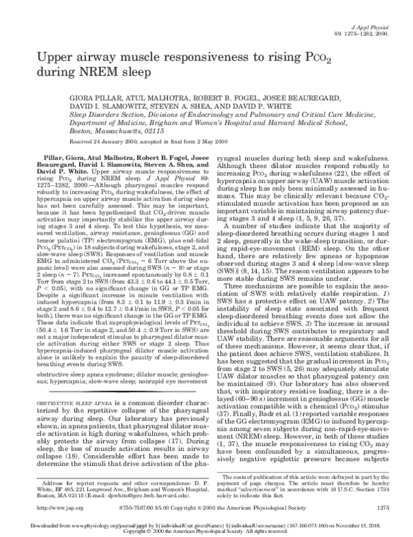
Fig. 1. Schematic diagram of the study protocol. First, data were collected during quiet wakefulness. After 5 min
of stable stage 2 sleep were observed, 5 min of data were recorded. Similarly, after 2 min of stable slow-wave sleep
(SWS) were observed, 5 min of data were recorded. Finally, data during externally administered CO2 were
collected. n, No. of subjects recorded.
TX) were inserted through this nostril and localized to measure choanal and epiglottic pressures. Before insertion, all
three pressure signals were calibrated simultaneously in a
rigid cylinder using a standard water manometer. These
three signals, plus flow, were demonstrated to be without
amplitude or phase lags at up to 2 Hz. Pharyngeal resistance
(the pressure difference between choanae and epiglottis divided by flow), nasal resistance (the pressure difference between mask and choanae divided by flow) and supraglottic
resistance (nasal resistance 1 pharyngeal resistance) were
determined at both peak flow and 0.2 l/s inspiratory flow.
CO2 administration and PETCO2 measurement. PETCO2 was
measured from expired air sampled within the mask using a
calibrated infrared CO2 analyzer (Capnograph Monitor, BCI,
Waukesha, WI). To assess the hypercapnic response, the
inspired fraction of CO2 was increased using a calibrated gas
source (25% CO2-21% O2-balance N2) fed into the inspiratory
line, to achieve an PETCO2 of 5–6 Torr above eucapnic basal
sleep levels. Once this level was reached and remained stable
for 3 min, data were recorded for 3 min.
Study Protocol
Subjects reported to the laboratory in the evening, having
abstained from food for at least 4 h. After informed consent
was obtained, all instrumentation was performed, and the
equipment was calibrated. Data were then recorded during
basal wakefulness (see Fig. 1) for a period of 5 min. Subjects
were then allowed to fall asleep. Once stable stage 2 sleep
was observed, 5 min of basal breathing were recorded. If
subjects awakened during the recordings, these data were
excluded and another 5-min period was recorded after stable
stage 2 sleep was again achieved. After subjects entered
SWS, an additional 5 min of recording took place. Two subjects did not reach SWS. Finally, CO2 was administered to
elevate PETCO2 to 5–6 Torr above baseline levels during sleep
(see Fig. 1). In 9 of the 18 subjects, CO2 administration was
performed during SWS, whereas CO2 was delivered to 7
subjects during stage 2 sleep. Recordings of supraphysiological hypercapnia were performed after a steady-state level of
PETCO2 with no arousals was reached. The time interval
between recording of baseline and CO2-stimulated muscle
activation was, on average, 31.7 min. In two subjects, CO2
administration could not be completed due to repetitive
awakenings.
Data Recording and Analysis
All signals (electroencephalogram, electrooculogram, submental EMG, inspiratory flow, PETCO2, GG EMG and TP
EMG) were recorded on a 16-channel Grass model 78 polygraph (Grass Instruments, Quincy, MA). Certain signals (VT,
V̇E, PETCO2, muscle EMG, and inspiratory flow) were also
recorded onto computer using signal-processing software
(Spike 2, Cambridge Electronic Design, Cambridge, UK).
Sampling frequency was 125 Hz.
For each recording period (awake, stage 2, SWS, CO2
administration) all breaths from each 5-min recording (3 min
in the administered CO2 portion) were signal averaged.
Thus, for each state, VT, PETCO2, GG EMG (tonic and peak
phasic) and TP EMG (tonic only) were determined from this
averaged breath. V̇E, as stated, was determined by summing
all VT values per minute.
All statistical analyses were performed with commercially
available software (Excel 97, Microsoft; and SigmaStat 1
Sigmaplot, SPSS, Chicago, IL). All data are presented as
means 6 SE unless otherwise stated. Repeated-measures
ANOVA with post hoc Student-Newman-Keuls testing was
used to assess state-dependent changes. Whenever data were
not normally distributed, Friedman repeated-measures
ANOVA on ranks was used. P , 0.05 was taken to indicate
significance.
RESULTS
Ventilation, PETCO2, UAW resistances, and activation levels of both dilator muscles in the three states
are shown in Table 1. V̇E decreased significantly from
wakefulness to stage 2 sleep, and further to SWS,
although this further decline was not statistically significant. Although PETCO2 increased significantly from
wakefulness to stage 2 sleep (P , 0.05), and further
Table 1. Ventilation, airway mechanics, and muscle
activation in different sleep stages
V̇E, l/min
Tidal volume, ml
Respiratory rate,
breaths/min
TI/Ttot, %
PETCO2, Torr
GG tonic, % of maximum
GG peak, % of maximum
TP, % of maximum
P choanal, cmH2O
P epiglottic, cmH2O
Peak flow, l/s
Resistance
Nasal
Pharyngeal
Supraglottic
Awake
Stage 2
SWS
9.4 6 0.4
595 6 22
8.6 6 0.3*
563 6 25
8.1 6 0.3*
543 6 31*
15.7 6 0.7
42.5 6 1.8
39.3 6 0.7
5.9 6 1.3
7.4 6 1.3
8.1 6 1.9
21.5 6 0.1
21.9 6 0.1
0.48 6 0.02
15.3 6 0.5
43.8 6 1.6
43.3 6 0.6*
5.9 6 1.2
7.4 6 1.3
4.8 6 1.0*
22.0 6 0.4
23.1 6 0.5*
0.50 6 0.03
14.8 6 0.6
43.3 6 1.5
44.1 6 0.5*†
5.6 6 1.2
7.7 6 1.5
4.2 6 1.0*
21.9 6 0.2
24.3 6 0.8*
0.53 6 0.02
1.2 6 0.2
0.7 6 0.2
1.9 6 0.2
2.3 6 0.8
2.5 6 0.8*
4.8 6 1.2*
1.5 6 0.4
4.8 6 1.6*
6.2 6 1.6*
Values are means 6 SE. SWS, slow-wave sleep; V̇E, minute ventilation; TI, inspiratory time; Ttot, total time; PETCO2, end-tidal PCO2;
GG, genioglossus muscle; TP, tensor palatini muscle; P, pressure.
* P , 0.05 vs. awake; † P , 0.05, stage 2 vs. SWS.
Downloaded from www.physiology.org/journal/jappl by ${individualUser.givenNames} ${individualUser.surname} (167.160.073.160) on November 15, 2018.
Copyright © 2000 the American Physiological Society. All rights reserved.
�1278
UPPER AIRWAY MUSCLE CO2 RESPONSE DURING SLEEP
Table 2. Comparison of ventilation, airway
mechanics and muscle activations between
baseline and elevated PCO2 conditions
Stage 2 (n 5 7)
V̇E, l/min
Tidal volume,
ml
Respiratory
rate,
breaths/min
TI/Ttot, %
PETCO2, Torr
GG tonic, % of
maximum
GG peak, % of
maximum
TP tonic, % of
maximum
Pchoanal,
cmH2O
Pepiglottic,
cmH2O
Peak flow, l/s
Resistance
Nasal,
flow 5 0.2 l/s
Pharyngeal,
flow 5 0.2 l/s
Nasal, peak
flow
Pharyngeal,
peak flow
Supraglottic,
peak flow
SWS (n 5 9)
Baseline
Externally
administrated
CO2
Baseline
Externally
administrated
CO2
8.3 6 0.4
11.9 6 0.3*
8.6 6 0.4
12.7 6 0.4*
509 6 34
742 6 59*
606 6 32
846 6 39*
16.2 6 0.8
42.2 6 3.3
43.1 6 0.4
16.5 6 0.9
46.3 6 2.0
50.4 6 1.6*
14.2 6 0.7
39.1 6 1.2
44.7 6 0.7
15.0 6 0.6*
43.8 6 1.5*
50.4 6 0.9*
5.7 6 0.4
4.7 6 1.2
6.3 6 2.0
4.9 6 1.2
7.7 6 0.6
7.8 6 1.4
7.9 6 2.1
8.8 6 1.9
6.5 6 0.6
5.9 6 1.9
4.2 6 1.4
5.2 6 1.5
22.8 6 0.9
23.1 6 0.6
21.7 6 0.2
22.2 6 0.2
23.7 6 0.9 25.5 6 1.3
24.9 6 1.4 25.9 6 1.2
0.45 6 0.04 0.55 6 0.05* 0.56 6 0.04 0.61 6 0.1
1.7 6 0.4
1.5 6 0.5
0.6 6 0.1
0.8 6 0.2
1.1 6 0.3
1.2 6 0.6
2.1 6 0.1
2.4 6 0.3
4.3 6 1.9
3.2 6 1.2
1.1 6 0.2
1.5 6 0.3
2.0 6 0.8
4.3 6 1.4
5.8 6 2.6
8.6 6 4.3
6.3 6 2.1
7.4 6 2.0
6.8 6 2.6
10.1 6 4.4
Nasal and pharyngeal resistances are given at 2 points in the
breath: at flow 5 0.2 l/s, which occurs during the linear part of the
pressure/flow curve, and at peak flow (which was also peak resistance, as flow limitation did not occur). n, No. of patients. * P , 0.05,
baseline vs. administered CO2.
from stage 2 to SWS (P , 0.05), no significant change
in GG EMG was observed. TP EMG decreased significantly from wake to stage 2 sleep but did not change
from stage 2 to SWS. Pharyngeal resistance significantly increased from wakefulness to sleep and tended
to increase further from stage 2 to SWS, although this
change did not reach statistical significance. There was
no correlation between the change in pharyngeal resistance and the change in ventilation from wakefulness
to stage 2 sleep, but there was a significant correlation
between the change in these variables from stage 2
sleep to SWS (r 5 0.55, P , 0.05). Nasal resistance did
not change significantly between conditions.
Despite significant increases in V̇E with induced hypercapnia (8.3 6 0.1 to 11.9 6 0.3 l/min in stage 2, and
8.6 6 0.4 to 12.7 6 0.4 in SWS, P , 0.05 for both), there
was no change in the GG EMG or the TP EMG (Table
2). One example, 30 s of raw data from stage 2 sleep,
SWS, and during hypercapnia in SWS, is shown in Fig.
2. As can be seen, hypercapnia was associated with
substantial increases in ventilation but no important
change in muscle activation. Figure 2 also demon-
strates the phasic nature of the GG and the tonic
nature of the TP.
As stated in METHODS, to ensure EMG signal stability,
CO2 responsiveness was assessed in three subjects,
awake at the beginning of the study, during stable
NREM sleep, and during wakefulness thereafter. Adequate data were obtained in two subjects. As shown in
Fig. 3 (data from one representative subject), both
ventilation and GG EMG increased in response to CO2
during wakefulness on both occasions (before and after
NREM sleep), but little to no GG EMG response was
observed during NREM sleep. This suggests stable
signals throughout the recordings. In addition, no consistent changes were observed in the response of either
the GG or TP to spontaneous swallows over the course
of the study. Average GG EMG during a swallow was
59.1 6 13% of maximum during the first 15 min vs.
57.5 6 12.2% during the last 15-min period (not significant). Average TP EMG was 56.8 6 17.7% during the
first 15 min vs. 55.2 6 20.3% of maximum during the
last 15-min period. Therefore, EMG responsiveness to
spontaneous swallows was as robust at the end of the
study as at the beginning. Thus we believe we had a
stable EMG signal. Finally, two subjects occasionally
snored, but no evidence of inspiratory flow limitation
was observed in the buffered (signal-averaged) breath.
DISCUSSION
This study suggests that, in normal subjects, pharyngeal dilator muscle activation is not importantly
modulated by CO2 during either SWS or stage 2 sleep.
Although PETCO2 did increase significantly from stage 2
sleep to SWS, it was not associated with an increase in
activation of either tonic or phasic pharyngeal dilator
muscles. Even with supraphysiological levels of CO2
that were clearly effective in increasing ventilation,
dilator muscle activation did not change significantly.
These data strongly suggest that hypercapnia alone is
not a strong stimulus to pharyngeal dilator muscle
activation during NREM sleep.
GG EMG did not change from wake to sleep; however, TP activation fell significantly. These observations are generally in agreement with previous studies
in normal humans, which have demonstrated a substantial fall in TP EMG with the change from wake to
sleep but highly variable changes in GG EMG with
state changes (18, 31, 32). Hypercapnia has previously
been shown to be a potent stimulator of ventilation (7)
and leads to increases in GG EMG during wakefulness
(22). However, GG responsiveness to CO2 has not been
tested during sleep in humans. As stated, we observed
that induced hypercapnia during sleep increased ventilation but failed to increase pharyngeal dilator activation. When this observation is added to the previous
reports that document negative pressure stimuli activating UAW muscles during wakefulness but not during sleep (11, 35), we have to conclude that pharyngeal
dilator muscles are generally unresponsive to either
mechanoreceptive or chemoreceptive stimuli during
sleep. In apnea patients, although increments in pha-
Downloaded from www.physiology.org/journal/jappl by ${individualUser.givenNames} ${individualUser.surname} (167.160.073.160) on November 15, 2018.
Copyright © 2000 the American Physiological Society. All rights reserved.
�UPPER AIRWAY MUSCLE CO2 RESPONSE DURING SLEEP
1279
Fig. 2. Representative example in a single
subject: 30 s of raw data collected during stage
2 sleep (left), SWS (middle), and CO2 administration (hypercapnia; right) during SWS are
shown. As can be seen in this case, end-tidal
PCO2 (PETCO2) increased from during stage 2
sleep, SWS, and with hypercapnia. This was
associated with a substantial increase in ventilation (only inspiratory tidal volume is
shown) but no change in genoglossus (GG) or
tensor palatini (TP) muscle activation. The
data also demonstrate the phasic nature of GG
and the tonic nature of TP. max, Maximum.
ryngeal dilator muscle activation have been observed
over the course of an apnea (as intrapharyneal pressure becomes progressively negative and hypoxia plus
hypercapnia develop), the majority of such muscle activation occurs with arousal at apnea termination (3,
19). This may explain the necessity for sleep apneics to
arouse to regain pharyngeal patency. In other words,
because pharyngeal dilators fail to adequately respond
to respiratory stimuli during sleep, arousal from sleep
is required to terminate sleep-disordered breathing
events.
Although individuals with sleep apnea were not
studied, the lack of response of pharyngeal dilator
muscles to hypercapnia (physiological and supraphysiological levels) does not support the hypothesis that
SWS-induced hypercapnia drives muscle activation
and thereby protects UAW patency. Although the
study of Basner et al. (2) previously reported increased
GG activation in five subjects during SWS compared
with stage 2 sleep (without significant change in
PETCO2 or ventilation), we could not replicate the results of that study. In fact, in our group of 16 subjects,
PETCO2 significantly increased from stage 2 to SWS
without activation of either GG or TP. However, the
subjects of Basner et al. (2) were studied in the supine
posture, whereas ours were in the lateral decubitus
posture, which may have influenced airflow resistance,
epiglottic negative pressure, and muscle activation.
Henke et al. (9) also reported an increase in PETCO2
from stage 2 sleep to SWS, in association with an
increase in the EMG of ventilatory muscles (diaphragm and scalene) in five snorers (measured with
surface electrodes). Flow limitation was noted in both
stage 2 and SWS, and the change from stage 2 to SWS
was associated with a significant increase in pharyngeal resistance. When patients were unloaded by con-
Downloaded from www.physiology.org/journal/jappl by ${individualUser.givenNames} ${individualUser.surname} (167.160.073.160) on November 15, 2018.
Copyright © 2000 the American Physiological Society. All rights reserved.
�1280
UPPER AIRWAY MUSCLE CO2 RESPONSE DURING SLEEP
Fig. 3. Representative example of raw data collected during 6 conditions in one subject: awake baseline breathing,
awake hypercapnia, sleep baseline breathing, sleep hypercapnia, and, again, awake baseline breathing and awake
hypercapnia at the end of the study (after 2–3 h of recording). GG EMG was similar during basal breathing and
similarly responded to hypercapnia in the 2 wakeful periods (before and after sleep). During sleep, however,
despite an increase in ventilation with induced hypercapnia, GG EMG remained unchanged. VT, tidal volume.
tinuous positive airway pressure application, both
PETCO2 and ventilatory muscle activation declined.
When CO2 was added to restore eucapnia (with continuous positive airway pressure in place), EMG increased toward baseline levels, suggesting some effect
of CO2 on scalene and diaphragm activity in snorers.
Pharyngeal dilator muscles, however, were not monitored in that study.
Interestingly, in animal models, induced hypercapnia resulted in decreased pharyngeal airflow resistance and increased EMG of the GG and ala nasi (27).
This decrease in resistance was also observed in cats,
independent of GG or strap muscle activation (25).
Other studies in animals have also found a reduction in
airway resistance with induced hypercapnia (20). However, these studies were not conducted in humans and
not during sleep, making it difficult to compare with
our observations. The one study that did measure GG
EMG in humans with induced hypercapnia found
highly variable responses. In the single subject from
that study for whom raw data were presented, esophageal pressure became extremely subatmospheric
(230 cmH2O, in the supine posture), and there was a
robust response of GG EMG (1). Thus hypercapnia and
airway negative pressure could potentially work in
combination to activate pharyngeal dilators.
The changes in ventilation and in UAW resistance
observed in the present study are generally in agreement with previous findings. We observed that the
change from wakefulness to sleep was associated with
an increase in UAW resistance, a decrease in ventilation, and an increase in PETCO2 (4, 5, 7, 9, 12, 13, 23,
26). Increased pharyngeal resistance is likely a substantial contributor to the fall in ventilation from wake
to sleep (9, 30). It is not surprising, however, that the
correlation between the change in pharyngeal resistance and ventilation in this transition was weak, as
many other changes in respiratory control likely occurred as well, thus making the isolated effect of pharyngeal resistance on V̇E difficult to detect. We observed a further increment in PETCO2 with the change
from stage 2 to SWS, although the trend toward decreases in ventilation and increases in UAW resistance
did not reach statistical significance. Several previous
studies have reported similar observations (5, 7, 9, 26,
33). Unlike the transition from wakefulness to stage 2
sleep, the decrement in V̇E from stage 2 to SWS was
significantly correlated with the increment in pharyn-
Downloaded from www.physiology.org/journal/jappl by ${individualUser.givenNames} ${individualUser.surname} (167.160.073.160) on November 15, 2018.
Copyright © 2000 the American Physiological Society. All rights reserved.
�UPPER AIRWAY MUSCLE CO2 RESPONSE DURING SLEEP
geal resistance. This suggests that, in the absence of
the behavioral influences present during wakefulness,
changes in pharyngeal resistance with state change
play a susbstantial role in determining the associated
change in ventilation (9, 30, 33).
Our observation that induced hypercapnia leads to
an increment in ventilation with no significant change
in pharyngeal resistance (Table 2) is in contrast to
previous studies that have reported hypercapnia to
reduce pharyngeal resistance (16, 28). However, Badr
et al. (1), using total pulmonary resistance as an index
of UAW patency, found no significant change in this
measure with 12, 14, and 16 Torr increments in PCO2
during NREM sleep in nine subjects, although there
was a trend toward a decrease in total pulmonary
resistance with PCO2 6 Torr above baseline (1). In
anesthetized animals, however, airway resistance decreased with induced hypercapnia (20, 25, 27). The
most plausible explanation for this observation is that
elevated PCO2 levels (in combination with negative
pharyngeal pressure) lead to increased pharyngeal dilator muscle activation or tracheal caudal displacement (1, 20, 34). Our finding that pharyngeal resistance did not decrease with induced hypercapnia may
be a result of our subjects’ sleeping in the lateral
decubitus posture. In the lateral position, airway resistance tends to be lower, and thus the negative pressure
generated by inspiratory muscles is reduced. If a combination of negative airway pressure and elevated PCO2
is required to activate the pharyngeal dilator muscle
during NREM sleep, one would expect more muscle
activity during hypercapnia in the supine posture.
However, we wanted to assess the isolated effect of
hypercapnia and observed little such effect in our subjects sleeping in the lateral posture.
There are several potential limitations to our study.
First, although our intention was to provide insights
into the pathogenesis of obstructive sleep apnea by
studying only normals, any conclusions regarding patients with obstructive sleep apnea are speculative.
However, because of the fragmented sleep seen in
individuals with apnea and their minimal SWS, assessment of muscle activation and chemosensitivity
during stable sleep states would have been exceedingly
difficult to accomplish. Second, because of the long time
constants of central chemoreceptors, hypercapnic stimulation cannot be meaningfully assessed during wakesleep transitions, which is why stable sleep was selected for this study. However, as stated, such stable
sleep is not commonly encountered in individuals with
apnea. Third, we did not directly measure lung volume
in this study, and it could be argued that changes in
lung volume may change the mechanics of the UAW
and pharyngeal dilator muscle activation. Fourth,
there is the possibility that, after 2–3 h of recording,
our electrode sensitivity was reduced and actual incremements in muscle activity with incremental PCO2
were not observed. However, the data in Fig. 3 suggest
that robust increments in GG EMG can be observed
with rising PCO2 hours after the electrodes were placed.
Our laboratory has similarly reported (using the same
1281
equipment in the same laboratory as the present
study) the muscle responsiveness to negative pressure
pulses to be easily demonstrable $3 h after electrode
placement (29), suggesting no deterioration in our ability to measure muscle responsiveness. Thus we do not
believe this represents a problem. Finally, although we
did not observe a substantial dilator muscle activation
in response to hypercapnia during SWS, we cannot
rule out the possibility that hypercapnia could protect
UAW patency during SWS via different mechanisms,
such as changes in parapharyngeal blood flow (21) or
lung volume (10).
In conclusion, we believe that our results demonstrate pharyngeal dilator muscles to be largely unresponsive to hypercapnia during NREM sleep (stage 2
and SWS). The lack of responsiveness of these muscles
to physiological CO2 levels, supraphysiological CO2
levels, and negative pharyngeal pressure during
NREM sleep may explain the necessity of apnea patients to arouse from sleep to terminate a sleep-disordered breathing event.
We thank Yvonne J. Gilreath for assistance.
This work was supported by National Heart, Lung, and Blood
Institute Grants HL-48531 and HL-60292 and National Center for
Research Resources Grant RR-02635. In addition, G. Pillar received
a Fulbright grant to conduct this research.
REFERENCES
1. Badr MS, Skatrud JB, Simon PM, and Dempsey JA. Effect
of hypercapnia on total pulmonary resistance during wakefulness and during NREM sleep. Am Rev Respir Dis 144: 406–414,
1991.
2. Basner RC, Ringler J, Schwartzstein RM, Weinberger SE,
and Weiss JW. Phasic electromyographic activity of the genioglossus increases in normals during slow-wave sleep. Respir
Physiol 83: 189–200, 1991.
3. Berry RB, McNellis MI, Kouchi K, and Light RW. Upper
airway anesthesia reduces phasic genioglossus activity during
sleep apnea. Am J Respir Crit Care Med 156: 127–132, 1997.
4. Brouillette R and Thach B. A neuromuscular mechanism
maintaining extrathoracic airway patency. J Appl Physiol 46:
772–779, 1979.
5. Bulow K. Respiration and wakefulness in man. Acta Physiol
Scand 59: 1–99, 1963.
7. Douglas NJ, White DP, Weil JV, Pickett CK, and Zwillich
CW. Hypercapnic ventilatory response in sleeping adults. Am
Rev Respir Dis 126: 758–762, 1982.
8. Guchu R, Findley L, Woodson H, Fabrizio M, and Suratt P.
Upper airway stability is increased during slow-wave sleep in
obstructive sleep apnea (Abstract). Am Rev Respir Dis 143: A796,
1991.
9. Henke K, Dempsey J, Badr M, Kowitz J, and Skatrud J.
Effect of sleep-induced increases in upper airway resistance on
respiratory muscle activity. J Appl Physiol 70: 158–168, 1991.
10. Hoffstein V, Zamel N, and Phillipson EA. Lung volume
dependence of pharyngeal cross-sectional area in patients with
obstructive sleep apnea. Am Rev Respir Dis 130: 175–178, 1984.
11. Horner R, Innes J, Morrell M, Shea S, and Guz A. The effect
of sleep on reflex genioglossus muscle activation by stimuli of
negative airway pressure in humans. J Physiol (Lond) 476:
141–151, 1994.
12. Hudgel D, Martin R, Johnson B, and Hill P. Mechanics of the
respiratory system and breathing pattern during sleep in normal
humans. J Appl Physiol 56: 133–137, 1984.
13. Issa F and Sullivan C. Upper airway closing pressures in
snorers. J Appl Physiol 57: 528–535, 1984.
Downloaded from www.physiology.org/journal/jappl by ${individualUser.givenNames} ${individualUser.surname} (167.160.073.160) on November 15, 2018.
Copyright © 2000 the American Physiological Society. All rights reserved.
�1282
UPPER AIRWAY MUSCLE CO2 RESPONSE DURING SLEEP
14. Jung R and Kuhlo W. Neurophysiological studies of abnormal
light sleep and the pickwickian syndrome. Prog Brain Res 18:
140–159, 1965.
15. Krieger J and Kurtz D. EEG Changes Before and After Apnea.
New York: Liss, 1978.
16. Meurice JC, Marc I, and Series F. Influence of sleep on
ventilatory and upper airway response to CO2 in normal subjects
and patients with COPD. Am J Respir Crit Care Med 152:
1620–1626, 1995.
17. Mezzanotte WS, Tangel DJ, and White DP. Waking genioglossal EMG in sleep apnea patients vs. normal controls (neuromuscular compensatory mechanisms). J Clin Invest 89: 1571–
1579, 1992.
18. Mezzanotte WS, Tangel DJ, and White DP. Influence of sleep
onset on upper-airway muscle activity in apnea patients vs. normal
controls. Am J Respir Crit Care Med 153: 1880–1887, 1996.
19. Okabe S, Chonan T, Hida W, Satoh M, Kikuchi Y, and
Takishima T. Role of chemical drive in recruiting upper airway
and inspiratory intercostal muscles in patients with obstructive
sleep apnea. Am Rev Respir Dis 147: 190–195, 1993.
20. Oliven A, Odeh M, and Gavriely N. Effect of hypercapnia on
upper airway resistance and collapsibility in anesthetized dogs.
Respir Physiol 75: 29–38, 1989.
21. Olson LG and Strohl KP. Nonmuscular factors in upper airway patency in the rabbit. Respir Physiol 71: 147–155, 1988.
22. Onal E, Lopata M, and O’Connor TD. Diaphragmatic and
genioglossal electromyogram responses to CO2 rebreathing in
humans. J Appl Physiol 50: 1052–1055, 1981.
23. Phillipson EA. Control of breathing during sleep. Am Rev
Respir Dis 118: 909–939, 1978.
24. Rechtschaffen A and Kales A. A Manual of Standardized
Terminology and Scoring System for Sleep Stages of Human
Subjects. Los Angeles, CA: Brain Information Services/Brain
Research Institute, University of California at Los Angeles,
1968.
25. Rowley J, Williams B, Smith P, and Schwartz A. Neuromuscular activity and upper airway collapsibility. Mechanisms of
action in the decerebrate cat. Am J Respir Crit Care Med 156:
515–521, 1997.
26. Schafer T. Variability of vigilance and ventilation: studies on
the control of respiration during sleep. Respir Physiol 114: 37–
48, 1998.
27. Schwartz A, Thut D, Brower R, Gauda E, Roach D, Permutt S, and Smith P. Modulation of maximal inspiratory
airflow by neuromuscular activity: effect of CO2. J Appl Physiol
74: 1597–1605, 1993.
28. Series F, Cormier Y, Desmeules M, and La Forge J. Influence of respiratory drive on upper airway resistance in normal
men. J Appl Physiol 66: 1242–1249, 1989.
29. Shea S, Edwards J, and White D. Effect of wake-sleep transitions and rapid eye movement sleep on pharyngeal muscle
response to negative pressure in humans. J Physiol (Lond) 520:
897–908, 1999.
30. Skatrud J, Dempsey J, Badr S, and Begle R. Effect of airway
impedance on CO2 retention and respiratory muscle activity
during NREM sleep. J Appl Physiol 65: 1676–1685, 1988.
31. Tangel DJ, Mezzanotte WS, Sandberg EJ, and White DP.
Influences of NREM sleep on the activity of tonic vs. inspiratory
phasic muscles in normal men. J Appl Physiol 73: 1058–1066,
1992.
32. Tangel D, Mezzanotte WS, and White DP. Influence of sleep
on tensor palatini EMG and upper airway resistance in normal
men. J Appl Physiol 70: 2574–2581, 1991.
33. Trinder J, Kay A, Kleiman J, and Dunai J. Gender differences in airway resistance during sleep. J Appl Physiol 83:
1986–1997, 1997.
34. Van de Graaff WB. Thoracic influence on upper airway patency. J Appl Physiol 65: 2124–2131, 1988.
35. Wheatley J, Mezzanotte W, Tangel D, and White D. Influence of sleep on genioglossus muscle activation by negative
pressure in normal men. Am Rev Respir Dis 148: 597–605, 1993.
36. Wheatley J, Tangel D, Mezzanotte W, and White D. Influence of sleep on response to negative airway pressure of tensor
palatini muscle and retropalatal airway. J Appl Physiol 75:
2117–2124, 1993.
37. Wiegand L, Zwillich CW, and White DP. Collapsibility of the
human upper airway during normal sleep. J Appl Physiol 66:
1800–1808, 1989.
Downloaded from www.physiology.org/journal/jappl by ${individualUser.givenNames} ${individualUser.surname} (167.160.073.160) on November 15, 2018.
Copyright © 2000 the American Physiological Society. All rights reserved.
�
 G. Pillar