Academia.edu no longer supports Internet Explorer.
To browse Academia.edu and the wider internet faster and more securely, please take a few seconds to upgrade your browser.
Faculty of 1000 evaluation for Orbitofrontal cortex neurons as a common target for classic and glutamatergic antipsychotic drugs
Faculty of 1000 evaluation for Orbitofrontal cortex neurons as a common target for classic and glutamatergic antipsychotic drugs
 Jonathan Ting
2008, F1000 - Post-publication peer review of the biomedical literature
Related Papers
Current Drug Discovery Technologies
On the Mechanism of Action of Antipsychotic Drugs: A Chemical Reaction Not Receptor Blockade2013 •
2006 •
Journal of Neural Transmission
Abstracts Second Congress of the European Society for Clinical Neuropharmacology1995 •
Biological Psychiatry
Neuroendocrine response to antipsychotics: effects of drug type and gender1999 •
2012 •
Pharmacology Biochemistry and Behavior
Evaluation of the potential of antipsychotic agents to induce catalepsy in rats: Assessment of a new, commercially available, semi-automated instrument2014 •
Archives of Psychiatry and …
Pharmacology of atypicality of antipsychotic drugs: status and perspectives2010 •
Orbitofrontal cortex neurons as a common target for
classic and glutamatergic antipsychotic drugs
Houman Homayoun and Bita Moghaddam1
Department of Neuroscience, University of Pittsburgh, A210 Langley Hall, Pittsburgh, PA 15260
Until recently, all known antipsychotic drugs were thought to
block the dopamine D2 receptor. New evidence that agonists of the
metabotropic glutamate 2/3 (mGlu2/3) receptors ameliorate psychotic and affective symptoms of schizophrenia suggests that
compounds with different molecular targets may act on a common
cellular target to treat schizophrenia. We hypothesized that normalizing the activity of neurons in the orbitofrontal cortex (OFC),
a region that is increasingly implicated in the pathophysiology of
schizophrenia, presents such a target. We disrupted OFC activity in
behaving rats with a use-dependent NMDA antagonist to model
the NMDA hypofunction state that may occur in schizophrenia.
This systemic treatment increased the activity of most pyramidal
cells while inhibiting the activity of putative inhibitory GABA
interneurons and increasing behavioral stereotypy. A similar pattern of OFC firing disruption was observed after amphetamine,
which models a dopamine hyperactivity state in schizophrenia and
which produces a pattern of firing disruption different from those
of NMDA antagonists in other prefrontal cortex regions. Antipsychotic drugs haloperidol and clozapine, which target monoamine
receptors, as well as an mGlu2/3 agonist and an mGlu5 receptor
modulator proposed to have antipsychotic efficacy, reversed the
impact of NMDA hypofunction on OFC cells and on behavior. A
similar pattern of normalization of OFC activity was observed
when treatments were given after amphetamine. Thus, proven or
putative antipsychotic drugs with different mechanisms of action
similarly reduced the impact of NMDA hypofunction and dopamine
hyperfunction on OFC neurons, suggesting that these neurons are
a candidate target for the therapeutic effects of antipsychotic
medications.
amphetamine 兩 NMDA 兩 dopamine 兩 prefrontal cortex 兩 schizophrenia
T
wo longstanding views about the pathophysiology of schizophrenia state that it is associated with a hyperactive dopamine system (1, 2) and a state of ‘‘hypofrontality,’’ the latter
referring to reduced activation of the dorsal prefrontal cortex
(PFC) during performance of cognitive tasks with a working
memory component (3, 4). The dopaminergic hyperactivity is
linked to the so-called positive symptoms, which include hallucinations and delusions, whereas hypofrontality is thought to
subserve the cognitive deficits associated with schizophrenia.
Several recent findings, however, have questioned these prevailing notions. Specifically, the principal finding that supports a role
for dopamine hyperactivity in schizophrenia has been that all
antipsychotic drugs, which are effective in treating these symptoms, inhibit the dopamine D2 receptors. A recent report that an
agonist of the metabotropic glutamate 2/3 (mGlu2/3) receptor
has comparable efficacy to the antipsychotic drug olanzapine in
treating positive and negative symptoms of schizophrenia (5)
suggests that blocking dopamine receptors is not necessary for
antipsychotic efficacy. The notion of a cortical hypoactivity that
is limited to dorsal PFC regions, in particular dorsolateral PFC,
has also been questioned by recent functional imaging, structural, and postmortem studies demonstrating hyperactivity as
well as hypoactivity in ventral regions of the PFC regions, in
particular the orbitofrontal cortex (OFC). Unlike the deficits
associated with dorsal regions of the PFC, which are selectively
www.pnas.org兾cgi兾doi兾10.1073兾pnas.0806669105
cognitive, abnormalities associated with the OFC correlate with
positive (6) and affective (7–10) symptoms, as well as cognitive
deficits of schizophrenia (11, 12).
The finding that compounds with different mechanisms of
action—that is, agonists of mGlu2/3 receptors and antagonists of
dopamine D2 receptors—have similar efficacies in treating
positive and negative symptoms of schizophrenia suggests that
these compounds may share a common cellular target. We
hypothesized that OFC neurons may be such a target. OFC
neurons are involved in sensory integration, feedback processing, and extradimensional set shifting, allowing this region to
play a key role in goal- and context-appropriate behavioral
planning (13–15). We reasoned that these are the functions that
are fundamentally disrupted in schizophrenia, leading to aberrant perception and deficient affective processing, which manifest as positive and negative symptoms of the disease, respectively. If OFC is a key region in the pathogenesis of
schizophrenia, then it would be expected that its activity is
disrupted in animal models of the disease and that it is a target
for antipsychotic agents. We first characterized the impact of
NMDA hypofunction, a model with predictive, construct, and
face validity for some aspects of schizophrenia (16), on the
spontaneous activity of OFC neurons in behaving animals.
Treatment with an NMDA antagonist increased the spontaneous activity of most pyramidal cells at the same time that it
inhibited the activity of putative inhibitory GABA interneurons
and increased behavioral stereotypy. We then examined the
effects of pretreatment with haloperidol, a D2 antagonist and a
typical antipsychotic drug; clozapine, an atypical antipsychotic
drug with a wide range of affinity on dopamine and serotonin
receptors; LY354740, a selective mGlu2/3 agonist (5, 17); and
CDPPB, a novel mGlu5 receptor-positive allosteric modulator
that has been proposed to have antipsychotic efficacy (18, 19), on
NMDA antagonist-induced disrupted OFC neuronal activity
and behavior. To further establish the clinical utility of our
results, we also examined whether normalization of activity by
antipsychotic drugs is observed after the induction of NMDA
receptor hypofunction. Finally, we noted that in the OFC (but
not in the medial PFC) NMDA receptor antagonists disrupt
neuronal activity similarly to another psychotomimetic compound and dopamine releaser, amphetamine (20). Therefore, we
also investigated whether the effects of antipsychotic drugs in
normalizing OFC neuronal activity generalize to amphetamineinduced OFC hyperactivity.
Results
Differential Response of Regular Firing and Fast Firing Neurons to
NMDA Receptor Blockade. Single units were classified as either
regular firing (RF; putative pyramidal neurons, n ⫽ 582) or fast
Author contributions: B.M. designed research; H.H. performed research; H.H. analyzed
data; and H.H. and B.M. wrote the paper.
The authors declare no conflict of interest.
This article is a PNAS Direct Submission.
1To
whom correspondence should be addressed. E-mail: bita@pitt.edu.
This article contains supporting information online at www.pnas.org/cgi/content/full/
0806669105/DCSupplemental.
© 2008 by The National Academy of Sciences of the USA
PNAS 兩 November 18, 2008 兩 vol. 105 兩 no. 46 兩 18041–18046
NEUROSCIENCE
Edited by L. L. Iversen, University of Oxford, Oxford, United Kingdom, and approved September 25, 2008 (received for review July 10, 2008)
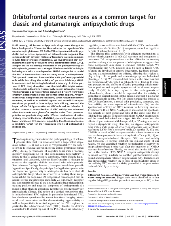
Fig. 1. Distinct effects of NMDA receptor inhibition on RF and FF neurons in
the OFC. (A) Average waveforms of an RF and an FF unit are compared. The
waveforms remained stable during the session. (B and C) Representative firing
rate histograms of individual OFC RF (B) and FF (C) units in response to MK801
(0.1 mg/kg i.p.). Note the sustained firing increase in RF units and sustained
firing decrease in FF units. Systemic injections were made at 0 and 20 min. (D
and E) Distribution of significant firing rate responses (increase, decrease, no
change) among RF and FF units. (F) Comparison of average (⫾SEM) firing rates
of all RF and FF units in response to MK801. Average response of all RF units
in vehicle/vehicle (Veh/Veh) group is also shown.
firing (FF; putative interneurons, n ⫽ 41) units. The fast-firing
units were characterized by faster firing rate (baseline, 13.2 Hz
vs. 3.8 Hz), narrower spike waveforms (peak-to-valley width,
283.6 s vs. 587.3 s; Fig. 1A), and high-frequency components
in their interspike interval and autocorrelation histograms.
Systemic treatment with the NMDA antagonist MK801 caused
sustained firing changes that were primarily excitatory in RF
neurons and inhibitory in FF units. This is consistent with
findings in the hippocampus (21) and medial PFC regions (22,
23). Examples are depicted in Fig. 1 B and C. More than 80% of
RF units displayed a sustained excitatory response (2 ⫽ 84.5,
P ⬍ 0.001 vs. vehicle/vehicle group; Fig. 1D), whereas more than
70% of FF units showed sustained inhibition (2 ⫽ 11.01, P ⬍
0.005; Fig. 1E) in response to MK801 treatment. Comparison of
the temporal profile of average firing rates of all RF units
between vehicle/MK801 and vehicle/vehicle groups (ANOVA
with time as repeated measure; Fig. 1F) revealed a significant
effect for both groups (F(1,224) ⫽ 121.63, P ⬍ 0.001) and time
(F(29,6496) ⫽ 54.21, P ⬍ 0.001) as well as for group ⫻ time
interaction (F(29,6496) ⫽ 69.67, P ⬍ 0.001). Within the vehicle/
MK801 group, FF units showed a significantly different time
course from RF units (P ⬍ 0.001).
Typical and Atypical Antipsychotic Drugs Reverse the Effects of NMDA
Antagonist Blockade on OFC Neurons. Systemic pretreatment with
haloperidol or clozapine decreased the sustained excitatory
effect of MK801 on OFC RF units (Fig. 2A). This was apparent
in both the relative number of responses (haloperidol, 2 ⫽
58.99, P ⬍ 0.001; clozapine, 2 ⫽ 41.39, P ⬍ 0.001) (Fig. 2B) and
changes in average firing rate (ANOVA, vehicle vs. haloperidol
pretreatment: group, F(1,188) ⫽ 46.42, P ⬍ 0.001; time, F(29,5452) ⫽
41.63, P ⬍ 0.001; group ⫻ time interaction, F(29,5452) ⫽ 33.09, P ⬍
0.001; vehicle vs. clozapine pretreatment: group, F(1,182) ⫽ 14.08,
18042 兩 www.pnas.org兾cgi兾doi兾10.1073兾pnas.0806669105
Fig. 2. Antipsychotic agents reversed the MK801 effects on OFC RF units. (A)
Superimposed firing rate histograms of individual neurons pretreated with
haloperidol (Hal; 0.1 mg/kg i.p.) or clozapine (Cloz; 10 mg/kg i.p.), followed 20
min later by MK801. (B) Both haloperidol and clozapine significantly decreased the proportion of neurons that showed an increase in firing response
after MK801 (shown for comparison). (C) The average (⫾SEM) firing rates of
all RF units pretreated with haloperidol or clozapine compared with the
vehicle pretreated group (Veh). Both antipsychotic drugs inhibited the sustained excitatory effects of MK801. (D) Distribution of FF unit responses to
MK801. Haloperidol significantly decreased the proportion of inhibitory responses to MK801 and increased the excitatory responses within this subset.
Note that the number of recorded units in the clozapine group (n ⫽ 3) was
insufficient for analysis.
P ⬍ 0.05; time, F(29,5278) ⫽ 40.06, P ⬍ 0.001; group ⫻ time
interaction, F(29,5278) ⫽ 21.42, P ⬍ 0.001) (Fig. 2C).
We recorded from a small subset of FF units during these
treatments [see supporting information (SI) Fig. S1 for examples
of individual FF neurons]. In the haloperidol-treated group, this
number was sufficient (n ⫽ 9) to perform statistical analysis.
Haloperidol reversed the inhibitory response of FF units to
MK801 (2 ⫽ 5.85, P ⬍ 0.05; Fig. 2D). In the clozapine group,
only 3 FF units were recorded, which showed a similar response
pattern.
Candidate Antipsychotic Drugs Reverse the Effects of NMDA Antagonist Blockade on OFC Neurons. Next, we examined the effects of
2 novel antipsychotic candidates with no known affinity for
dopamine or other monoamine receptors but with affinities for
distinct subtypes of metabotropic glutamate receptors on
MK801-induced activation of OFC neurons. These included the
mGlu2/3 receptor agonist LY354740 and the mGlu5 receptorpositive allosteric modulator CDPPB. Both compounds significantly reduced the excitatory response of RF units to MK801
(Fig. 3A, individual neuronal responses; Fig. 3B, LY354740, 2
⫽ 62.26, P ⬍ 0.001, and CDPPB, 2 ⫽ 86.35, P ⬍ 0.001). Notably,
LY354740 caused sustained inhibition in a subset of units. This
effect was also reflected in the population response in this group
(vs. vehicle: group, F(1,160) ⫽ 98.39, P ⬍ 0.001; time, F(29,4640) ⫽
23.17, P ⬍ 0.001; group ⫻ time interaction, F(29,4640) ⫽ 35.13, P ⬍
Homayoun and Moghaddam
Fig. 3. Metabotropic glutamate receptor modulators blocked MK801 effects
on OFC RF units. (A) Superimposed firing rate histograms of individual neurons pretreated with LY354740 (LY; 10 mg/kg i.p.) or CDPPB (10 mg/kg i.p.),
followed 20 min later by MK801. (B and C) Both LY354740 and CDPPB inhibited
the excitatory influence of MK801 on OFC units. LY354740 but not CDPPB also
caused lasting inhibitory responses in average firing rate. All conventions are
as in Fig. 2. (D) Distribution of FF unit responses to MK801. Similarly to
haloperidol, CDPPB decreased the proportion of inhibitory responses to
MK801. Veh indicates vehicle.
0.001) (Fig. 3C). CDPPB blocked the excitatory effect of MK801
over time (group, F(1,262) ⫽ 150.4, P ⬍ 0.001; time, F(29,7598) ⫽
76.64, P ⬍ 0.001; group ⫻ time interaction, F(29,7598) ⫽ 80.14, P ⬍
0.001) without causing inhibition. A relatively small subset of
units in the CDPPB-treated group were characterized as FF
units (see example in Fig. S1). In this group, CDPPB produced
an effect similar to haloperidol, reversing the inhibitory effects
of NMDA receptor blockade on this subset of neurons (2 ⫽
11.59, P ⬍ 0.005; Fig. 3D).
To better simulate conditions in which antipsychotic medications are administered in the context of already present NMDA
receptor hypofunction, we also examined the impact of posttreatment with drugs after MK801 administration. Again, the
excitatory effect of MK801 was reversed by posttreatment with
haloperidol, LY354740, and CDPPB, an effect that was reflected
both in the proportion of cells with excitatory responses to
MK801 and in the average firing rates of neurons in each
treatment group (Fig. S2).
Reversal of NMDA Antagonist-Induced Behavioral Stereotypy by Established and Candidate Antipsychotic Drugs. During the recording
sessions, behavioral stereotypy was measured as an index of
behavioral activation by MK801. The temporal profile and
average post-MK801 stereotypy scores are shown on Fig. 4 A and
B, respectively. Haloperidol, clozapine, LY354740, and CDPPB
similarly reversed the MK801 stereotypy (ANOVA, F(5,25) ⫽
32.45, P ⬍ 0.001; post hoc analysis of each group vs. vehicle, P ⬍
0.05).
Disruption of OFC Neuronal Activity by Amphetamine Is Reversed by
Antipsychotic Drugs. The psychotomimetic compound amphet-
amine produces an excitatory influence on OFC RF neurons
Homayoun and Moghaddam
similar to that observed here with MK801, which is in contrast
to the effect of amphetamine in the medial PFC (20). We
reasoned that if normalization of hyperactive OFC neurons is a
key mechanism of action for antipsychotic drugs, then it should
generalize to amphetamine-induced OFC neuron hyperactivation. Thus, we treated animals with a dose of amphetamine that
produced levels of activation of OFC RF neurons similar to those
of MK801 and then compared the effects of posttreatment with
vehicle, a classic antipsychotic drug (haloperidol), and a candidate compound (CDPPB). Amphetamine caused an excitatory
response in the majority of OFC RF units (example in Fig. 5A).
Posttreatment with either haloperidol (0.1 mg/kg) or CDPPB (10
mg/kg) reversed this excitatory effect (examples in Fig. 5A).
Among the population of RF units, the predominantly excitatory
effect of amphetamine was reversed by both haloperidol (2 ⫽
43.92, P ⬍ 0.001; Fig. 5B) and CDPPB (2 ⫽ 28.68, P ⬍ 0.001).
Comparing the population responses among RF units in each
treatment group, 2-way ANOVA revealed significant effects for
both posttreatment groups compared with vehicle (haloperidol:
group, F(1,102) ⫽ 32.38, P ⬍ 0.001; time, F(29,2958) ⫽ 24.39, P ⬍
0.001; group ⫻ time interaction, F(29,2958) ⫽ 14.28, P ⬍ 0.001;
CDPPB: group, F(1,108) ⫽ 5.49, P ⬍ 0.05; time, F(29,3132) ⫽ 58.25,
P ⬍ 0.001; group ⫻ time interaction, F(29,3132) ⫽ 22.01, P ⬍ 0.001)
(Fig. 5C). This reversal of neuronal activity was also associated
with reversal of amphetamine-induced stereotypy by both haloperidol and CDPPB (P ⬍ 0.01; Fig. 5D).
Discussion
Use-dependent blockade of NMDA receptors, which provides a
pharmacological model of schizophrenia (16, 24), profoundly
increased the spontaneous activity of putative pyramidal cells in
the OFC, and at the same time inhibited the activity of putative
inhibitory GABAergic interneurons and increased behavioral
stereotypy. The pattern of activation mimicked the effect of
amphetamine on OFC pyramidal neurons (20). Amphetamine is
a dopamine releaser and psychotomimetic drug (25) that is
commonly used to model a hyperdopaminergic state in schizophrenia (16). The similar effects of 2 psychotomimetic compounds with different mechanisms of action on OFC neurons are
consistent with clinical studies demonstrating hyperactivity of
OFC regions in individuals with schizophrenia (26–28). Treatment with 4 compounds with distinct mechanisms of action that
are either well-established antipsychotic compounds or novel
candidates for treatment of schizophrenia ameliorated the impact of NMDA hypofunction on OFC neurons and on behavior.
These compounds included (i) haloperidol, a D2 antagonist and
a typical antipsychotic drug; (ii) clozapine, an atypical antipsyPNAS 兩 November 18, 2008 兩 vol. 105 兩 no. 46 兩 18043
NEUROSCIENCE
Fig. 4. Behavioral stereotypy. (A) Average stereotypy scores during recording sessions (5-min bins) are shown for vehicle/vehicle (Veh/Veh), vehicle/
MK801, and CDPPB/MK801 groups. The results for other groups were not
demonstrated for clarity. (B) Average stereotypy scores for the post-MK801
period (minutes 10 –120) are compared between all groups. Both antipsychotic agents as well as LY354740 (LY) and CDPPB blocked MK801-induced
stereotypy. Hal indicates haloperidol; Cloz, clozapine. *, P ⬍ 0.05 compared
with Veh/Veh; #, P ⬍ 0.05 compared with Veh/MK801.
Fig. 5. Response of OFC RF units to amphetamine (Amp). (A) Representative
firing rate histograms of individual OFC RF units treated with amphetamine
followed 50 min later by vehicle (Veh), haloperidol (Hal; 0.1 mg/kg i.p.), or
CDPPB (10 mg/kg i.p.). All 3 units showed an excitatory response to MK801 that
was sustained in the vehicle posttreated unit but was reversed in the haloperidol or CDPPB posttreated units. (B) Amphetamine (Amph) caused an
excitatory response in the majority of OFC units. This response was reversed in
the majority of neurons in the haloperidol and CDPPB groups. (C) The mean
(⫾SEM) firing rates of all RF units in each group. (D) Stereotypy score in the
amphetamine group. Stereotypy was scored based on the percentage of time
spent on stereotypical behavior (rearing, up and down sniffing, turning, and
ambulating) during 30-s windows assessed every 5 min. Average scores for
minutes 0 –50 (post amphetamine) and minutes 50 –120 (post haloperidol or
post-CDPPB) are shown separately. In all 3 groups, amphetamine caused
significant stereotypy. This effect was reversed by haloperidol and CDPPB
posttreatments. C and D are color coded as in A and B.
chotic drug with a wide range of affinity at dopamine and
serotonin receptors; (iii) LY354740, a selective mGlu2/3 agonist
with no affinity for dopamine or serotonin receptors that has
been suggested recently to have antipsychotic efficacy (5); and
(iv) CDPPB, a novel mGlu5 receptor-positive allosteric modulator that has also been proposed as a potential antipsychotic
agent (18, 19). In a similar way, haloperidol and CDPPB reversed
amphetamine-induced hyperactivation of OFC neurons. Together, these findings suggest that normalization of OFC neuronal activity, whether caused by NMDA receptor hypofunction
or excess dopamine neurotransmission, may be a common target
for different classes of antipsychotic drugs. Thus, determining
the ability of compounds to normalize OFC activity may provide
an evaluation of antipsychotic potential.
OFC Dysfunction and Schizophrenia. The OFC receives extensive
input from sensory, limbic, and basal ganglia regions and plays
a critical role in sensory integration and feedback processing
(29). Disruption in OFC function may lead to inappropriate
integration of relevant sensory stimuli and previously learned
associations, and failure in suppression of irrelevant stimuli and
associations (14, 30, 31). This mode of disruption may be a
critical component of positive and negative symptoms as well as
cognitive deficits of schizophrenia. Multidisciplinary lines of
evidence have, in fact, reported various OFC abnormalities in
18044 兩 www.pnas.org兾cgi兾doi兾10.1073兾pnas.0806669105
schizophrenia that are associated with different symptoms of the
disease (6–12, 32, 33). For example, MRI studies have reported
volume deficits in association with thought disorder (6) and
severity of negative symptoms (9, 34, 35). Longitudinal MRI
studies in individuals at risk to develop schizophrenia have
shown reduction in OFC gray matter in those who develop
psychosis (36). These latter findings, together with another study
that reported OFC volume deficits in drug-naive patients (37),
strongly suggest that OFC anomalies are components of the
disease process and not a consequence of chronic antipsychotic
drug treatment. Accordingly, individuals with schizophrenia
show significant performance deficits in cognitive paradigms,
such as a probabilistic reversal learning task and the Iowa
Gambling Task, which involve feedback processing and which
are classically used to assess OFC function (12, 38, 39). In related
functional imaging studies, hyperactivation of OFC is reported in
patients during performance of cognitive tasks that generally
occur concomitantly with hypoactivation of dorsal PFC regions
(26, 27). Finally, in one of the few studies reporting regional
metabolic activity in actively hallucinating individuals with
schizophrenia, significant hyperactivity of OFC was reported
(28). This is consistent with our findings that in 2 animal models
of schizophrenia, the NMDA hypofunction model (the present
study) and the amphetamine model (20), there is general overactivation of OFC neurons.
Although the major focus of postmortem work in schizophrenia has been on dorsal regions of the PFC, a few interesting
findings have been reported in the OFC. These include increases
in glial fibrillary acidic protein, changes in the density of NMDA
and kainate receptor subunits (40), and dramatic decreases in
the subtypes of dopamine receptors localized within the OFC
that are not observed in other PFC regions or the striatum (41).
Significant postmortem changes in the distribution of DisruptedIn-Schizophrenia (DISC1) protein also seem to be selective to
OFC regions (42). DISC1 has been identified recently as a
susceptibility gene for schizophrenia (43, 44), and a DISC1
polymorphism has been correlated with severity of positive
symptoms (45).
Different Pharmacological Agents with Antipsychotic Efficacies Similarly Normalize OFC Neuronal Firing. Causes of schizophrenia
remain elusive, but recent genetic studies suggest that multiple
mutations (46), including rare mutations (47), may contribute to
the pathophysiology of the disease. This suggests that changes in
the function of a variety of molecules may lead to disruption
in the function of a common group of neurons and cellular
networks. Thus, although the focus of research for treating the
disorder has been on normalizing the function of a single protein
(such as the D2 receptor), it is reasonable to suggest that this
focus should be expanded to include normalizing the function of
groups of neurons and their associated networks. Until recently,
the dopamine D2 receptor was the only established target for
treatment of schizophrenia. New findings that an mGlu2/3
receptor agonist that is devoid of activity at the D2 receptor has
a profile of antipsychotic efficacy similar to that of an approved
antipsychotic drug (5) provides an important tool for discovering
neuronal networks that serve as common targets for established
and novel antipsychotic agents and may be critical for treatment
of schizophrenia. Although cortical dysfunction has long been
acknowledged as the main site of pathology in schizophrenia,
this research has been focused mostly on the dorsal and medial
regions of the PFC (48, 49). Neurons in these regions are likely
to play a role in aspects of cognitive deficits associated with the
disease; however, it is unlikely that they account for the whole
spectrum of the therapeutic properties of antipsychotic drugs;
functional alterations in these regions are generally not associated with noncognitive symptoms of schizophrenia, and antipsychotic drugs do not consistently improve the cognitive deficits
Homayoun and Moghaddam
1. Seeman P (1987) Dopamine receptors and the dopamine hypothesis of schizophrenia.
Synapse 1:133–152.
2. Carlsson A (1988) The current status of the dopamine hypothesis of schizophrenia.
Neuropsychopharmacology 1:179 –186.
3. Weinberger DR, Berman KF (1988) Speculation on the meaning of cerebral metabolic
hypofrontality in schizophrenia. Schizophr Bull 14:157–168.
4. Goldman-Rakic P (1994) Cerebral cortical mechanisms in schizophrenia. Neuropsychopharmacology 10:22S–27S.
5. Patil ST, et al. (2007) Activation of mGlu2/3 receptors as a new approach to treat
schizophrenia: A randomized Phase 2 clinical trial. Nat Med 13:1102–1107.
6. Nakamura M, et al. (2008) Orbitofrontal volume deficit in schizophrenia and thought
disorder. Brain 131:180 –195.
7. Fahim C, et al. (2005) Brain activity during emotionally negative pictures in schizophrenia with and without flat affect: An fMRI study. Psychiatry Res 140:1–15.
8. Baas D, et al. (2008) Evidence of altered cortical and amygdala activation during social
decision-making in schizophrenia. Neuroimage 40:719 –727.
9. Lacerda AL, et al. (2007) Morphology of the orbitofrontal cortex in first-episode
schizophrenia: Relationship with negative symptomatology. Prog Neuropsychopharmacol Biol Psychiatry 31:510 –516.
10. Sanfilipo M, et al. (2000) Volumetric measure of the frontal and temporal lobe
regions in schizophrenia: Relationship to negative symptoms. Arch Gen Psychiatry
57:471– 480.
11. Waltz JA, Frank MJ, Robinson BM, Gold JM (2007) Selective reinforcement learning
deficits in schizophrenia support predictions from computational models of striatalcortical dysfunction. Biol Psychiatry 62:756 –764.
12. Sevy S, et al. (2007) Iowa gambling task in schizophrenia: A review and new data in
patients with schizophrenia and co-occurring cannabis use disorders. Schizophr Res
92:74 – 84.
13. Rolls ET (1998) The orbitofrontal cortex. The Prefrontal Cortex, eds Roberts AC, Robbins
TW, Weiskrantz L (Oxford Univ Press, New York), pp 67– 86.
14. Bechara A, Damasio H, Damasio AR (2000) Emotion, decision making and the orbitofrontal cortex. Cereb Cortex 10:295–307.
15. Schoenbaum G, Chiba A, Gallagher M (1998) Orbitofrontal cortex and basolateral
amygdala encode expected outcomes during learning. Nat Neurosci 1:155–159.
16. Geyer M, Moghaddam B (2002) Animal models relevant to schizophrenia disorder.
Psychopharmacology: The Fifth Generation of Progress, eds Davis KL, Charney C, Coyle
JT, Nemeroff C (Lippincott Williams and Wilkins, Philadelphia), pp 689 –702.
17. Schoepp DD, et al. (1997) LY354740 is a potent and highly selective group II metabotropic glutamate receptor agonist in cells expressing human glutamate receptors.
Neuropharmacology 36:1–11.
18. Conn P (2003) Physiological roles and therapeutic potential of metabotropic glutamate
receptors. Ann NY Acad Sci 1003:12–21.
19. Lecourtier L, Homayoun H, Tamagnan G, Moghaddam B (2007) Positive allosteric
modulation of metabotropic glutamate 5 (mGlu5) receptors reverses N-Methyl-Daspartate antagonist-induced alteration of neuronal firing in prefrontal cortex. Biol
Psychiatry 62:739 –746.
20. Homayoun H, Moghaddam B (2006) Progression of cellular adaptations in medial
prefrontal and orbitofrontal cortex in response to repeated amphetamine. J Neurosci
26:8025– 8039.
Homayoun and Moghaddam
nisms by which different classes of compounds can have similar
downstream effects on OFC pyramidal and GABAergic neurons. Although the present study is limited to reporting a
dynamic cellular phenomenon, it demonstrates that NMDA
receptor hypofunction and dopamine hyperfunction, both of
which are suspected to occur in schizophrenia, leads to a similar
disruption in the activity of OFC neurons. Furthermore, compounds that work on distinct proteins and receptor classes can
have comparable effects in reversing these disruptions. These
findings support the notion that the key to understanding the
pathophysiology of schizophrenia and the design of better
treatments is to identify the common group of neurons, and the
functional networks influenced by these neurons, that are similarly affected by the diverse, and often rare mutations, that are
associated with this brain disorder.
Materials and Methods
Detailed methods are included as SI Experimental Procedures. Sprague–
Dawley male adult rats were used. All experimental protocols were approved
by the University of Pittsburgh Institutional Animal Care and Use Committee
and were conducted in accordance with the National Institutes of Health
Guide for the Care and Use of Laboratory Animals (53). Details of surgery,
recording, and isolation of single units have been published previously
(20, 50).
ACKNOWLEDGMENTS. This study was supported by the National Institute of
Mental Health and the Pittsburgh Life Sciences Greenhouse.
21. Grunze HC, et al. (1996) NMDA-dependent modulation of CA1 local circuit inhibition.
J Neurosci 16:2034 –2043.
22. Homayoun H, Moghaddam B (2007) NMDA receptor hypofunction produces opposite
effects on prefrontal cortex interneurons and pyramidal neurons. J Neurosci 27:11496 –
11500.
23. Lisman JE, et al. (2008) Circuit-based framework for understanding neurotransmitter
and risk gene interactions in schizophrenia. Trends Neurosci 31:234 –242.
24. Javitt DC, Jotkowitz A, Sircar R, Zukin SR (1987) Non-competitive regulation of phencyclidine/sigma-receptors by the N-methyl-D-aspartate receptor antagonist D-(⫺)-2amino-5-phosphonovaleric acid. Neurosci Lett 78:193–198.
25. Snyder S (1973) Amphetamine psychosis: A ‘‘model’’ schizophrenia mediated by catecholamines. Am J Psychiatry 130:61– 67.
26. Tan HY, et al. (2006) Dysfunctional prefrontal regional specialization and compensation in schizophrenia. Am J Psychiatry 163:1969 –1977.
27. Ragland JD, et al. (2004) Event-related fMRI of frontotemporal activity during word
encoding and recognition in schizophrenia. Am J Psychiatry 161:1004 –1015.
28. Silbersweig DA, et al. (1995) A functional neuroanatomy of hallucinations in schizophrenia. Nature 378:176 –179.
29. Rolls E, Critchley H, Mason R, Wakeman E (1996) Orbitofrontal cortex neurons: Role in
olfactory and visual association learning. J Neurophysiol 75:1970 –1981.
30. Rolls ET (2000) The orbitofrontal cortex and reward. Cereb Cortex 10:284 –294.
31. Schoenbaum G, Setlow B, Saddoris MP, Gallagher M (2003) Encoding predicted outcome and acquired value in orbitofrontal cortex during cue sampling depends upon
input from basolateral amygdala. Neuron 39:855– 867.
32. Davatzikos C, et al. (2005) Whole-brain morphometric study of schizophrenia revealing
a spatially complex set of focal abnormalities. Arch Gen Psychiatry 62:1218 –1227.
33. Hoptman MJ, et al. (2005) Quantitative MRI measures of orbitofrontal cortex in
patients with chronic schizophrenia or schizoaffective disorder. Psychiatry Res
140:133–145.
34. Gur RE, et al. (2000) Reduced dorsal and orbital prefrontal gray matter volumes in
schizophrenia. Arch Gen Psychiatry 57:761–768.
35. Sapara A, et al. (2007) Prefrontal cortex and insight in schizophrenia: A volumetric MRI
study. Schizophr Res 89:22–34.
36. Pantelis C, et al. (2003) Neuroanatomical abnormalities before and after onset of
psychosis: A cross-sectional and longitudinal MRI comparison. Lancet 361:281–288.
37. Crespo-Facorro B, Kim J, Andreasen NC, O’Leary DS, Magnotta V (2000) Regional
frontal abnormalities in schizophrenia: A quantitative gray matter volume and cortical
surface size study. Biol Psychiatry 48:110 –119.
38. Ritter LM, Meador-Woodruff JH, Dalack GW (2004) Neurocognitive measures of prefrontal cortical dysfunction in schizophrenia. Schizophr Res 68:65–73.
39. Waltz JA, Gold JM (2007) Probabilistic reversal learning impairments in schizophrenia:
Further evidence of orbitofrontal dysfunction. Schizophr Res 93:296 –303.
40. Toro CT, et al. (2006) Glial fibrillary acidic protein and glutamine synthetase in
subregions of prefrontal cortex in schizophrenia and mood disorder. Neurosci Lett
404:276 –281.
41. Meador-Woodruff J, et al. (1997) Dopamine receptor transcript expression in striatum
and prefrontal and occipital cortex. Focal abnormalities in orbitofrontal cortex in
schizophrenia. Arch Gen Psychiatry 54:1089 –1095.
PNAS 兩 November 18, 2008 兩 vol. 105 兩 no. 46 兩 18045
NEUROSCIENCE
of this disorder (36, 46, 49). Here, we postulated that the OFC
is a key cortical component of the distributed networks targeted
by antipsychotic drugs because functional changes in this region
have been observed during active psychosis (28) and are associated with the negative symptoms and cognitive deficits of
schizophrenia (see above). We found that NMDA antagonists,
as with another psychotomimetic compound, amphetamine,
similarly increased the activity of the majority of cortical pyramidal neurons. Although a similar pattern of response to NMDA
antagonists (50) and antipsychotic drugs has been reported in the
medial and dorsal regions of the PFC (51, 52), it is important to
emphasize that amphetamine leads to a primarily inhibitory
response in other cortical regions (20). Thus, the present finding
is in line with OFC activation reported during active psychosis
(28) and suggests that both of these pharmacological treatments
provide comparable dynamic models of the OFC hyperactivity
that may be present in schizophrenia. That compounds with
antipsychotic efficacy (or potential) with very different mechanisms of action, and different effects on OFC neuronal activity
when administered alone, similarly reduce the impact of NMDA
receptor hypofunction and amphetamine on OFC neurons further suggests that normalizing the disrupted balance of excitatory and inhibitory activity in OFC may be critical for treatment
of schizophrenia.
Further work is required to examine the molecular mecha-
42. Sawamura N, Sawamura-Yamamoto T, Ozeki Y, Ross CA, Sawa A (2005) A form of DISC1
enriched in nucleus: Altered subcellular distribution in orbitofrontal cortex in psychosis
and substance/alcohol abuse. Proc Natl Acad Sci USA 102:1187–1192.
43. Hikida T, et al. (2007) Dominant-negative DISC1 transgenic mice display schizophreniaassociated phenotypes detected by measures translatable to humans. Proc Natl Acad
Sci USA 104:14501–14506.
44. Ishizuka K, Paek M, Kamiya A, Sawa A (2006) A review of Disrupted-In-Schizophrenia-1
(DISC1): Neurodevelopment, cognition, and mental conditions. Biol Psychiatry
59:1189 –1197.
45. Szeszko PR, et al. (2008) DISC1 is associated with prefrontal cortical gray matter and
positive symptoms in schizophrenia. Biol Psychol 79:103–110.
46. Harrison P, Weinberger D (2005) Schizophrenia genes, gene expression, and neuropathology: On the matter of their convergence. Mol Psychiatry 10:40 – 68.
47. Walsh T, et al. (2008) Rare structural variants disrupt multiple genes in neurodevelopmental pathways in schizophrenia. Science 320:539 –543.
18046 兩 www.pnas.org兾cgi兾doi兾10.1073兾pnas.0806669105
48. Weinberger DR, et al. (2001) Prefrontal neurons and the genetics of schizophrenia. Biol
Psychiatry 50:825– 844.
49. Lewis DA, Hashimoto T, Volk DW (2005) Cortical inhibitory neurons and schizophrenia.
Nat Rev Neurosci 6:312–324.
50. Homayoun H, Jackson ME, Moghaddam B (2004) Activation of metabotropic glutamate 2/3 (mGlu2/3) receptors reverses the effects of NMDA receptor hypofunction on
prefrontal cortex unit activity in awake rats. J Neurophysiol 93:1989 –2001.
51. Kargieman L, Santana N, Mengod G, Celada P, Artigas F (2007) Antipsychotic drugs
reverse the disruption in prefrontal cortex function produced by NMDA receptor
blockade with phencyclidine. Proc Natl Acad Sci USA 104:14843–14848.
52. Homayoun H, Moghaddam B (2007) Fine-tuning of awake prefrontal cortex neurons
by clozapine: comparison with haloperidol and N-desmethylclozapine. Biol Psychiatry
61:679 – 687.
53. Institute of Laboratory Animal Resources (1996) Guide for the Care and Use of
Laboratory Animals (Natl Acad Press, Washington, DC).
Homayoun and Moghaddam
RELATED PAPERS
Social Science Research Network
The Impact of Agricultural Credit Guarantee Scheme Fund on Agricultural Sector Development in Nigeria2017 •
2013 •
Journal of Advances in Medical and Pharmaceutical Sciences
Assessment of Some Brain Antioxidants Profile in Plasmodium berghei berghei Infected Mice Treated with Ethanol Leaf Extract of Musa paradisiacaRevista Ler , nº 170
A Trilogia do Amor (Eros, Agape, Philia) e a Identidade da Cidade de Coimbra2024 •
Anales de geografía de la Universidad Complutense
Cartografía de la respuesta sísmica local: una contribución a la gestión del riesgo en la zona metropolitana del Valle de Toluca2022 •
Eikasía: revista de filosofía
La responsabilidad hacia el extraño: una propuesta para la crisis migratoria en la filosofía social de Zygmunt Bauman2024 •