Aquatic Toxicology 161 (2015) 154–169
Contents lists available at ScienceDirect
Aquatic Toxicology
journal homepage: www.elsevier.com/locate/aquatox
Combined toxicity of two crystalline phases (anatase and rutile) of
Titania nanoparticles towards freshwater microalgae: Chlorella sp
V. Iswarya a , M. Bhuvaneshwari a , Sruthi Ann Alex a , Siddharth Iyer a , Gouri Chaudhuri a ,
Prathna Thanjavur Chandrasekaran b , Gopalkrishna M. Bhalerao c , Sujoy Chakravarty c ,
Ashok M. Raichur b , N. Chandrasekaran a , Amitava Mukherjee a,∗
a
Centre for Nanobiotechnology, VIT University, Vellore, India
Department of Materials Engineering, Indian Institute of Science, Bangalore, India
c
UGC-DAE CSR, Kalpakkam Node, Kokilamedu, India
b
a r t i c l e
i n f o
Article history:
Received 13 December 2014
Received in revised form 9 February 2015
Accepted 11 February 2015
Available online 13 February 2015
Keywords:
Binary mixture
Bio uptake
Crystalline phases
Titania NPs
Toxicity
a b s t r a c t
In view of the increasing usage of anatase and rutile crystalline phases of titania NPs in the consumer
products, their entry into the aquatic environment may pose a serious risk to the ecosystem. In the
present study, the possible toxic impact of anatase and rutile nanoparticles (individually and in binary
mixture) was investigated using freshwater microalgae, Chlorella sp. at low exposure concentrations
(0.25, 0.5 and 1 mg/L) in freshwater medium under UV irradiation. Reduction of cell viability as well as
a reduction in chlorophyll content were observed due to the presence of NPs. An antagonistic effect was
noted at certain concentrations of binary mixture such as (0.25, 0.25), (0.25, 0.5), and (0.5, 0.5) mg/L, and
an additive effect for the other combinations, (0.25, 1), (0.5, 0.25), (0.5, 1), (1, 0.25), (1, 0.5), and (1, 1)
mg/L. The hydrodynamic size analyses in the test medium revealed that rutile NPs were more stable in
lake water than the anatase and binary mixtures [at 6 h, the sizes of anatase (1 mg/L), rutile NPs (1 mg/L),
and binary mixture (1, 1 mg/L) were 948.83 ± 35.01 nm, 555.74 ± 19.93 nm, and 1620.24 ± 237.87 nm,
respectively]. The generation of oxidative stress was found to be strongly dependent on the crystallinity
of the nanoparticles. The transmission electron microscopic images revealed damages in the nucleus and
cell membrane of algal cells due to the interaction of anatase NPs, whereas rutile NPs were found to
cause chloroplast and internal organelle damages. Mis-shaped chloroplasts, lack of nucleus, and starchpyrenoid complex were noted in binary-treated cells. The findings from the current study may facilitate
the environmental risk assessment of titania NPs in an aquatic ecosystem.
© 2015 Elsevier B.V. All rights reserved.
1. Introduction
Titanium dioxide (TiO2 ) nanoparticles were extensively used
in various industrial applications and consumer products such
as water treatment, medicine, cosmetics, and engineering (ICIS
Chemical Report, 2010). Excessive usage of TiO2 nanoparticles have
led to their exposure to the aquatic environment and their consequent hazards to the ecosystem (Furman et al., 2013). There
are mainly three crystalline phases of TiO2 viz., anatase (tetragonal), rutile (tetragonal), and brookite (orthorhombic) (Cho et al.,
2013). Among these, rutile is the most common and natural form
of TiO2 , and it is an integral part of heavy minerals. It is employed in
∗ Corresponding author. Centre for Nanobiotechnology, VIT University Vellore,
632014 Tamil Nadu. Tel.: +91 416 2202620; fax: +91 416 2243092.
E-mail address: amit.mookerjea@gmail.com (A. Mukherjee).
http://dx.doi.org/10.1016/j.aquatox.2015.02.006
0166-445X/© 2015 Elsevier B.V. All rights reserved.
optical elements due to its highest refractive indices and also used
as a construct for refractory ceramics, pigments, etc. (Winkler,
2003; Yu et al., 2013). Anatase is extensively used in organic
photovoltaics as an electron collecting layer (Small et al., 2012).
Anatase is also applied as a catalytic support for the production
of nanotubes and nanoribbons (Gregory et al., 2008). Both rutile
and anatase phases are being extensively used in sunscreens (due
to their high-energy-absorbing property), paints, plastics, paper,
foods, electronics, and other applications (Ferguson et al., 2005;
Mueller and Nowack 2008; Wang et al., 2006; Winkler, 2003). Since
there is scarcity of brookite in nature, this form does not have significant economic importance (Allen et al., 2009).
The released nanomaterials from different industries, consumer
products may inevitably end up in the water bodies. They may
potentially exert adverse impacts on the aquatic ecosystem due
to their unique physical and chemical characteristics such as high
reactivity and the photoactivity (Cardinale et al., 2012). Kaegi et al.
�V. Iswarya et al. / Aquatic Toxicology 161 (2015) 154–169
(2008) mentioned about the direct release of TiO2 NPs of about
16 g/L into the surface water from aged paints. The predicted environmental concentration (PEC) of TiO2 NPs in surface water has
been stated to be less than 1 g/mL (Gottschalk et al., 2009). Hence,
it becomes necessary to evaluate the toxicity of TiO2 NPs at its environmentally relevant concentrations. In our previous studies (Dalai
et al., 2013; Pakrashi et al., 2013), we have evaluated the toxicity of
TiO2 and Al2 O3 NPs under environmentally relevant low-exposure
concentrations, i.e., 0.05, 0.5, and 1 mg/L, towards freshwater algae.
Microalgae are of great importance for the maintenance of aquatic
ecosystem. It can be used as a model for the studies of aquatic
risk assessment of the nanomaterials (Aruoja et al., 2009). Lubick
(2008) and Navarro et al. (2008) reported that the interaction of
nanoparticles with algae influenced the aquatic toxicity of nanomaterials. Recently, Ji et al. (2011) noted that ZnO and TiO2 (anatase)
nanoparticles caused severe damage to freshwater green algae.
Cardinale et al. (2012) evaluated the toxicity of TiO2 nanoparticles (Degussa, 82% anatase/18% rutile) on three algal species viz.,
Chlorella vulgaris, Scenedesmus quadricauda, and Chlamydomonas
moewusii. They observed that the gross primary production of these
algae were reduced, and the reduction rate varied depending on the
species type. Dalai et al. (2013) reported the photoinduced toxicity of TiO2 anatase NPs on Scenedesmus obliquus at low-exposure
doses (≤1 g/mL). They observed reduced cell viability, increased
reactive oxygen species (ROS) generation, and membrane damage.
Miller et al. (2012) observed the increased toxicity of TiO2 NPs on
marine algae under UV-A irradiation than non-irradiation condition due to their increased photocatalytic activity.
A number of prior reports strongly indicated that titanium
dioxide nanoparticles caused severe toxicity towards freshwater
microalgae species. However, information on their fate, behavior,
and mechanism of uptake (pathways) based on crystallinity, shape,
and other properties of materials is still lacking. Ji et al. (2011) stated
that TiO2 NP toxicity varied with respect to the crystalline structure
of TiO2 NPs. The two allotropic forms of TiO2 NPs viz., anatase and
rutile have different surface properties and reactivity. Prior studies
have demonstrated that anatase phases are more cytotoxic than
those of rutile phases (Hirakawa et al., 2004). Braydich-Stolle et al.
(2009) have observed that rutile TiO2 NPs were capable of initiating apoptosis through the formation of ROS, whereas pure anatase
TiO2 NPs caused cell necrosis and membrane leakage in cells. Most
of the previous toxicity studies on microalgae dealt with the anatase
phase and P25 form of TiO2 (Chen et al., 2012; Clement et al., 2013;
Dalai et al., 2013; Lee and An, 2013; Wang et al., 2008) and only a
handful studies are available with the rutile phase (Ji et al., 2011).
Therefore, it is pertinent to study the other crystalline phase, i.e.,
rutile TiO2 NPs, which is also commonly employed in commercial
products/applications (Winkler, 2003; Yu et al., 2013).
As UV-C radiation is a shorter and higher energized radiation
than UV-A and UV-B, the photocatalytic action of TiO2 NPs was
reported to be enhanced significantly under UV-C irradiation than
other UV radiations (Termtanun, 2013). Due to its greater photolytic activity and energy, UV-C irradiation is being widely used
for water disinfection and in the photodegradation studies, along
with TiO2 NPs (Bushnaq et al., 2004). Since, UV-C radiation gets
absorbed by the earth’s atmosphere, not much attention was given
in evaluating its effects on the environment and the transformations caused by it (Holzinger and Lütz, 2006; Basti et al., 2009).
McGivney (2007) studied the combined effect of UV-C, vacuum
UV, and TiO2 on freshwater algae, Pseudokirchneriella subcapitata,
and marine algae, Tetraselmis suecica, in a ballast water treatment
system. They noticed that UV-C/TiO2 exerted a higher mortality
compared to UV irradiation alone. Most of the algal toxicity studies
on TiO2 NPs till date were carried out under different irradiation
conditions such as visible light and UV light especially, UV A and
UV B (Lee and An, 2013; Ji et al., 2011). To the best of our knowledge,
there are limited reports highlighting the photocatalytic effects of
UV-C on the toxicity of TiO2 NPs. Vileno et al. (2007) studied the
effect of TiO2 NPs on the stiffness of human skin fibroblasts in the
presence of UV-A and UV-C. Hence, it is crucial to evaluate the risk
of TiO2 NPs in the presence of UV-C on Chlorella sp.
In the natural ecosystem, various toxicants are expected to be
present in the mixed form rather than as individuals. The toxicity
assessment of a single toxicant alone does not adequately reflect
the actual impact in the aquatic environment. The mode of action
may vary for individual toxicants in the mixture; they may mask
the effect of each other. It is indispensable to study the effect of their
mixture in addition to the individual toxicants to adequately assess
the environmental toxicity of different forms of toxicants (Jak et al.,
1996). Zou et al. (2014) studied the toxicity of silver (Ag) NPs in
the presence of TiO2 NPs on a ciliated protozoan, Tetrahymena
pyriformis. Increased ecotoxicity was noted due to the coexistence
of TiO2 NPs and Ag NPs. This elucidates that the level of toxicity
increases in the presence of a mixture of nanoparticles rather than
the individual forms (Utgikar et al., 2004) and provides an understanding of the complex interaction between different substances.
The present investigation is the first of its nature to evaluate
the combined toxicity of anatase and rutile NPs towards freshwater microalgae in a freshwater matrix. It may be hypothesized
that there are inherent differences in the toxic effects of the two
different crystal phases of titania NPs (anatase and rutile). Their
binary combination would be more toxic than the respective individual phases. The aim of the present study was to elucidate the
toxic effects of the two crystalline phases of titania nanoparticles
i.e., anatase and rutile, as well as their binary mixture towards
freshwater algae, Chlorella sp. at environmentally relevant low concentration levels (0.25, 0.5 and 1 mg/L) in the lake water matrix
under UV-C irradiation.
2. Materials and methods
2.1. Chemicals
Dry titanium(IV) dioxide (TiO2 ) nanopowder (anatase, <25 nm,
CAS No: 1317-70-0, 99.7% trace metal basis; and rutile, <100 nm
(∼10 nm Diam. × 40 nm L), CAS No: 1317-80-2, 99.5% trace metals basis) were purchased from Sigma–Aldrich, Missouri, USA, and
their supplier information was summarized in Table 1. BG-11
broth was purchased from Himedia Labs Pvt., Ltd. (Mumbai, India).
N, N-dimethylformamide was procured from SD fine chemicals
Table 1
Information about the physicochemical parameters of two different types of TiO2 NPs has been represented in the table as per the supplier.
Assay
Form
Particle size
CAS No
Surface area
Density
Bulk density
155
Anatase NPs
Rutile NPs
99.7% trace metal basis
Nanopowder
<25 nm
1317–70–0
Spec. surface area 45–55 m2 /g
3.9 g/mL at 25 ◦ C
0.04–0.06 g/mL
99.5% trace metals basis
Nanopowder
<100 nm (∼10 nm Diam. × 40 nm L)
1317–80–2
Spec. surface area 130–190 m2 /g
4.17 g/mL at 25 ◦ C (lit.)
0.06–0.10 g/mL
�156
V. Iswarya et al. / Aquatic Toxicology 161 (2015) 154–169
(Mumbai, India). 2′ ,7′ -dichlorofluorescein diacetate (DCFH-DA),
and propidium iodide were obtained from Sigma–Aldrich (St. Louis,
MO, USA). All the chemicals used in the study were of analytical
grade.
2.2. Stock preparation of TiO2 nanoparticles
A stock solution of TiO2 NPs (anatase and rutile) at a concentration of 100 mg/L was prepared in Milli-Q water. TiO2 NP suspension
in Milli-Q water was sonicated for about 10 min in an ultrasonicator
(130 W, 20 kHz, Sonics, USA) and further used for the characterization and toxicity studies. A stock suspension of TiO2 NPs was freshly
prepared every time prior to the experiment. Homogeneity of the
stock solution was ensured through the DLS analysis of the upper,
middle, and bottom layers of the stock suspension.
of algal species. Illumination was provided by white fluorescent
lights (TL-D Super 80 Linear fluorescent tube, Philips, India) with
an intensity of 3000 Lux. After 15 days, the algal species were examined under an optical microscope (Zeiss Axiostar Microscope, USA)
to confirm the presence of green algae. Then, the pure algal cultures
were obtained by the streak plate method. The obtained pure algal
cultures were maintained in BG-11 broth in a day/night rhythm of
16 h/8 h under white fluorescent light at 23 ◦ C. The dominant algal
species were isolated as the most occurring algal species and identified as Chlorella sp. through morphology identification (Fig. S1,
Supplementary information). Chlorella sp., a single-celled, spherical green alga was further used for the toxicity assessment of NPs as
per OECD guidelines (Organisation for Economic Cooperation and
Development, 2011).
2.5. Characterization of lake water matrix used for the study
2.3. Characterization of nanoparticles (anatase and rutile)
2.3.1. Primary characterization of nanoparticles
Surface morphology, primary particle size, and shape of TiO2 NPs
were analyzed using transmission electron microscopy (Field Emission TEM, Libra Model 200, Zeiss, Germany) and scanning electron
microscopy (SEM, Model S400, HITACHI, Japan). The hydrodynamic
size of titania NPs (0.25, 0.5 and 1 mg/L) was analyzed in Milli-Q
water using a Dynamic Light Scattering analyzer (90 Plus Particle Size Analyzer, Brookhaven Instruments Corp., USA) in order
to determine their size in the aqueous solution. The specific surface areas of anatase and rutile nanoparticles were determined
using Brunauer-Emmett-Teller (BET) method (Micrometrics, Tristar II 3020, USA). The UV-Visible absorption and diffuse reflectance
spectra (DRS) of TiO2 nanoparticles (anatase and rutile) were evaluated to confirm the photoactivity of TiO2 NPs. X-Ray diffraction
pattern (XRD) was analyzed to confirm the difference in the crystalline pattern of anatase and rutile nanoparticles. The surface
functional groups of TiO2 NPs were analyzed for both anatase and
rutile phases with the help of Fourier Transform Infra Red (FT-IR)
Spectroscopy (IR Affinity 1, Shimadzu, Kyoto, Japan).
2.3.2. Solubility analysis
The solubility of TiO2 nanoparticles was analyzed to evaluate
the effect of dissolution in lake water (as detailed in our previous study by Dalai et al., 2012). TiO2 NPs (1 mg/L) dispersed in
sterile filtered lake water was incubated for 72 h at room temperature under UV light condition (UV-C, Philips, 15 W, Wavelength
<280 nm). After incubation, the dispersion was subjected to centrifugation at 12,000 rpm for 20 min, followed by filtration through
a 0.1-m membrane filter and 3-kDa filter. The hydrodynamic size
analysis was performed to ensure the complete removal of NPs in
the filtrate. The concentration of Ti4+ ions retained in the filtrate
was measured at a wavelength of 334.94 nm using inductively coupled plasma-optical emission spectrometry (ICP-OES, PerkinElmer
Optima 5300 DV, USA) with a detection limit of 0.003 mg/L. The
concentration of dissolved Ti4+ ions in the NP dispersion at 72 h
was found to be below the detection limit of the instrument (ICPOES) for both anatase and rutile NPs. Thus, toxicity due to Ti4+ ion
dissolution was negligible.
2.4. Isolation and identification of freshwater algal species
The freshwater samples collected from different sites of VIT
Lake, VIT University, Vellore, Tamilnadu, India was used for the
isolation of freshwater green algal species. A standard isolation protocol was followed for the isolation of microalgae as described by
Sadiq et al. (2011). The freshwater sample collected from VIT Lake
was inoculated into sterile BG-11 broth and cultured in a growth
chamber (I.L.E Co., India) at 23 ◦ C, for about 15 days for the growth
Freshwater collected from the VIT Lake was used as an experimental matrix without any nutrient supplements in order to
mimic the environmental conditions for the toxicity studies. The
physicochemical parameters of the lake water were analyzed and
found to be, pH: 7.76 ± 0.16, temperature: 26 ± 1.2 ◦ C, conductance: 2.145 ± 0.085 mS/cm, total dissolved solids: 775 ± 50 mg/L,
total carbon: 26.795 ± 0.185 mg/L, and total organic carbon (TOC):
13.89 ± 0.72 mg/L. The lake water was also found to contain trace
amounts of some metal ions (ICP-OES) such as Cu2+ , Zn2+ , Mn2+ ,
Cr6+ , and Al3+ . It was also found to contain some other inorganic
ions (as detailed in our previous study by Pakrashi et al., 2011).
Freshwater collected from the VIT Lake was immediately filtered, sterilized, and stored for further toxicity studies. The lake
water was first coarse filtered through a blotting paper and then
by Whatman No. 1 (pore size: 11 m). It was further sterilized for
about 15 min, at 121 ◦ C, 15 psi and filtered again with Whatmann
No. 1 to remove the cell debris. This sterile lake water was further
used for all the experimental studies.
2.6. Binary mixture toxicity study (UV light conditions)
Algal cells in the exponential phase were harvested from the cultures, which were grown in media with a day and night rhythm of
about 16 h:8 h in visible light (Philips, 18 W, intensity of 3000 Lux),
by centrifugation at 7000 rpm, 0 ◦ C for 10 min. Then, an algal cell
suspension with an initial cell population of 5 × 10⁵ cells/mL was
prepared in sterile lake water and further used for the toxicity studies. The algal cell suspension (5 mL) prepared in sterile lake water
was interacted with an appropriate concentration of anatase and
rutile NPs (NPs concentration per cell: 0.05 g NPs per 105 cells for
0.25 mg/L, 0.1 g NPs per 105 cells for 0.5 mg/L, 0.2 g NPs per 105
cells for 1 mg/L). Toxicity studies were carried out with continuous
UV irradiation (UV-C, Philips, 15 W, Wavelength <280 nm) for about
72 h, at 23 ◦ C, under static condition, i.e., without any mechanical shaking. Preliminary experiments on UV-C revealed that UV-C
had a negligible effect on the growth of algal cells (93.22% growth
in comparison with visible light). Hence, all the toxicity experiments were continued under UV-C irradiation. Individual toxicity
of anatase and rutile NPs was evaluated over a range of concentrations (0.25, 0.5, 1, 2, 4, 8, 16, and 32 mg/L) with the help of a cell
enumeration method. Their effective concentration values such as
EC10, EC50, and EC90 were determined using the EPA Probit Analysis
Program, Version 1.5. The concentration of the TiO2 NPs (anatase,
rutile) used for the binary toxicity experiments was tabulated in
Table 2. In the table, (0.25, 0.5) represents that 0.25 mg/L of anatase
(A) NPs were interacted with 0.5 mg/L of rutile (R) NPs. Similarly,
the toxicity experiments were also conducted for anatase and rutile
nanoparticles individually at various concentrations, i.e., 0.25, 0.5,
and 1 mg/L.
�V. Iswarya et al. / Aquatic Toxicology 161 (2015) 154–169
Table 2
Matrix represents the concentration of TiO2 NPs (A, R) used for the binary mixture
toxicity study.
Anatase (A) NPs (mg/L)
Rutile (R) NPs (mg/L)
0.25
0.5
1
0.25
(0.25, 0.25)
(0.5, 0.25)
(1, 0.25)
0.5
(0.25, 0.5)
(0.5, 0.5)
(1,0.5)
1
(0.25, 1)
(0.5, 1)
(1,1)
2.6.1. Cell viability assessment
2.6.1.1. Cell enumeration. The algal growth inhibition was evaluated by the cell enumeration method in order to determine the
effect of TiO2 NPs on the algal growth after 72 h under UV-C irradiation. Aliquots of algal cell suspension (TiO2 NPs treated and
untreated cells) were loaded into the Neubauer chamber. The number of intact cells, i.e., without any distortion in the size and shape
of algal cells was counted. The percentage growth inhibition of the
NP-treated cells was calculated with respect to untreated cells.
2.6.1.2. Chlorophyll estimation. Chlorophyll is the primary pigment
that plays a significant role in the photosynthesis of algae. Hence, it
is measured to analyze the direct impact of nanoparticles on algal
cells. The chlorophyll pigments were extracted with N, N, dimethylformamide (DMF) and quantified according to the protocol (Suzuki
and Ishimaru 1990). After 72 h interaction, the algal cells were centrifuged at 7000 rpm for 10 min at 4 ◦ C. To the pellet containing only
the algal cells, one mL of N, N, dimethylformamide was added and
incubated for about 30 min at 4 ◦ C in dark condition. After a 30-min
incubation, the dissolved pellet was centrifuged again at 7000 rpm
for 10 min (4 ◦ C). The supernatant containing chlorophyll extract
was subjected to chlorophyll analysis using a UV–vis spectrophotometer (Model U2910, HITACHI, Japan) at the wavelengths, 649
and 665 nm. Then, the reduction in the chlorophyll yield was calculated after its normalization with the chlorophyll yield of untreated
cells.
The type of nanoparticle interactions in the binary mixture such
as synergism, antagonism, or addition was evaluated by the Abott’s
statistical model (Teisseire et al., 1999). Abott’s modelling is widely
used to estimate the effect of toxicants in the presence of a natural mortality source (Bliss, 1939). It is also a best way to compare
the observed inhibitions with the expected growth inhibitions, in
the case of binary mixtures (Chesworth et al., 2004). From the
cell enumeration and chlorophyll results, the ratio of inhibition
(RI ) was calculated for the assessment of binary mixture toxicity
using the Abott’s formula. For this binary toxicity approach, the
expected toxicity (Cexp ) of the binary mixture was computed using
Eq. (1) from the inhibitions caused by the individual nanoparticles
(anatase and rutile NPs). The expected toxicity is the percentage
growth inhibition predicted from the growth inhibition observed
for the individual NPs (anatase and rutile NPs) using the endpoints
such as % growth inhibition (from cell enumeration) and reduction
in chlorophyll yield (%).
C exp = A + B − (AB/100)
(1)
where, A and B are the inhibitions caused by the individual anatase
and rutile NPs, respectively, which was observed from the endpoints such as, % growth inhibition (from cell enumeration) and
reduction in the chlorophyll yield (%).
Then, the expected toxicity was compared with the observed
toxicity using Eq. (2), and RI values were calculated. The ratio of
inhibition (RI ) is the ratio of observed toxicity to expected toxicity.
RI = observedtoxicity/expectedtoxicity(C exp )
(2)
where, the observed toxicity is the percentage growth inhibition
observed for binary mixtures after interaction with algae for 72 h
157
using the end points such as % growth inhibition (from cell enumeration) and reduction in chlorophyll yield (%).
The interactive effects of the binary mixture were evaluated by
comparing the RI with 1. If RI < 1, the toxic action is said to be antagonistic, if RI > 1, it is synergism, and if RI = 1, it is additive. The mean
RI values calculated from the triplicates of the treatment should be
greater or lower than standard deviation (SD) from 1, i.e. 1 ± SD,
such that the interactive effect was assumed to be statistically different from additivity. Statistical differences were analyzed with
two-way ANOVA at p < 0.01 using a graph pad prism, Version 5.
2.6.2. Stability of TiO2 nanoparticles in lake water matrix
The hydrodynamic size of the nanoparticles was evaluated in
the sterile filtered lake water under UV-C irradiation to determine
the stability of nanoparticles individually and in a binary mixture
over a period (0, 6, and 72 h).
The morphological variations between the nanoparticles in a
binary mixture was evaluated in an abiotic system. After interaction
with anatase and rutile nanoparticles (equal ratio) devoid of algal
cells under UV light condition for about 72 h, the NP suspension
was subjected to TEM and SEM analysis.
2.6.3. Uptake/internalization of Ti (ICP-OES)
Bioavailability of titania in the algal cells was analyzed to predict
their uptake by the algae. TiO2 NP-treated algal samples were centrifuged at 7000 rpm for 10 min. The pellet containing only algal
cells were further washed with PBS(1X) to remove the loosely
bound NPs by centrifuging it once again at 7000 rpm for 10 min.
Then, the pellet obtained was stored to evaluate the intracellular metal content (as detailed in our previous study by Dalai
et al., 2013). Then, the samples were acid digested using concentrated HNO3 and subjected to ICP-OES analysis at a wavelength
of 334.94 nm for Ti analysis. Relative Ti uptake was calculated by
normalizing the Ti uptake obtained with the initial total metal (Ti)
concentrations available in their respective concentrations for individual NPs, as well as binary combinations. The initial total metal
concentration is the Ti content present in their respective concentrations of TiO2 NPs.
2.6.4. Microscopic studies
2.6.4.1. Scanning
electron
microscopy. Scanning
electron
microscopy helps to reveal the changes that occurred in the
algal cell surface due to the nanoparticles. Aliquots of untreated
and NP-treated algal cells (Anatase (1 mg/L), rutile (1 mg/L), and
binary mixture (1, 1 mg/L)) were coated on a thin glass piece
and air dried (Dalai et al., 2013). Then, it was subjected to gold
sputtering and analyzed under a scanning electron microscope.
2.6.4.2. Confocal laser scanning microscopy. Confocal laser scanning
microscopy (CLSM) helps to reveal the three-dimensional structure
of the organisms (Pawley, 2006). Nanoparticle-treated (anatase,
rutile and (1, 1) mg/L) and untreated algal cells were stained with
500 L of propidium iodide for about 10 min. Stained algal cells
were washed with 2X saline-sodium citrate (SSC) buffer thrice (as
detailed in our previous study by Pakrashi et al., 2013). The stained
algal cells were observed using confocal laser scanning microscopy
(Zeiss LSM 510 META Confocal system, Germany) by employing the
emission filter BP, 565–615 nm, and the excitation filter, LP 543 nm.
2.6.4.3. Transmission electron microscopy. The ultrastructural
changes in algal cells due to the interaction with nanoparticles
(anatase and rutile (1 mg/L) and binary mixture (1, 1) mg/L) were
determined with the help of TEM (Dalai et al., 2014). Ultrathin
sections of algal cells (untreated and treated cells) were prepared
with a microtome and placed on the copper grids. Then, the copper
�158
V. Iswarya et al. / Aquatic Toxicology 161 (2015) 154–169
Fig. 1. Transmission electron microscopy images of (A) anatase and (B) rutile nanoparticles.
grids were observed under a transmission electron microscope
(Philips CM12, Netherlands).
2.6.5. Oxidative stress assay-ROS
Reactive oxygen species (ROS) generation is one of the most
important mechanisms of cell death. It also acts as a bioindicator of
the stress caused by some external factors on biological organisms
(Mittler, 2002; Sevcu et al., 2011). DCFH-DA, a non-fluorescent cell
membrane-permeable dye reacts with the ROS produced intracellularly and becomes fluorescent, and this can be further quantified
by a standard protocol as described by Wang and Joseph (1999)
with minor modifications. 5 mL of treated algal culture was incubated for about 30 min at room temperature under dark after the
addition of 5 L of DCFH-DA (100 M). The fluorescence intensity
of DCFH-DA was analyzed at excitation and emission wavelengths
of 530 and 485 nm using a fluorescence spectrophotometer (Model
G9800A, Cary Eclipse fluorescence spectrophotometer, Agilent
technologies, USA).
2.7. Statistical analysis
All the experiments were conducted in triplicates, results were
represented as mean ± SE. Significant differences between the individual NPs and the binary mixture were calculated using a graph
pad prism, Version 5. Two-way ANOVA (p < 0.01) was performed
for the stability study of TiO2 NPs, cell viability assessment, and
chlorophyll assays. One-way ANOVA (Tukey multiple comparison
test, p < 0.05) was used for oxidative stress assay and internalization
studies.
3. Results and discussion
3.1. Primary characterization of nanoparticles
Literature reports on TiO2 NPs revealed that TiO2 nanoparticles
were more reactive than its bulk form. They have enhanced physical, chemical, and electrical properties due to their size and large
surface area per given mass (Karakoti et al., 2006). Nevertheless,
the composition and phase of the material were still considered
as a determining factor in the toxicological studies (Gojova et al.,
2007; Sayes et al., 2006). Thus, the preliminary characterizations
of as received nanoparticles were carried out. Anatase NPs were
found to have cubical- and spherical-shaped particles, in the size
range of about 2–8 nm (TEM, Fig. 1A). Aggregates of nanoparticles in a size range of 60–100 nm were also observed. Rutile NPs
were found to be rod-shaped (Fig. 1B) and in a bunch of bundles
to form spherical structures with a roughened surface as observed
in SEM micrograph (Fig. S2, Supplementary information). The size
of rod-shaped rutile NPs ranged from 20–100 nm in length and
2–14 nm in breadth. Their colloidal size was evaluated at 0 h in
Milli-Q water using dynamic light scattering analysis. The effective diameters of the anatase NPs in MilliQ-water were found to be
445.48 ± 7.3 nm, 409.44 ± 4.87 nm, and 407.63 ± 5.57 nm for 0.25,
0.5, and 1 mg/L, respectively. Whereas, the effective diameters of
the rutile NPs in Milli-Q water were noted to be 206.99 ± 6.99 nm,
199.41 ± 0.49 nm, and 205.02 ± 4.32 nm for 0.25, 0.5, and 1 mg/L,
respectively. From the DLS analysis, it can be confirmed that the
size of the nanoparticles did not vary significantly with increase in
concentration (p > 0.05), for both anatase and rutile nanoparticles
in Milli-Q water at 0 h.
The surface areas of anatase and rutile NPs were found to be
94.90 and 90.46 m2 /g, respectively. The X-ray diffraction pattern of
both crystalline phases of TiO2 NPs showed dominant characteristic crystalline peaks at (1 0 1) and (1 1 1) for anatase and rutile NPs,
respectively (Fig. S3, Supplementary information). The FT-IR spectra of both anatase and rutile NPs showed a strong, broad peak
at 3406.29 cm−1 due to O H stretching (Fig. S4, Supplementary
information). It also showed the O H and C H bending at 1629.85
and 1402.25 cm−1 for anatase and 1631.78 and 1435.04 cm−1 for
rutile NPs, respectively. Ti O O stretching vibration peaks were
noted at 428.20 and 460.99 cm−1 for anatase and rutile NPs, respectively.
The absorption spectra of TiO2 NPs revealed the characteristic
absorption peaks at 337 and 296 nm for anatase and rutile NPs,
respectively, in the UV region (Fig. S5, Supplementary information).
The DRS spectra revealed absorption peak edges at 390 (UV region)
and 407 nm (visible region) corresponding to the band gap energy
(Eg ) of about 3.18 and 3.05 eV for anatase and rutile NPs, respectively (Fig. S6, Supplementary information). Marcone et al. (2012)
and Reyes-Coronado et al. (2008) reported similar band gap energy
values for anatase and rutile NPs. The higher band gap energy of
anatase NPs clearly represents its higher photocatalytic activity
than that of the rutile NPs (Scanlon et al., 2013). TiO2 NPs (anatase
and rutile) exhibit differential photocatalytic action on different
irradiation conditions, which might play a significant role in the
cytotoxicity of NPs (Marcone et al., 2012). The preliminary characterizations of TiO2 NPs revealed that the properties of anatase and
rutile NPs varied with their hydrodynamic size, shape, and photocatalytic activity. Their primary sizes and BET surface areas were
found to be almost similar.
�159
V. Iswarya et al. / Aquatic Toxicology 161 (2015) 154–169
Table 3
The effective concentration (EC) of anatase and rutile NPs for the growth inhibition
of green algae, Chlorella sp. tested over a range of concentrations of NPs (0, 0.25,
0.5, 1, 2, 4, 8, 16, and 32 mg/L) under UV irradiation for 72 h. The median effective
concentration (EC50 ) of rutile NPs was found to be 6.255 which is double than the
EC50 of anatase NPs (3.362 mg/L).
EC10
EC50
EC90
Anatase NPs (mg/L)
Rutile NPs (mg/L)
0.053
3.362
213.177
0.067
6.255
585.961
3.2. Toxicity of TiO2 NPs as individual and binary toxicants
3.2.1. Cell viability assessment
EC10 , EC50 , and EC90 values of anatase and rutile NPs as individual toxicants towards green algae, Chlorella sp. were represented
in Table 3. The median effective concentrations (EC50 ) were about
3.36 and 6.25 mg/L for anatase and rutile NPs, respectively. A few
recent studies on Chlorella sp. reported an EC50 value of about
16.12 g/mL (Sadiq et al., 2011) and 4.9 g/mL (Lin et al., 2012) for
anatase nanoparticles with a particle size of <25 nm and 5–10 nm,
respectively. Similarly, Sadiq et al. (2011) reported an EC50 value
of 35.50 mg/L for micron-sized titania on Chlorella sp. The Ti4+
LC50 value was noted to be around 1404 g Ti/L in the freshwater
amphipod, Hyalella azteca (Malhi, 2012). To the best of our knowledge, EC50 and LC50 values for rutile NPs and dissolved Ti4+ ion
on microalgae have not been reported so far. Individual toxicity of
both the NPs was analyzed for low exposure concentrations (0.25,
0.5, and 1 mg/L), and the results were presented in Table 4. The
algal growth rates were found to be reduced by about 18.41 ± 1.98%,
27.24 ± 1.53%, and 38.59 ± 1.28% for 0.25, 0.5, and 1 mg/L anatase
NPs, respectively. Rutile NPs also showed a similar concentrationdependent growth inhibition of about 17.91 ± 0.38%, 24.21 ± 3.54%,
and 29.76 ± 5.90% for 0.25, 0.5, and 1 mg/L, respectively. Though, a
concentration-dependent growth inhibition was observed for both
anatase and rutile NPs individually, the differences between them
were statistically insignificant (p > 0.05) at low exposure concentrations (≤1 mg/L). Braydich-Stolle et al. (2009) reported a similar
insignificant cytotoxic effect on HEL-30 mouse keratinocyte cell
lines between the anatase and rutile TiO2 NPs at low concentrations
(5 to 50 mg/L).
Similarly, the algal growth inhibition was studied for the
binary mixtures (Table 5). As the concentration of rutile NPs was
increased at a fixed concentration of anatase NPs and vice versa,
a concentration-dependent inhibition was observed. In the presence of 0.25 mg/L of anatase NPs, the algal growth inhibition
was observed to be 15.89 ± 1.12%, 23.83 ± 0.5%, and 35.43 ± 1.77%
at increasing concentrations of rutile NPs, 0.25, 0.5, and 1 mg/L,
respectively. Similarly, concentration-dependent increments in
the growth inhibition of about 29.89 ± 2.33%, 32.92 ± 0.67%, and
38.71 ± 1.22% were noted for 0.25, 0.5, and 1 mg/L rutile NPs,
respectively, on addition of 0.5 mg/L anatase NPs to the respective mixture. Algal growth was found to be significantly reduced
by about 39.6 ± 2.1%, 45.14 ± 0.76%, and 50.69 ± 2.56%, for 0.25, 0.5
and 1 mg/L rutile NPs, respectively, with 1 mg/L anatase NPs. A maximum growth inhibition of about 50.69 ± 2.56% was noted for (1,
1) mg/L, indicating an additive effect of the binary mixture. The
enhanced toxic action of the binary mixture may be due to their
increased photocatalytic activity under UV irradiation.
The algal growth inhibition of the binary mixtures was compared in terms of total concentration of TiO2 NPs (A, R) in a mixture
with its equivalent concentration of individual TiO2 NPs. The binary
combination, (0.25, 0.25) mg/L, showed an increase in the algal
growth with its equivalent concentration of TiO2 NPs, i.e., 0.5 mg/L
of individual anatase or rutile NPs. Similarly, a significant reduction in growth inhibition, i.e., lesser toxicity was observed for the
mixture (0.5, 0.5) mg/L, in comparison with its equivalent concentration, 1 mg/L of anatase NPs. In contrast, higher toxicity was noted
in comparison with 1 mg/L of rutile NPs and here an antagonistic
effect of binary mixtures could be observed.
The additive and antagonistic effects of the binary mixtures
were further confirmed by Abott’s statistical modeling (Table 5).
The ratio of inhibition (RI ) of binary mixtures was calculated from
the observed algal growth inhibition with the expected growth
inhibition (Cexp ). RI values were observed within the range of 0.48
to 0.90 for all the binary mixtures. At (1, 1) mg/L, the ratio of inhibition was calculated as 0.90 ± 0.08. However, this RI value was
not statistically significant (p > 0.05) from 1, indicating the additive
type of action. A similar additive effect was observed in different
concentrations of binary mixtures, (0.25, 1), (0.5, 0.25), (0.5, 1), (1,
.25), and (1, 0.5) mg/L. For all these combinations, RI values were
found to be below 1 and were statistically insignificant indicating
the additive effect. On the other hand, antagonistic effect was noted
for the binary mixtures, (0.25, 0.25), (0.25, 0.5), and (0.5, 0.5) mg/L.
3.2.2. Chlorophyll estimation
Since chlorophyll is a primary photosynthetic pigment necessary for the algal cell function, it was used as an indicator for
toxicity assessment. Reduction in the chlorophyll yield was compared between the anatase and rutile NPs individually (Table 4)
as well as for the binary mixtures (Table 5). Both the NPs showed
a concentration-dependent decrease in the chlorophyll content
with respect to untreated cells. At 1 mg/L, anatase and rutile
NP-treated cells showed a reduction in the chlorophyll yield
by 31.64 ± 6.22% and 54.26 ± 5.43%, respectively. Significant differences in the reduction of chlorophyll content (p < 0.05) were
noticed between the anatase and rutile NPs. Hartmann et al. (2010)
reported that entrapment of particles by the cells (direct shading
effect) results in decreased light availability, which in turn leads
to disturbance in the energy transduction processes. As a result,
oxidative stress occurs and creates an impact on the algal growth
and chlorophyll content of the algal cells. Thus, rutile NPs showed
a higher decrement in the chlorophyll content than anatase NPs. It
was found to be contradictory to the growth inhibition (%) results.
A significant decrease in the chlorophyll yield was observed for
binary mixtures with respect to untreated cells, similar to cytotoxicity results (p < 0.05). The maximum reduction in the chlorophyll
yield was observed for the binary mixture (1, 1) mg/L of about
76.06 ± 4.45%. At a fixed concentration of anatase NPs (0.25, 0.5,
or 1 mg/L) and varied concentrations of rutile NPs (0.25, 0.5 and
1 mg/L), no concentration-dependent effect was observed for the
binary combinations, except at the combination of (0.5, 0.25), (0.5,
Table 4
Individual toxicity (%) of anatase and rutile NPs towards the green algae Chlorella sp. after 72 h was assessed using cell enumeration and chlorophyll yield. * denotes a
statistical significance with respect to untreated cells at p < 0.05, n = 3.
Anatase NPs
Rutile NPs
Concentration of TiO2 NPs
Growth inhibition (%)
Reduction in chlorophyll yield (%)
Growth inhibition (%)
Reduction in chlorophyll yield (%)
0.25 mg/L
0.5 mg/L
1 mg/L
18.41 ± 1.98*
27.24 ± 1.53*
38.59 ± 1.28*
1.30 ± 0.001
17.15 ± 3.92
31.64 ± 6.22*
17.91 ± 0.38*
24.21 ± 3.54*
29.76 ± 5.9*
48.01 ± 1.94*
52.30 ± 4.51*
54.26 ± 5.43*
�160
V. Iswarya et al. / Aquatic Toxicology 161 (2015) 154–169
Table 5
Binary mixture toxicity of anatase and rutile NPs towards a freshwater algae, Chlorella sp. and are represented as observed toxicity. The ratio of inhibition (RI ) for the binary
mixture toxicity representing the antagonistic/synergistic model calculated using Abott’s formula.
Concentration of A, R (mg/L)
Observed toxicitya
Expected toxicity
(Cexp )b
Ratio of inhibition
(RI )c
Statistically significant
(p ≤ 0.05)
Type of binary action
(A) Cell enumeration
(0.25, 0.25)
(0.25, 0.5)
(0.25, 1)
(0.5, 0.25)
(0.5, 0.5)
(0.5, 1)
(1, 0.25)
(1, 0.5)
(1, 1)
15.89
23.83
35.43
29.89
32.91
38.71
39.6
45.14
50.64
±
±
±
±
±
±
±
±
±
1.12
0.5
0.7
2.33
0.67
1.22
2.1
0.76
2.96
33.03
38.5
42.76
40.26
44.96
48.87
49.58
53.49
56.75
±
±
±
±
±
±
±
±
±
1.4
4.26
4.59
1.51
1.5
4.59
1.15
1.96
4.34
0.48
0.64
0.85
0.75
0.73
0.8
0.8
0.84
0.9
±
±
±
±
±
±
±
±
±
0.04
0.06
0.09
0.08
0.01
0.07
0.05
0.02
0.08
Yes
Yes
No
No
Yes
No
No
No
No
Antagonistic
Antagonistic
Additive
Additive
Antagonistic
Additive
Additive
Additive
Additive
(B) Chlorophyll yield
(0.25, 0.25)
(0.25, 0.5)
(0.25, 1)
(0.5, 0.25)
(0.5, 0.5)
(0.5, 1)
(1, 0.25)
(1, 0.5)
(1, 1)
6.88
22.75
12.85
8.15
55.89
66.79
53.92
51.90
76.06
±
±
±
±
±
±
±
±
±
6.8
5.16
2.95
8.99
4.34
5.54
4.92
1.77
4.45
48.69
52.92
54.86
56.93
60.48
62.11
64.46
67.39
68.73
±
±
±
±
±
±
±
±
±
2.04
4.63
5.6
3.19
4.32
3.42
1.79
0.98
3.65
0.14
0.43
0.23
0.14
0.92
1.07
0.84
0.77
1.10
±
±
±
±
±
±
±
±
±
0.14
0.10
0.05
0.16
0.07
0.09
0.08
0.03
0.06
Yes
Yes
Yes
Yes
No
No
No
No
No
Antagonistic
Antagonistic
Antagonistic
Antagonistic
Additive
Additive
Additive
Additive
Additive
a
Observed toxicity – percentage growth inhibition observed after interaction with algae for 72 h using the end points–% growth inhibition (cell enumeration) and reduction
in chlorophyll yield (%).
b
Expected toxicity – percentage growth inhibition predicted from the inhibition observed for the individual toxicants (anatase and rutile NPs).
c
Ratio of inhibition – ratio of observed toxicity to expected toxicity.
0.5), and (0.5, 1) mg/L. When the concentration of anatase NPs
was varied (0.25, 0.5 and 1 mg/L) at a fixed concentration of rutile
NPs (either 0.25 or 1 mg/L), a concentration-dependent effect was
observed for the binary combinations. On the other hand, in the
presence of 0.5 mg/L of rutile NPs, the chlorophyll yield was reduced
by 22.75 ± 5.16%, 55.89 ± 4.34%, and 51.90 ± 1.77% at increasing
concentrations of anatase NPs, 0.25, 0.5, and 1 mg/L, respectively.
The reduction in the chlorophyll yield of the binary mixtures
was compared with its equivalent concentration of individual TiO2
NPs. It was observed that the reduction in the chlorophyll yield was
lesser for the mixture (0.25, 0.25) in comparison with 0.5 mg/L of
individual anatase or rutile NPs. In contrast, the highest reduction
in the chlorophyll yield was observed for the mixture (0.5, 0.5) mg/L
in comparison with 1 mg/L of individual anatase NPs. In addition, no
difference was found as compared to 1 mg/L of individual rutile NPs.
This proved the antagonistic effect of the binary mixtures towards
Chlorella sp.
Similar to cytotoxicity results, the ratio of inhibition was calculated based on the reduction in the chlorophyll yield (Table 5) and
found to be in the range of 0.14-1.1. Additive effect was obtained for
most of the binary combinations, (0.5, 0.5), (0.5, 1), (1, 0.25), (1, 0.5),
and (1, 1) mg/L. Antagonistic effect was noted for the binary combinations, (0.25, 0.25), (0.25, 0.5), (0.25, 1), and (0.5, 0.25) mg/L.
Fargasova (2001) investigated the interaction of various metals
with freshwater algae, Scenedesmus quadricauda. He stated that the
metal-metal interactions reduced the unfavorable effects of metals
Fig. 2. Stability of TiO2 nanoparticles (anatase and rutile) in sterile and filtered lake water as individual and binary mixture. Rutile NPs were more stable than anatase NPs.
Particle size of the NPs in the binary mixture were increased owing to the aggregation of NPs after 6 h. Asterisk (*) represents that the size of the NPs at 6 h were statistically
significant with respect to particle size at 0 h (p < 0.01). The symbol, # represents that the size of the NPs at 72 h were statistically significant with respect to particle size at
6 h (p < 0.01).
�V. Iswarya et al. / Aquatic Toxicology 161 (2015) 154–169
Fig. 3. TEM micrograph of the binary mixture (A, R) showing the aggregation of NPs
under UV irradiation. Rutile NPs were surrounded by the anatase NPs and created
an interaction between them owing to the aggregation of NPs.
on the algal growth and its photosynthetic activity, which resulted
in antagonism. Thus, the anatase–rutile interaction may have influenced the mode of binary action (antagonism, additive) based on
its concentration in the mixture apart from the shading effect and
stress induced by nanoparticles.
3.3. Stability of anatase and rutile nanoparticles in the test
system as individual and binary toxicants
The colloidal stability of nanoparticles depends mainly on the
test medium used for the study, which in turn has an impact on
its reactivity and toxicity (Panessa-Warren et al., 2009). Therefore,
the colloidal size of nanoparticles was evaluated in the sterile filtered lake water, in order to reveal the size effect of nanoparticles
at 0, 6, and 72 h, especially in binary mixtures (Fig. 2). The effec-
161
tive diameter of anatase NPs (0.25 mg/L) in sterile filtered lake
water was noted to be 524.49 ± 0.09 nm, 761.07 ± 93.48 nm, and
830.47 ± 4.54 nm at 0, 6, and 72 h, respectively. However, there
were only insignificant variations in the size (p > 0.01) by comparing the time ranges from 0-6 h and 6–72 h. The effective diameter
for 0.5 mg/L anatase NPs was found to be about 574.76 ± 30.02 nm,
838.19 ± 78.82 nm, 1164.205 ± 14.08 nm at 0, 6 and 72 h, respectively. Similar to the lower concentration (0.25 mg/L), the size
differences for the time range 0–6 h and 6–72 h were found to be
statistically insignificant (p > 0.01). The effective diameter of the
anatase NPs, 1 mg/L was found to be significantly increased from
522.94 ± 18.54 (0 h) to 948.83 ± 35.01 and 1723.27 ± 51.18 nm at 6
and 72 h, respectively.
The effective diameter of rutile NPs (0.25 mg/L) in sterile filtered lake water was found to be 311.95 ± 2.4, 485.15 ± 34.52 and
737.2 ± 16.76 nm at 0, 6, 72 h, respectively (Fig. 2). However, no significant size differences were observed for the time periods (0–6 h
and 6–72 h). Similarly, the effective diameter (0.5 mg/L) was found
to be increased from 333.10 ± 19.13 (0 h) to 508.85 ± 29.06 and
1228.79 ± 75.61 nm at 6 and 72 h, respectively. The effective diameter of the rutile NPs (1 mg/L) in sterile filtered lake water was found
to be 301.34 ± 2.59, 555.74 ± 19.93 and 1745.84 ± 89.5 nm at 0, 6,
72 h, respectively. There was no significant increase in the particle size of rutile NPs until 6 h for 0.5 and 1 mg/L. However, both
concentrations showed a significant increase (p < 0.01) in their size
between 6 h and 72 h. This confirmed the aggregation of nanoparticles, which may be due to the interaction between the nanoparticles
and the natural colloids (<200 nm) present in the lake water. Natural colloids of the lake water such as natural organic matter (NOM)
derived from geochemical as well as microbial processes form complexes with the nanoparticles. These complexes in turn alter the
colloidal stability of the nanoparticles in the aquatic environment
(Ghosh et al., 2008; Hyung et al., 2007). The aggregation profile
of the NPs revealed that rutile NPs were more stable in the sterile and filtered lake water up to 6 h than anatase NPs under UV-C
irradiation. Thus, rutile NPs had a higher impact on the chlorophyll
content of algal cells than anatase NPs owing to its stability.
When rutile NPs are added to anatase NPs, the effective hydrodynamic size for the mixture in lake water was found to be less
than the effective hydrodynamic size of individual NPs at 0 h
(Fig. 2). A substantial increase in the effective diameter of the
Fig. 4. Relative Ti uptake by the algal cells after interaction with anatase and rutile NPs, both individually and as a binary mixture for 72 h. Highest Ti internalization was
observed at the binary mixture, (0.5, 0.5) mg/L. Asterisk (*) denotes that NP uptake were of statistically insignificant (p > 0.05) in comparison with individual and binary
mixture.
�162
V. Iswarya et al. / Aquatic Toxicology 161 (2015) 154–169
Fig. 5. Scanning electron microscopic images of Chlorella sp. (A) Untreated cells with uncompromised cell membrane; (B–D) Anatase NP-treated cells; (E and F) Rutile
NP-treated cells and (G–I) binary mixture-treated cells (1, 1) mg/L.
nanoparticles in a binary mixture was noticed over a period (0,
6, 72 h). At (1, 1) mg/L, the effective hydrodynamic size rapidly
increased from 386.56 ± 8.7 nm (0 h) to 1620.24 ± 237.87 nm (6 h)
and 3169.59 ± 223.38 nm (72 h). A significant increase in the effective diameter was noted for all the concentrations of the binary
mixture used in the study from 0 to 6 h. After 6 h, a significant
increase in the effective diameter was observed for the combinations, (0.5, 1), (1, 0.5), and (1, 1) mg/L. For the combinations,
(0.25, 0.25) (0.25, 0.5), (0.25, 1), (0.5, 0.25), (0.5, 0.5), and (1, 0.25)
mg/L, the increase in effective diameter from 6 to 72 h was found
to be statistically insignificant (p > 0.01). From the hydrodynamic
size analysis, it was noted that the nanoparticles tend to aggregate
rapidly in the aqueous solution (lake water) when the anatase and
rutile NPs coexist. The aggregation of nanoparticles was quite pos-
sibly influenced by the interacting forces acting between the two
different nanoparticles. In addition, the natural colloids available
in the lake water exacerbated the aggregation of NPs. In addition,
the pH, ionic composition, and ionic strength of the aqueous suspension may influence the aggregation of NPs in the lake water
(Sharma, 2009; Keller et al., 2010).
Under abiotic conditions, the surface interactions of nanoparticles in a binary mixture were observed using SEM and TEM analysis
in order to reveal the type of interaction between them. The SEM
micrographs (Fig. S7, Supplementary information) revealed that
coexistence of anatase and rutile nanoparticles in Milli-Q water
under UV light (after 72 h interaction) induced the aggregation
of NPs (Fig. S7). The final size range of the aggregates reached
1–20 m. Sun and Smirniotis (2003) reported that the agglomer-
�V. Iswarya et al. / Aquatic Toxicology 161 (2015) 154–169
163
Fig. 6. Confocal laser scanning micrographs of untreated and treated cells. (A) Untreated cell; (B–D) Anatase-treated cells (1 mg/L) with two to three nuclei (white arrows)
and few are with scattered nucleus (yellow arrows); (E and F) Rutile-reated cells (1 mg/L) showed nuclear damage, smeared nucleus, and release of nuclear contents into
the cytoplasm; G–J) Figure & Table captions 2 binary-treated cells (1, 1 mg/L) showing both multi-nuclei cells (white arrows) and smeared nucleus (yellow arrows). (For
interpretation of the references to colour in this figure legend, the reader is referred to the web version of this article).
ation was one of the conditions in which the anatase and rutile
titania nanoparticles can interact. They also stated that the interactions between anatase and rutile and also that its effect were
dependent on their relative fermi levels and particle shape. TEM
analysis also elucidated the similar aggregation of NPs in an abiotic
system (Fig. 3) and substantiated the interparticle interactions. The
spherically shaped, aggregated anatase NPs were noted to be surrounded by the rod-shaped rutile NPs. Even though both the NPs
were hydrophilic (Creutzenberg, 2013), an interfacial interaction
between the anatase and rutile NPs was observed and responsible for the additive toxicity of the binary mixture. Zou et al. (2014)
reported that the Ag NP toxicity was enhanced in Tetrahymena pyriformis by the formation of activated TiO2 –Ag NPs complexes under
continuous light condition. They also stated that the surface chemistry of Ag NPs was changed in the existence of TiO2 NPs under
various illumination modes, which led to different toxicity effects.
3.4. Uptake/Internalization of Ti
When two or more toxicants were present in the test system,
their interaction with the algal cells might follow several pathways.
Internalization or uptake of nanoparticles is one of the possible
pathways apart from the cell membrane damage. Thus, the uptake
of Ti by the algal cells was evaluated to understand the action of
anatase and rutile NPs both individually as well as a binary mixture upon UV irradiation using ICP-OES analysis (Fig. 4). The highest
Ti uptake of about 76% was observed at both 0.25 and 0.5 mg/L
of rutile NP-treated cells. A significant decrease in the Ti uptake
was noticed from 0.5 to 1 mg/L for both anatase and rutile NPs.
Anatase NP-treated cells showed lesser Ti uptake (%) than rutile NPtreated cells. However, the differences in Ti uptake between anatase
and rutile NPs were found to be statistically insignificant for 0.25,
0.5 and 1 mg/L with p > 0.05. Ekstrand-Hammarström et al. (2012)
stated that the primary agglomeration of nanoparticles determines
their availability and plays a significant role in the cellular uptake.
Anatase NPs were found to be agglomerated in the sterile filtered
lake water as observed from the DLS stability results, which in turn
reduced their bioavailability for the cellular uptake. Thus, the highest Ti uptake was observed for rutile NPs owing to their higher
bioavailability than the anatase NPs. NP adhesion on the algal cell
surface facilitated the NP uptake that was further validated with
the SEM micrograph.
For a binary mixture of NPs (Fig. 4), the highest Ti uptake (%) was
observed to be around 57% at (0.5, 0.5) mg/L. Based on the concentration of anatase and rutile NPs in a mixture, total Ti uptake was
found to vary. A concentration-dependent increase in the relative
�164
V. Iswarya et al. / Aquatic Toxicology 161 (2015) 154–169
Fig. 7. Transmission electron micrograph of untreated and anatase-treated cells (1 mg/L). (A and B) Untreated cells with the intact features: compact nucleus, cup-shaped
chloroplast, and starch-pyrenoid complex; (C and D) Anatase-treated cells showing the destruction of starch grains in the starch-pyrenoid complex. (D and F) Nucleus
were surrounded by the black colored particles (red-colored arrows); (E) NPs were accumulated in a vacuole space region that confirmed the uptake of NPs along with
lipid globules formation (yellow arrows). (F) Nucleus damage along with mis-shaped chloroplast (inset: NP accumulation around the nucleus); (G) Lipid globules formation
(yellow arrows); (H) Mis-shaped chloroplast. N-Nucleus, SP-Starch Pyrenoid complex, C-Chlorophyll, L-Lipid production, P-Pyrenoid. (For interpretation of the references to
colour in this figure legend, the reader is referred to the web version of this article).
Ti uptake was observed for all the binary combinations, as the concentration of rutile NPs increased with a constant concentration of
anatase NPs (0.25, 0.5 or 1 mg/L), except the combination, (0.5, 1)
mg/L. As the concentration of anatase NPs was increased with a
constant concentration of rutile NPs, the total Ti uptake was found
to be reduced for all the binary mixtures except at (0.5, 0.5) mg/L.
It might be due to the adsorption of rutile NPs over the anatase NPs
(TEM, Fig. 3) in addition to the agglomeration of NPs, and this in turn
resulted in less bioavailability of TiO2 NPs. Nur et al. (2014) stated
that the agglomeration of nanoparticles leads to the sedimentation
of nanoparticles and in turn results in less bioavailability of NPs in
the suspension, thereby altering the dose-response relationship in
the toxicity analysis. Differences in the NP uptake were found to
be statistically insignificant (p > 0.05) when comparing individual
with binary mixture. Thus, the crystalline behavior of anatase and
rutile NPs does not have any significant effect on the Ti uptake by
the algal cells.
From these results, it can be concluded that Ti uptake in a binary
mixture was found to be dependent on the bioavailability of NPs.
In turn, it was also influenced by various factors such as, agglomeration of particles, concentrations of anatase and rutile NPs in the
mixture, and interaction between nanoparticles.
3.5. Microscopic studies
Microscopic examination was performed for the individual as
well as binary mixture NPs at the highest toxic concentration, in
order to visualize the internal damages in the cell by NPs.
�V. Iswarya et al. / Aquatic Toxicology 161 (2015) 154–169
165
Fig. 8. Transmission electron micrograph of rutile-treated cells (1 mg/L). (A) Complete destruction of the internal organelles; (B) Necrotized cell with more production of
starch grains (blue arrows); (C and E) Undamaged starch-pyrenoid complex with destroyed chloroplasts and other organelles (yellow arrows); (D) Black putative body
formation in the cell (orange-colored arrows); (E–G) Disrupted thylakoids, absence of nucleus, and lack of mitochondrial organelles. N-Nucleus, SP-Starch Pyrenoid complex,
C-Chlorophyll. (For interpretation of the references to colour in this figure legend, the reader is referred to the web version of this article).
3.5.1. Scanning electron microscopy
The changes on the algal cell surface by the action of the
nanoparticles were observed using SEM analysis (Fig. 5). The
untreated cells were of round shape with a smooth surface showing
an uncompromised cell membrane (Fig. 5A). The anatase NPtreated cells showed aggregation as well as attachment of cells
(Fig. 5B) forming a network-like rough surface with altered morphology (Fig. 5C). The internalization of the NPs and the distorted
cell membrane were clearly visible from the SEM micrograph
(Fig. 5D). Rutile-treated cells also demonstrated similar aggregation of cells with distorted morphology and the release of exudates
from the cells (Fig.5E and F). A differential distortion in the cell surface morphology was observed with anatase- and rutile-treated
cells. Whereas, enhanced aggregation and grouping of cells with
typically altered surface morphology (Fig. 5G–I) were noted for the
binary mixture-treated cells (1, 1) mg/L. The exudates released by
the algal cells helped in the aggregation and attachment of cells
together, as a result of NP stress. Dalai et al. (2013) reported a similar
agglomeration of cells on interaction with anatase NPs.
3.5.2. Confocal laser scanning microscopy
Confocal laser scanning microscopic (CLSM) images (Fig. 6) further helped us to confirm the toxic behavior of anatase and rutile
NPs both individually and in the binary mixture. The untreated
cells (Fig. 6A) were not stained owing to the intact cell membrane. The treated cells showed red fluorescence due to the uptake
of the nuclear-specific stain, propidium iodide, as a result of the
compromised cell membrane. It was also observed that anatase
and rutile-treated cells showed differential toxicity as compared
to individual NPs. In anatase-treated cells, the nucleus was divided
into two to three nuclei (Fig. 6B–D), and few are smeared (Fig. 6D)
clearly indicating its nucleus (DNA) specific action on the algal cells.
Similar observations were reported by Dalai et al. (2013) and Jin
et al. (2011) for anatase-treated cells. The nuclear contents of the
cell were released into the cytoplasm in the rutile-treated cells.
This appeared as smeared or scattered nucleus (Fig. 6E and F, yellow arrows) as a result of the interaction with rutile NPs in the
nucleus of the cell revealing their probable genotoxicity. Thus, both
anatase and rutile NPs induced DNA damage as a result of NP entry
into the cell, which further caused the cell membrane damage and
NP uptake into the cell.
Binary mixture-treated cells (1, 1 mg/L) revealed the combined
action of anatase NPs (micronuclei formation) and rutile NPs (diffused nucleus) (Fig. 6G–J). This was found to have more toxic effects
in comparison with the individual TiO2 NP (anatase and rutile)treated cells. Gurr et al. (2005) and Hund-Rinke and Simon (2006)
noted lipid peroxidation and micronuclei formation on treatment
with different phases of TiO2 NPs in various organisms, such as
human bronchial epithelial cells, algae, and Daphnia sp. The aggregation of algal cells was also noted as a result of binary action of
NPs.
3.5.3. Transmission electron microscopy
Similarly, TEM images also confirmed the substantial damage
due to the internalization of titania NPs into the algal cells. The
untreated cells were compact and round shaped with its typical
characteristic organelles, i.e., starch-pyrenoid complex centered
along the nucleus and the well-packed cup-shaped chlorophyll
(Fig. 7A and B). The anatase NP-treated cells displayed specific interactions of the NPs with the starch-pyrenoid complex and nucleus,
which showed altered cell membrane. In the anatase-treated cells,
starch grains were found to be destroyed in the starch-pyrenoid
complex, leaving pyrenoid alone (Fig. 7C and D). The nucleus was
found to be surrounded by black-colored particles (Fig. 7D and F,
red pointed arrows). It might be due to the stress produced by the
nanoparticles at the early stage necrosis of the cells. Lipid glob-
�166
V. Iswarya et al. / Aquatic Toxicology 161 (2015) 154–169
Fig. 9. Transmission electron micrograph of binary mixture (A, R)-treated cells (1, 1mg/L). (A) Complete destruction of the cells; (B) Nanoparticle accumulation in the cell
(red colored arrows); (C and E) Separation of starch grains (blue arrows) from starch-pyrenoid complex; (C, D, F) Disrupted thylakoids, absence of nucleus, and lack of
mitochondrial organelles (yellow arrows); N-Nucleus, C-Chlorophyll. (For interpretation of the references to colour in this figure legend, the reader is referred to the web
version of this article).
ules formation (Fig. 8E and G, yellow pointers) and mis-shaped
chloroplast (Fig.7F and H) were also observed as a result of nanoparticle stress. Kang et al. (2014) reported that lipid production was
enhanced in the Chlorella vulgaris by the TiO2 anatase NPs under
UV-A irradiation through oxidative stress. In addition, NP internalization was very clearly observed in cells where the NPs were
accumulated in a vacuole space region (Fig. 8E).
In the rutile-treated cells, a distorted cell membrane with
the clear changes in the intracellular organelles (Fig. 8) were
noted. Rutile NPs were found to have interacted with the internal organelles, mainly mitochondria and chloroplasts. The cells
were found to be necrotized with more production of starch grains
(Fig. 8B). Cell membrane damage occurred with complete destruction of the internal organelles (Fig. 8A) leaving the starch-pyrenoid
complex without any damage to it (Fig. 8C and D). In some cells,
the black putative bodies were observed (Fig. 8D–G) along with
disrupted thylakoids, absence of a nucleus, and lack of mitochondrial organelles. Therefore, rutile action was more specific to the
chloroplasts, and other cellular organelles apart from the nucleus
that varied its action from anatase NPs. Chloroplasts, together with
mitochondria are the prime sites of reactive oxygen species production. Therefore, disruption of the chloroplasts and mitochondria
may generate intermediate signals involved in programmed cell
death and induces the apoptosis of cells (Apel and Hirt, 2004; Van
Breusegem and Dat, 2006).
The destabilized cell membrane, absence of nucleus and starchpyrenoid complex, complete destruction of mitochondria and other
internal organelles, mis-shaped as well as distorted chlorophylls
were noted in the binary NP-treated cells (Fig. 9A–F). Starch grains
were found to be separated from the starch–pyrenoid complex
(Fig. 9E) as of anatase-treated cells and putative oil bodies were
also observed as a result of NP stress. Cell membrane injury and
complete destruction of the cells indicated that the binary NPtreated cells underwent a necrotic process, which in turn induced
cell death. Several previous reports suggested that ROS generation
induces damage to DNA (Dick et al., 2003; Olmedo et al., 2005; Yeo
and Kang 2006). However, the damage is not only to the DNA, but
also to the proteins, lipids and other metabolites in cells (Hirakawa
et al., 2004; Tucci et al., 2013) during the TiO2 photocatalysis. All
these may lead to the release of proapoptotic factors and cause
programmed cell death (Jin et al., 2011).
3.6. Oxidative stress assessment
ROS generation is one of the most important mechanisms inducing cell death. A concentration-dependent ROS generation was
observed for both anatase and rutile NP- treated cells (Fig. 10)
and was statistically significant at p < 0.05 with respect to the
untreated cells. Anatase-treated cells showed ROS generation of
about 65.23 ± 0.22%, 77.53 ± 3.2%, and 101.54 ± 2.82% at increasing concentrations of 0.25, 0.5, and 1 mg/L, respectively. Similarly,
rutile-treated cells showed an increase in ROS generation of about
27.01 ± 0.44%, 113 ± 3.72%, and 161.49 ± 4.71% for 0.25, 0.5, and
1 mg/L, respectively. Rutile-treated cells showed a higher ROS generation than anatase-treated cells. ROS generation was found to
have a direct correlation with the decrement in the chlorophyll con-
�V. Iswarya et al. / Aquatic Toxicology 161 (2015) 154–169
167
Fig. 10. Oxidative stress assessment of individual toxicants and binary mixture with the help of ROS assay. * denotes a statistical significance with respect to untreated cells
(p < 0.05).
tent for both anatase and rutile NP-treated cells. Varela-Valencia
et al. (2014) noted that the physicochemical characteristics of TiO2
NPs varied the CAT, GST, and SOD transcript levels in O. niloticus,
which were treated with the NPs. They also reported that ROS
generation was higher for the rutile phase than that of anatase
phase. Hirakawa et al. (2004) and Braydich-Stolle et al. (2009)
also described that anatase NPs induced cell necrosis through cell
membrane damage and rutile NPs initiated apoptosis through ROS
formation. Thus, the crystalline phase impacts the type of mechanism (cell necrosis or apoptosis) persuading the cell death.
Similarly, ROS generated by the binary mixture of NPs was
found to be significant with respect to the untreated cells for
all the binary combinations. ROS generation was observed to
be higher (156.05 ± 4.05%) at (0.5, 0.5) mg/L. While, the highest
combination, (1, 1) mg/L showed a less ROS generation of about
77.02 ± 2.97%. Upon the addition of 0.25 mg/L rutile NPs to 0.25, 0.5,
and 1 mg/L of anatase NPs, a concentration-dependent ROS generation was obtained and the levels were 56.02 ± 1.43%, 68.37 ± 9.79%,
and 102.33 ± 5.16%, respectively. In contrast, no concentrationdependent ROS generation was observed for any other combination
of binary mixtures, i.e., at the fixed concentration of rutile NPs
(0.5 and 1 mg/L) with varying concentration of anatase NPs (0.25,
0.5, and 1 mg/L). ROS production was significantly lower at high
concentration of anatase (1 mg/L) with increasing concentration of
rutile (0.25, 0.5, and 1 mg/L). Braydich-Stolle et al. (2009) also indicated that the concentration and the level of rutile phase in the NP
mixture play a major role in the ROS production. Nel et al. (2006)
and Xia et al. (2006) illustrated that oxidative stress occurs when
there is an imbalance between the oxidative pressure and antioxidant defense due to the ROS generation in the mitochondria. Thus,
decreased ROS generation might be due to the antioxidant defense
mechanisms induced by the cell in response to the NP stress.
The decrease in the chlorophyll content, generation of reactive oxygen species, and cell membrane integrity was strongly influenced
by the crystallinity of the nanoparticles. The surface interactions
between the NPs and the algal cell wall might have facilitated
the uptake of the NPs. The NP interaction with the algal cells
and substantial morphological damages were confirmed through
microscopic analysis (SEM, CLSM, and TEM). In summary, the current study explored the possible underlying mechanism(s) of the
toxicity of two different TiO2 crystalline phases as well as their
combined form towards freshwater microalgae paving the way for
further in-depth studies along similar directions in nanoecotoxicology.
Acknowledgements
We acknowledge Life Science Research Board-Defense Research
and Development Organization (LSRB-DRDO), Government of India
for providing the grant throughout this research work. We would
also like to thank Sophisticated Analytical Instrumentation Facility
(SAIF), Department of Science & Technology (DST) at Indian Institute of Technology, Madras, for the Scanning electron microscopy
(SEM) and ICP-OES analysis facility and Christian Medical College,
Vellore, India, for the Transmission Electron Microscopy facilities
used in our study.
Appendix A. Supplementary data
Supplementary data associated with this article can be
found, in the online version, at http://dx.doi.org/10.1016/j.aquatox.
2015.02.006.
References
4. Conclusion
The present study clearly supports the hypothesis that the
two crystalline phases of TiO2 NPs showed different toxic impacts
towards the freshwater microalgae, Chlorella sp., and their combination demonstrated both antagonistic and additive toxicity
effects, when compared to the individual phases in the freshwater
system. The difference in reduction of cell viability caused by the
two crystalline phases of titania NPs was statistically insignificant.
Allen, N.S., Edge, M., Verran, J., Caballero, L., Abrusci, C., Stratton, J., Maltiby, J.,
Bygott, C., 2009. Photocatalytic surfaces: environmental benefits of
Nanotitania. Open Mater. Sci. J. 3, 6–27.
Apel, K., Hirt, H., 2004. Reactive oxygen species: metabolism oxidative stress and
signal transduction. Annu. Rev. Plant Biol. 55, 373–399.
Aruoja, V., Dubourguier, H.C., Kasemets, K., Kahru, A., 2009. Toxicity of
nanoparticles of CuO, ZnO and TiO2 to microalgae Pseudokirchneriella
subcapitata. Sci. Total Environ. 407 (4), 1461–1468.
Basti, D., Bricknell, I., Beane, D., Bouchard, D., 2009. Recovery from a near-lethal
exposure to ultraviolet-C radiation in a scleractinian coral. J. Inv. Path. 101,
43–48.
�168
V. Iswarya et al. / Aquatic Toxicology 161 (2015) 154–169
Bliss, C.I., 1939. The toxicity of poisons applied jointly. Ann. Appl. Biol. 26, 585–615.
Braydich-Stolle, L.K., Schaeublin, N.M., Murdock, R.C., Jiang, J., Biswas, P., John, J.,
Saber, M.H., 2009. Crystal structure mediates mode of cell death in TiO2
nanotoxicity. J. Nanopart. Res. 11, 1361–1374.
Bushnaq, Z., Othma, M.Z., Roddick, F.A., 2004. Evaluation of Uva, Uvb and Uvc
Photolysis for the Removal of Atrazine from Polluted Water. In: Nadebaun, P.
(Ed.), Environ. 04 Proceedings. Australian Water Association, Sydney.
Cardinale, B.J., Bier, R., Kwan, C., 2012. Effects of TiO2 nanoparticles on the growth
and metabolism of three species of freshwater algae. J. Nanopart. Res. 14 (8),
1–8.
Chen, L., Zhou, L., Liu, Y., Deng, S., Wu, H., Wang, G., 2012. Toxicological effects of
nanometer titanium dioxide (nano-TiO2 ) on Chlamydomonas reinhardtii.
Ecotoxicol. Environ. Saf. 84, 155–162.
Chesworth, J.C., Donkin, M.E., Brown, M.T., 2004. The interactive effects of the
antifouling herbicides Irgarol 1051 and Diuron on the seagrass Zostera marina
(L.). Aquat. Toxicol. 66, 293–305.
Cho, W.S., Kang, B.C., Lee, J.K., Jeong, J., Che, J.H., Seok, S.H., 2013. Comparative
absorption, distribution, and excretion of titanium dioxide and zinc oxide
nanoparticles after repeated oral administration. Part Fibre Toxicol. 10 (9).
Clement, L., Hurel, C., Marmier, N., 2013. Toxicity of TiO2 nanoparticles to
cladocerans, algae, rotifers and plants–effects of size and crystalline structure.
Chemosphere 90 (3), 1083–1090.
Creutzenberg, O., 2013. Toxic effects of various modifications of a nanoparticle
following inhalation. (1. eds), 404. Project number: F 2246.
Dalai, S., Pakrashi, S., Joyce Nirmala, M., Chaudhri, A., Chandrasekaran, N., Mandal,
A.B., Mukherjee, A., 2013. Cytotoxicity of TiO2 nanoparticles and their
detoxification in a freshwater system. Aquat. Toxicol. 138, 1–11.
Dalai, S., Pakrashi, S., Kumar, R.S., Chandrasekaran, N., Mukherjee, A., 2012. A
comparative cytotoxicity study of TiO2 nanoparticles under light and dark
conditions at low exposure concentrations. Toxicol. Res. 1 (2), 116–130.
Dalai, S., Pakrashi, S., Bhuvaneshwari, M., Iswarya, V., Chandrasekaran, N.,
Mukherjee, A., 2014. Toxic effect of Cr(VI) in presence of n-TiO2 and n-Al2 O3
particles towards freshwater microalgae. Aquat. Toxicol. 146, 28–37.
Dick, C.A., Brown, D.M., Donaldson, K., Stone, V., 2003. The role of free radicals in
the toxic and inflammatory effects of four different ultrafine particle types.
Inhal. Toxicol. 15, 39–52.
Ekstrand-Hammarström, B., Akfur, C.M., Andersson, P.O., Lejon, C., Österlund, L.,
Bucht, A., 2012. Human primary bronchial epithelial cells respond differently
to titanium dioxide nanoparticles than the lung epithelial cell lines A549 and
BEAS-2B. Nanotoxicology 6 (6), 623–634.
Fargasova, A., 2001. Interactive effect of manganese, molybdenum, nickel, copper I
and II: and vanadium on the freshwater alga Scenedesmus quadricauda. Bull.
Environ. Contam. Toxicol. 67, 688–695.
Ferguson, M.A., Hoffmann, M.R., Hering, J.G., 2005. TiO2 -photocatalyzed As(III)
oxidation in aqueous suspensions: reaction kinetics and effects of adsorption.
Environ. Sci. Technol. 39 (6), 1880–1886.
Furman, O., Usenko, S., Lau, B.L.T., 2013. Relative importance of the humic and
fulvic fractions of natural organic matter in the aggregation and deposition of
silver nanoparticles. Environ. Sci. Technol. 47, 1349–1356.
Ghosh, S., Mashayekhi, H., Pan, B., Bhowmik, P., Xing, B., 2008. Colloidal behavior of
aluminum oxide nanoparticles as affected by pH and organic matter. Langmuir
24, 12385–12391.
Gojova, A., Guo, B., Kota, R.S., Rutledge, J.C., Kennedy, I.M., Barakat, A.I., 2007.
Induction of inflammation in vascular endothelial cells by metal oxide
nanoparticles: effect of particle composition. Environ. Health Perspect. 115,
403–409.
Gottschalk, F., Sonderer, T., Scholz, R.W., Nowack, B., 2009. Modeled environmental
concentrations of engineered nanomaterials (TiO2 ZnO, Ag, CNT, fullerenes) for
different regions. Environ. Sci. Technol. 43, 9216–9222.
Gregory, M., Chen, Q., Kleinhammes, A., Wu, Y., 2008. The structure of multilayered
titania nanotubes based on delaminated anatase. Chem. Phys. Lett. 460,
517–520.
Gurr, J.R., Wang, A.S.S., Chen, K.C.H., Jan, Y., 2005. Ultrafine titanium dioxide
particles in the absence of photoactivation can induce oxidative damage to
human bronchial epithelial cells. Toxicology. 213, 66–73.
Hartmann, N.B., Von der Kammer, F., Hofmann, T., Baalousha, M., Ottofuelling, S.,
Baun, A., 2010. Algal testing of titanium dioxide NPs-testing considerations:
inhibitory effects and modification of cadmium bioavailability. Toxicology.
269, 190–197.
Hirakawa, K., Mori, M., Yoshida, M., Oikawa, S., Kawanishi, S., 2004.
Photo-irradiated titanium dioxide catalyzes site specific DNA damage via
generation of hydrogen peroxide. Free Radical Res. 38 (5), 439–447.
Holzinger, A., Lütz, C., 2006. Algae and UV irradiation: effects on ultrastructure and
related metabolic functions. Micron 37, 190–207.
Hund-Rinke, K., Simon, M., 2006. Ecotoxic effect of photocatalytic active
nanoparticles (TiO2 ) on algae and daphnids. Environ. Sci. Pollut. Res. 3,
225–232.
Hyung, H., Fortner, J.D., Hughes, J.B., Kim, J.H., 2007. Natural organic matter
stabilizes carbon nanotubes in aqueous phase. Environ. Sci. Technol. 41,
179–184.
ICIS, 2010. ICIS Chemical Report. ICIS.
Jak, R.G., Maas, J.L., Scholten Th, M.C., 1996. Evaluation of laboratory derived toxic
effect concentrations of a mixture of metals by testing fresh water plankton
communities in exposures. Water Res. 30, 1215–1227.
Ji, J., Long, Z., Lina, D., 2011. Toxicity of oxide nanoparticles to the green algae
Chlorella sp. Chem. Eng. J. 170, 525–530.
Jin, C., Tang, Y., Yang, F.G., Li, X.L., Xu, S., Fan, X.Y., Huang, Y.Y., Yang, Y.J., 2011.
Cellular toxicity of TiO2 nanoparticles in anatase and rutile crystal phase. Biol.
Trace Elem. Res. 141, 3–15.
Kaegi, R., Ulrich, A., Sinnet, B., Vonbank, R., Wichser, A., Zuleeg, S., Simmler, H.,
Brunner, S., Vonmont, H., Burkhardt, M., Boller, M., 2008. Synthetic TiO2
nanoparticle emission from exterior facades into the aquatic environment.
Environ. Pollut. 156, 233–239.
Kang, N.K., Lee, B., Choi, G.G., Moon, M., Park, M.S., Lim, J., Yang, J.W., 2014.
Enhancing lipid productivity of Chlorella vulgaris using oxidative stress by TiO2
nanoparticles. Kor. J. Chem. Eng. 31 (5), 861–867.
Karakoti, A.S., Hench, L.L., Seal, S., 2006. The potential toxicity of
nanomaterials—the role of surfaces. JOM 58, 77–82.
Keller, A.A., Wang, H., Zhou, D., Lenihan, H.S., Cherr, G., Cardinal, B.J., Miller, R., Ji, Z.,
2010. Stability and aggregation of metal oxide nanoparticles in natural
aqueous matrices. Environ. Sci. Technol. 44, 1962–1967.
Lee, W.M., An, Y.J., 2013. Effects of zinc oxide and titanium dioxide nanoparticles on
green algae under visible, UVA, and UVB irradiations: no evidence of enhanced
algal toxicity under UV pre-irradiation. Chemosphere 91 (4), 536–544.
Lubick, N., 2008. Nanosilver toxicity: ions, nanoparticles- or both? Environ. Sci.
Technol. 42 (23), 8617–8618.
Malhi, G.S., 2012. The Chronic Toxicity of Titanium Dioxide Nanoparticles to the
Freshwater Amphipod Hyalella azteca. Theses and Dissertations
(Comprehensive). Paper 839.
Marcone, G.P., Oliveira, Á.C., Almeida, G., Umbuzeiro, G.A., Jardim, W.F., 2012.
Ecotoxicity of TiO2 to Daphnia similis under irradiation. J. Hazard. Mater. 211,
436–442.
McGivney, E., 2007. Comparison of UV-C and Vacuum-UV induced advanced
oxidation technology on the acture mortality of microalgae. TRITA-LWR
Degree Project 13:24 p. 31.
Miller, R.J., Bennett, S., Keller, A.A., Pease, S., Lenihan, H.S., 2012. TiO2 nanoparticles
are phototoxic to marine phytoplankton. PloS One 7 (1), e30321.
Mittler, R., 2002. Oxidative stress, antioxidants and stress tolerance. Trends Plant
Sci. 7 (9), 405–410.
Mueller, N.C., Nowack, B., 2008. Exposure modeling of engineered nanoparticles in
the environment. Environ. Sci. Technol. 42, 4447–4453.
Navarro, E., Piccapietra, F., Wagner, B., Marconi, F., Kaegi, R., Odzak, N., Sigg, L.,
Behra, R., 2008. Toxicity of silver nanoparticles to Chlamydomonas reinhardtii.
Environ. Sci. Technol. 42 (23), 8959–8964.
Nel, A., Xia, T., Madler, L., Li, N., 2006. Toxic potential of materials at the nanolevel.
Science 311 (5761), 622–627.
Nur, Y., Lead, J.R., Baalousha, M., 2014. Evaluation of charge and agglomeration
behavior of TiO2 nanoparticles in ecotoxicological media. The Science of the
Total Environment http://dx.doi.org/10.1016/j.scitotenv.2014.11.057
Olmedo, D.G., Tasat, D.R., Guglielmotti, M.B., Cabrini, R.L., 2005. Effects of titanium
dioxide on the oxidative metabolism of alveolar macrophages: an
experimental study in rats. J. Biomed. Mater. Res. A 73, 142–149.
Organisation for Economic Cooperation and Development (OECD), 2011,
Freshwater Alga and Cyanobacteria, Growth Inhibition Test. OECD Guidelines
for the Testing of Chemicals, Test No. 201. OECD Publishing, Paris, France,
10.1787/9789264069923-en.
Pakrashi, S., Dalai, S., Prathna, T.C., Trivedi, S., Myneni, R., Raichur, A.M.,
Chandrasekaran, N., Mukherjee, A., 2013. Cytotoxicity of aluminium oxide
nanoparticles towards fresh water algal isolate at low exposure
concentrations. Aquat. Toxicol. 132, 34–45.
Pakrashi, S., Dalai, S., Sabat, D., Singh, S., Chandrasekaran, N., Mukherjee, A., 2011.
Cytotoxicity of Al2 O3 nanoparticles at low exposure levels to a freshwater
bacterial isolate. Chem. Res. Toxicol. 24 (11), 1899–1904.
Panessa-Warren, B.J., Maye, M.M., Warren, J.B., Crosson, K.M., 2009. Single walled
carbon nanotube reactivity and cytotoxicity following extended aqueous
exposure. Environ. Pollut. 157 (4), 1140–1151.
Pawley, J.B., 2006. Handbook of Biological Confocal Microscopy. Springerlink
Publishers, Midtown Manhattan, New York City USA, pp. 20–42.
Reyes-Coronado, D., Rodriguez-Gattorno, G., Espinosa-Pesqueira, M.E., Cab, C., De
Coss, R., Oskam, G., 2008. Phase-pure TiO2 nanoparticles: anatase, brookite and
rutile. Nanotechnology 19 (14), 145605.
Sadiq, I.M., Pakrashi, S., Chandrasekaran, N., Mukherjee, A., 2011. Studies on
toxicity of aluminum oxide (Al2 O3 ) nanoparticles to microalgae species:
Scenedesmus sp and Chlorella sp. J. Nanopart. Res. 13,
3287–3299.
Sayes, C.M., Wahi, R., Kurian, P.A., Liu, Y., West, J.L., Ausman, K.D., Warheit, D.B.,
Colvin, V.L., 2006. Correlating nanoscale titania structure with toxicity: a
cytotoxicity and inflammatory response study with human dermal fibroblasts
and human lung epithelial cells. Toxicol. Sci. 92 (1), 174–185.
Scanlon, D.O., Dunnill, C.W., Buckeridge, J., Shevlin, S.A., Logsdail, A.J., Woodley,
S.M., Catlow, C.R.A., Powell, M.J., Palgrave, R.G., Parkin, I.P., Watson, G.W., Keal,
T.W., Sherwood, P., Walsh, A., Sokol, A.A., 2013. Band alignment of rutile and
anatase TiO2 . Nat. Mater. 12, 798–801.
Sevcu, A., El-Temsah, Y.S., Joner, E.J., Cernik, M., 2011. Oxidative stress induced in
microorganisms by zero-valent iron nanoparticles. Microbes Environ. 26,
271–281.
Sharma, V.K., 2009. Aggregation and toxicity of titanium dioxide nanoparticles in
aquatic environment—a review. J. Environ. Sci. Health A Tox. Hazard. Subst.
Environ. Eng. 44, 1485–1495.
Small, C., Chen, S., Subbiah, J., Amb, C., Tsang, S., Lai, S., Reynolds, J., So, F., 2012.
High-efficiency inverted dithienogermole-thienopyrrolodione-based polymer
solar cells. Nat. Photonics 6, 115–120.
�V. Iswarya et al. / Aquatic Toxicology 161 (2015) 154–169
Sun, B., Smirniotis, P.G., 2003. Interaction of anatase and rutile TiO2 particles in
aqueous photooxidation. Catal Today 88, 49–59.
Suzuki, R., Ishimaru, T., 1990. An improved method for the determination of
phytoplankton chlorophyll using N,N-dimethylformamide. J. Oceanogr. Soc.
Jpn. 46, 190–194.
Teisseire, H., Couderchet, M., Vernet, G., 1999. Phytotoxicity of diuron alone and in
combination with copper or folpet on duckweed (Lemna minor). Environ.
Pollut. 106, 39–45.
Termtanun, M., 2013. Photocatalytic degradation of pesticides using TiO2
nanoparticles. In: Doctoral Dissertation. University of Nottingham, Available
online at: http://eprints.nottingham.ac.uk /13827/2/Thesis MUTSEE2 pdf.
Tucci, P., Porta, G., Agostini, M., Dinsdale, D., Iavicoli, I., Cain, K., Finazzi-Agró, A.,
Melino, G., Willis, A., 2013. Metabolic effects of TiO2 nanoparticles, a common
component of sunscreens and cosmetics, on human keratinocytes. Cell Death
Dis. 4, 549.
Utgikar, V.P., Chaudhary, N., Koeniger, A., Tabak, H.H., Haines, J.R., Govind, R., 2004.
Toxicity of metals and metal mixtures: analysis of concentration and time
dependence for zinc and copper. Water Res. 38, 3651–3658.
Van Breusegem, F., Dat, J.F., 2006. Reactive oxygen species in plant cell death. Plant
Physiol. 141, 384–390.
Varela-Valencia, R., Gómez-Ortiz, N., Oskam, G., De Coss, R., Rubio-Piña, J., Del
Río-García, M., Albores-Medina, A., Zapata-Perez, O., 2014. The effect of
titanium dioxide nanoparticles on antioxidant gene expression in tilapia
(Oreochromis niloticus). J. Nanopart. Res. 16 (4), 1–12.
Vileno, B., Lekka, M., Sienkiewicz, A., Jeney, S., Stoessel, G., Lekki, J., Forró, L.,
Stachura, Z., 2007. Stiffness alterations of single cells induced by UV in the
presence of nanoTiO2 . Environ. Sci. Technol. 41, 5149–5153.
169
Wang, B.Q., Jing, L.Q., Qu, Y.C., Li, S.D., Jiang, B.J., Yang, L.B., Xin, B.F., Fu, H.G., 2006.
Enhancement of the photocatalytic activity of TiO2 nanoparticles by
surface-capping DBSgroups. Appl. Surf. Sci. 252, 2817–2825.
Wang, H., Joseph, J.A., 1999. Quantifying cellular oxidative stress by
Dichlorofluorescein assay using microplate reader. Free Radical Bio. Med. 27,
612–616.
Wang, J., Zhang, X., Chen, Y., Sommerfeld, M., Hu, Q., 2008. Toxicity assessment of
manufactured nanomaterials using the unicellular green alga Chlamydomonas
reinhardtii. Chemosphere 73 (7), 1121–1128.
Winkler, J., 2003. Production of Titanium Dioxide Pigments, European Coatings
Literature. Vincentz, pp. 37–40.
Xia, T., Kovochich, M., Brant, J., Hotze, M., Sempf, J., Oberley, T., Sioutas, C., Yeh, J.I.,
Wiesner, M.R., Nel, A.E., 2006. Comparison of the abilities of ambient and
manufactured nanoparticles to induce cellular toxicity according to an
oxidative stress paradigm. Nano Lett. 6 (8), 1794–1807.
Yeo, M.K., Kang, M., 2006. Photodecomposition of bisphenol A on nanometer-sized
TiO2 thin film and the associated biological toxicity to zebrafish (Danio rerio)
during and after photocatalysis. Water Res. 40, 1906–1914.
Yu, H., Pan, J., Bai, Y., Zong, X., Li, X., Wang, L., 2013. Hydrothermal synthesis of a
crystalline rutile TiO2 nanorod based network for efficient dye-sensitized solar
cells. Chemistry 19 (40), 13569–13574.
Zou, X., Shi, J., Zhang, H., 2014. Coexistence of Silver and Titanium Dioxide
Nanoparticles: Enhancing or Reducing Environmental Risks? Aquat. Toxicol
154, 168–175.
�
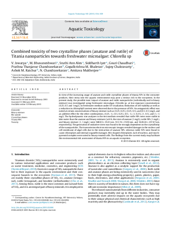
 18M157 - SIDDHARTH KRISHNAN IYER