Clinical review
Science, medicine, and the future
Functional magnetic resonance imaging in neuropsychiatry
Catherine Longworth, Garry Honey, Tonmoy Sharma
The ability of functional magnetic resonance imaging
to provide high quality imaging of brain function without the need for radioactive tracers is rapidly making it
the technique of choice for research into neuropsychiatric disorders and their treatment. The future is likely
to bring a closer involvement in clinical practice, with
the technique being used for early detection of
dysfunction, assessing the clinical efficacy of drug treatments, and as an alternative to invasive preoperative
procedures requiring localisation of function.
Functional magnetic resonance imaging
The development of anatomical neuroimaging enabled the in vivo visualisation of neuropathology in
conditions such as stroke, facilitating differential
diagnoses and early treatment. Since then scanning
techniques have gone beyond structural detail to
provide images relating to human brain function, and
in the past decade these techniques have been joined
by an impressive new imaging tool, functional
magnetic resonance imaging (functional MRI). This
has a spatial resolution within the millimetre scale and
can capture responses in the brain occurring over a few
seconds, although reconstruction and processing of
the raw data commonly occur after scanning.
Functional MRI is non-invasive and safe. It does not
require radioactive tracer substances, unlike positron
emission tomography (PET) or single photon emission
tomography (SPET), and uses the brain’s natural
haemodynamic response to neural activity as an
endogenous tracer. It can be carried out during the
same session as routine magnetic resonance imaging
in a clinical scanner. These features are making it
increasingly popular in neuropsychiatric research.
The commonest form of functional MRI is blood
oxygenation level dependent (BOLD) imaging.1 The
BOLD signal depends on the ratio of oxygenated to
deoxygenated haemoglobin. In regions of neuronal
activity this ratio changes as increased flow of oxygenated blood temporarily surpasses consumption,
decreasing the level of paramagnetic deoxyhaemoglobin. These localised changes cause increases in
magnetic resonance signal, which are used as markers
of functional activation (fig 1). Ultrafast scanning can
measure these changes in signal, which are mapped
directly onto a high resolution scan of the subject’s
anatomy. In addition, data from several subjects can be
BMJ VOLUME 319 11 DECEMBER 1999 www.bmj.com
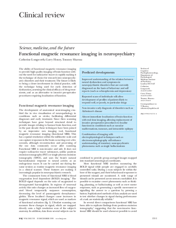
Predicted developments
Improved understanding of the relation between
neural dysfunction and symptoms in
neuropsychiatric disorders that are currently
diagnosed on the basis of behaviour and self
reports (such as schizophrenia and depression)
Repeated scans of individuals will allow
development of profiles of patients likely to
respond well, or poorly, to particular drugs
Non-invasive early diagnosis of disorders such as
Alzheimer’s disease
Section of
Cognitive
Psychopharmacology,
Department of
Psychological
Medicine, Institute
of Psychiatry,
London SE5 8AF
Catherine
Longworth
research worker
Garry Honey
research worker
Tonmoy Sharma
senior lecturer
Correspondence to:
T Sharma
t.sharma@iop.kcl.ac.uk
BMJ 1999;319:1551–4
Almost immediate localisation of brain function
with real time imaging, allowing replacement of
invasive preoperative procedures to localise
functions in conditions such as vascular
malformations, tumours, and intractable epilepsy
Combination of imaging with
electrophysiological techniques such as
electroencephalography will enhance
understanding of transitory neuropsychiatric
phenomena such as single hallucinations
combined to provide group averaged images mapped
into standard neurological coordinates.
Most functional MRI involves measuring the
BOLD signal while people are engaged in carefully
controlled tasks. During a scan subjects lie within the
bore of the magnet, and their behavioural responses to
presented stimuli are monitored. A wide range of
stimuli can be presented across sensory modalities. It is
possible to examine covert phenomena such as thinking, planning, or hallucinating as well as overt motor
responses, such as generating a specific movement or
signalling the answer to a question by pressing a
button. Sophisticated methods of data analysis are used
to test whether changes in signal during performance
of a task are statistically reliable.2
In several direct comparisons functional MRI has
been able to replicate findings from positron emission
tomography,3 suggesting that the non-invasive functional MRI should be used whenever possible to avoid
1551
�Clinical review
candidates for a scan can tolerate the noise of the
scanner and close confinement within the magnet
bore, as well as being free of metallic implants.
Applications to neuropsychiatric
disorders
Fig 1 Principles involved in converting neuronal activity into a blood oxygenation level
dependent (BOLD) signal, which can be measured with functional magnetic resonance imaging
exposure to radiation and the need for an expensive
cyclotron unit on site. Unlike positron emission
tomography, functional MRI is not limited in the
number of scans that can safely be performed on a
single person, which means that repeated scans of the
same patient can track the course of a disorder and,
potentially, its response to treatment. The safety of the
technique also facilitates the recruitment of research
subjects and enhances compliance, as well as extending
the range of people who can be scanned to vulnerable
groups such as children.
Like all neuroimaging methods, functional MRI
has limitations. Movement of subjects during scanning
can produce artefacts, although these can be resolved
to a certain extent by corrective data procedures.4 The
magnetic resonance properties of the anterior skull
base and petrous bone are another source of artefacts,
causing a relative loss of signal in the medial inferior
frontal lobe and inferior temporal lobe.5 This problem
can be reduced through careful choice of orientation
of the scan, but it must be considered when interpreting results. There are also issues of a practical nature,
such as the careful screening necessary to ensure that
Fig 2 Functional MRI images showing reduced activation of language areas during a
linguistic task in patients with schizophrenia (from Honey et al8)
1552
The infrastructure necessary for conducting functional
MRI is already available in the magnetic resonance
imaging departments of district general hospitals. It
can be carried out on standard clinical MRI scanners
with upgraded software. However, as with any new
technology, established findings and standardised
techniques will be required before functional MRI can
make the transition from research to routine use in
clinical practice. Its main applications to neuropsychiatry at present are to increase understanding of a wide
range of disease states and the effects of treatment.
Functional MRI can provide a window into disease
states, such as depression or schizophrenia, that,
because of the lack of biological markers, are currently
diagnosed on the basis of behavioural signs and self
reported symptoms such as auditory hallucinations.
Functional MRI has the potential to change our
understanding of these conditions by demonstrating
how neural dysfunction manifests itself in behaviour
and symptoms.
Unipolar depression
One study compared depressed patients and healthy
volunteers in their neural response to film clips
designed to evoke transient sadness.6 The brain activation recorded during emotionally neutral film clips was
compared with that occurring during sad films. This
revealed that, although many brain regions were
activated similarly by both groups, the depressed
subjects activated additional regions, namely the left
medial prefrontal cortex and the right anterior cingulate gyrus, during the processing of transient sadness.
These brain structures are thought to be involved in
the attribution of emotional importance and the
conscious experience of emotion. The investigators
postulated that in depression abnormal frontal activity
might disconnect the limbic system from normal
modulatory influences.
Schizophrenia
Patients with schizophrenia show specific deficits in
language processing, which are classically considered a
cardinal feature of the illness. Functional MRI has
begun to reveal the neural dysfunction underlying
these deficits.7 We found that patients performing a
language task showed a broadly similar pattern of neural activation, though with an attenuated power of
response, compared with controls.8 However, we
observed specific regions of hypoactivity in the frontotemporal cortex (fig 2). These may be related to deficits
in language processing that can be observed at a
cognitive level.
The extrapyramidal symptoms and neurological
“soft signs” prevalent in schizophrenia have prompted
the use of functional MRI to investigate brain function
during psychomotor tasks. For example, Wenz et al
reported functional abnormalities associated with
motor processing during performance of a sequential
BMJ VOLUME 319 11 DECEMBER 1999 www.bmj.com
�Clinical review
thumb to digit task in patients with schizophrenia
compared with controls.9 These results suggested that
interhemispheric communication is disturbed in
schizophrenia. The concept of anomalous cerebral
asymmetry in schizophrenia is supported by results
from other studies using functional MRI (fig 3).10 11
An alternative approach has used functional MRI
to investigate temporary states such as specific
symptoms rather than comparing patients with healthy
volunteers. Howard et al found that photic stimulation
of a patient experiencing visual hallucinations produced a significantly less extensive pattern of response
in the visual cortex than when the patient was
rescanned after successful resolution of symptoms with
risperidone treatment.12 Similarly, patients who are
experiencing auditory hallucinations show inhibited
activation of the auditory association cortex in
response to external auditory stimuli.13 These studies
indicate that processing of endogenous and exogenous
stimuli may compete for common neural resources.
The potential of functional MRI for conducting
repeated scanning of an individual patient has important clinical applications. Characterisation of the functional neuroanatomy of cognitive processes will
provide a framework for research into the longitudinal
effects of pharmacological treatments on cognitive
function. We have followed drug induced changes in
the brain function of patients with schizophrenia after
switching them to newer atypical antipsychotic
drugs.14 15 Such research raises the possibility of developing profiles of patients likely to respond well to particular drugs, allowing doctors to assess the probability
of a positive response before embarking on lengthy
and expensive courses of treatment. It could also be
used to develop treatment profiles outlining which disease related cognitive deficits are enhanced by particular drugs. Repeat scanning with functional MRI would
also allow physicians to track changes in a patient’s
brain function during the course of an illness. For
example, schizophrenia is characterised by psychotic
episodes and periods of remission. Repeat scanning
could be used to differentiate between those neural
deficits underlying the illness and those associated with
exacerbation of symptoms during acute psychotic
episodes.
Alzheimer’s disease
In disorders where neural correlates have been identified, such as Alzheimer’s disease and epilepsy, research
has focused on establishing that functional MRI can
adequately replicate existing clinical findings from
more invasive techniques. For example, Sandson et al
used a variant of functional MRI to investigate cerebral
hypoperfusion in patients with Alzheimer’s disease.16
They replicated previously demonstrated temperoparietal hypoperfusion and found it to correlate with the
severity of the dementia. Indeed, Harris et al reported
that, with a non-radioactive magnetic contrast agent,
functional MRI could detect such hypoperfusion at an
early stage in the disorder when symptoms were still
mild.17 Together, these studies indicate that functional
MRI shows promise as a clinical tool for the early
detection of Alzheimer’s disease.
BMJ VOLUME 319 11 DECEMBER 1999 www.bmj.com
Fig 3 Functional MRI images showing abnormal cerebral asymmetry during a psychomotor
task performed by people with schizophrenia (Honey et al11)
Epilepsy
Another potential use of functional MRI is in the presurgical testing of patients with intractable epilepsy. In
cases where temporal lobe resection is considered
patients undergo lateralisation testing of temporal lobe
functions to establish the risk of permanent neurological damage. This is commonly achieved by testing language and memory abilities after an injection of
sodium amylobarbitone into an internal carotid artery
to anaesthetise one hemisphere or by direct electrical
stimulation. Research has shown that functional MRI
can replicate the results of these tests, raising the possibility of replacing distressing and potentially harmful
procedures.18 In the United States, functional MRI of
sensorimotor and language functions has been used to
assess whether a patient is a candidate for surgery and
to guide surgical planning in cases of vascular malformations, tumours, intractable epilepsy, and lesions near
critical cortical areas.19
Clinical implications of technological
advances
Functional MRI is still in its infancy. This decade we
have seen many technical developments, and we can
expect to see further improvements. Currently,
functional MRI is mainly used in neuropsychiatry to
investigate static aspects of disorders. Improving the
temporal resolution of scanning extends the range of
disease processes that can be investigated to include
even momentary phenomena such as individual
psychotic hallucinations. Researchers have begun to
achieve this by combining functional MRI with electrophysiological techniques such as electroencephalography and magnetoencephalography.20
Another new development, real time functional
MRI, displays the course of neurological activation
during the scan rather than processing the data after
scanning. This is particularly useful for clinical practice
as it allows immediate assessment of brain activation
and movement within the scanner, thus adding to the
potential of functional MRI as a useful presurgical
tool.21 It might also be possible to use real time
scanning in treatments based on biofeedback—that is,
the self modulation of physiological parameters in
1553
�Clinical review
response to simultaneous feedback of biological information. For example, in cases of intractable epilepsy it
has been found that training patients to alter the
pattern of their electroencephalogram reduced seizure
rates over a six month period.22 With real time
functional MRI, it might become possible to show
patients images of their own brain function while they
are in the scanner in order to facilitate biofeedback.
Competing interests: None declared.
1
2
3
4
5
6
7
8
Ogawa S, Lee TM, Kay DW, Tank DW. Brain magnetic resonance imaging
with contrast dependent on blood oxygenation. Proc Natl Acad Sci USA
1990;87:9868-72.
Bullmore ET, Williams SCR, Rabe-Hesketh S, Janot N, David A, Mellers J,
et al. Statistical methods of estimation and inference for functional MR
image analysis. Magn Reson Med 1996;35:261-77.
Paulesu E, Connelly A, Frith CD, Friston KJ, Heather J, Myers R, et al.
Functional MR imaging correlations with positron emission tomography.
Initial experience using a cognitive activation paradigm on verbal working memory. Neuroimaging Clin North Am 1995;5:207-25.
Bullmore ET, Brammer MJ, Rabe-Hesketh S, Curtis VA, Morris RE, Williams SCR, et al. Methods for diagnosis and treatment of stimulus correlated motion in generic brain activation studies using fMRI. Hum Brain
Map 1999;7:38-48.
Ojemann JG, Kbudak E, Snyder AZ, McKinstry RC, Racihle ME, Conturo
TE. Anatomic localization and quantitative analysis of gradient refocused
echo-planar fMRI susceptibility artifacts. Neuroimage 1997;6:156-67.
Beauregard M, Leroux JM, Bergman S, Arzoumanian Y, Beaudoin G,
Bourgouin P, et al. The functional neuroanatomy of major depression: an
fMRI study using an emotional activation paradigm. Neuroreport
1998;9:3253-8.
Curtis VA, Bullmore ET, Brammer MJ, Wright IC, Williams SC, Morris
RG, et al. Attenuated frontal activation during a verbal fluency task in
patients with schizophrenia. Am J Psychiatry 1998;155:1056-63.
Honey GD, Soni W, Bullmore ET, Varatheeson M, Williams SCR, Andrew
C, et al. Dissecting the components of linguistic processing in schizophrenia using functional MRI. Schizophr Res 1998;29:65.
9
10
11
12
13
14
15
16
17
18
19
20
21
22
Wenz F, Schad LR, Knopp MV, Baudendistel KT, Flomer F, Schroder J, et
al. Functional magnetic resonance imaging at 1.5 T: activation pattern in
schizophrenic patients receiving neuroleptic medication. Magn Reson
Imaging 1994;12:975-82.
Mattay VS, Callicott JH, Bertolino A, Santha AK, Tallent KA, Goldberg
TE, et al. Abnormal functional lateralization of the sensorimotor cortex
in patients with schizophrenia. Neuroreport 1997;8:2977-84.
Honey GD, Soni W, Bullmore ET, Varatheesan M, Williams SCR, Andrew
CM, et al. Evidence of abnormal lateralisation of motor systems in schizophrenia using functional MRI. Schizophr Res 1998;29:70.
Howard R, David A, Woodruff P, Mellers I, Wright J, Brammer M, et al.
Seeing visual hallucinations with functional magnetic resonance
imaging. Dementia Geriatr Cogn Disord 1995;8:73-7.
Woodruff PW, Wright IC, Bullmore ET, Brammer M, Howard RJ, Williams
SC, et al. Auditory hallucinations and the temporal cortical response to
speech in schizophrenia: a functional magnetic resonance imaging study.
Am J Psychiatry 1997;154:1676-82.
Honey GD, Bullmore ET, Soni W, Varatheesan M, Williams SCR, Sharma
T. Risperidone restores fronto-parietal activation by a working memory
task in patients with schizophrenia [abstract]. Schizophr Res 1999;36:223.
Honey GD, Bullmore ET, Soni W, Varatheesan M, Williams SCR, Sharma
T. Investigation of the effect of typical versus atypical antipsychotics on
motor function using functional MRI [abstract]. Schizophr Res
1999;36:223.
Sandson TA, O’Connor M, Sperling RA, Edelman RR, Warach S. Noninvasive perfusion MRI in Alzheimer’s disease: a preliminary report.
Neurology 1996;47:1339-42.
Harris GJ, Lewis RF, Satlin A, English CD, Scott TM, Yurgelun-Todd DA,
et al. Dynamic susceptibility contrast MRI of regional cerebral blood
volume in Alzheimer’s disease. Am J Psychiatry 1996;153:721-4.
Bookheimer SY. Functional MRI applications in clinical epilepsy.
Neuroimage 1996;4(3 Pt 3):S139-46.
Buchbinder BR, Cosgrove GR. Cortical activation MR studies in brain
disorders. Magn Reson Imaging Clin North Am 1998;6:67-93.
George JS, Aine CJ, Mosher JC, Schmidt DM, Ranken DM, Schlitt HA, et
al. Mapping function in the human brain with magnetoencephalography,
anatomical magnetic resonance imaging, and functional magnetic
resonance imaging. J Clin Neurophysiol 1995;12:406-31.
Van Muiswinkle AMC, van den Brink JS, Folkers PJM. Real-time fMRI on
a clinical MR scanner. Neuroimage 1999;9(2):S212.
Kotchoubey B, Blankenhorn V, Froscher W, Strehl U, Birbaumer N.
Stability of cortical self-regulation in epilepsy patients. Neuroreport
1997;8:1867-70.
Lesson of the week
Hyponatraemic seizures and excessive intake of hypotonic
fluids in young children
P Bhalla, F E Eaton, J B S Coulter, F L Amegavie, J A Sills, L J Abernethy
Afebrile seizures
in young
children may be
caused by
hyponatraemia
—take a dietary
history and
measure serum
electrolytes
Royal Liverpool
Children’s NHS
Trust, Liverpool
L12 2AP
P Bhalla
specialist registrar
J B S Coulter
senior lecturer
L J Abernethy
consultant radiologist
continued over
BMJ 1999;319:1554–7
1554
The differential diagnosis of afebrile seizures in
children with normal development includes epilepsy
and metabolic disorders. Children admitted to hospital
with seizures (febrile or afebrile) of unknown cause are
often treated with antibiotics and antiviral agents for
suspected infection of the central nervous system while
investigations are undertaken. Afebrile seizures caused
by hyponatraemia associated with excessive intake of
hypotonic fluids was first reported in 1967.1 This is a
common problem in the United States,2–8 but it has
rarely been reported in the United Kingdom.9 10 We
describe four cases (table).
Case reports
Case 1
A 20 month old girl presented with a short history of
vomiting, cough, and anorexia. She had attended the
accident and emergency department on four
occasions—with a viral illness, urinary tract infection,
pertussis, and breath holding. She was admitted for
observation, and a provisional diagnosis of viral illness
was made. The girl refused solid food but took fluids
well over the next 48 hours. At this time she had a tonic
seizure associated with apnoea but responded to treatment with rectal diazepam. Biochemical investigations
showed serum sodium concentration 116 mmol/l,
chloride 84 mmol/l, potassium 2.8 mmol/l, urea 2.8
mmol/l, and creatinine 35 mmol/l.
The patient’s fluid intake was restricted to 60% of
the maintenance requirement, but four hours later she
had a further tonic seizure associated with decerebrate
posturing. She was intubated and ventilated and given
intravenous mannitol and phenytoin. Computed
tomograms of the brain showed diffuse cerebral
oedema (figure). Her urine output over the next 12
hours was approximately 12 ml/kg per hour, and with
fluid restriction her serum electrolyte values returned
to normal. Repeat computed tomography 24 hours
later showed appreciable improvement, with normal
basal cisterns and ventricles (figure).
The girl was considered to have encephalitis and
was ventilated for six days, during which time her electrolyte values remained normal. However, analysis of
cerebrospinal fluid removed by lumbar puncture was
normal, blood cultures were sterile, and viral serology
failed to show infection. Dietary inquiry showed that
BMJ VOLUME 319 11 DECEMBER 1999 www.bmj.com
�
 Tonmoy Sharma