African Journal of Microbiology Research Vol. 6(18), pp. 4006-4012, 16 May, 2012
Available online at http://www.academicjournals.org/AJMR
DOI: 10.5897/AJMR11.1472
ISSN 1996-0808 ©2012 Academic Journals
Full Length Research Paper
The effect of a continuous mercury stress on mercury
reducing community of some characterized bacterial
strains
Ashraf M. M. Essa
Botany Department, Faculty of Science, Fayoum University, El Fayoum, Egypt. E-mail: ashraf.essa@yahoo.com.
Accepted 29 December, 2011
Active resistance to the mercuric ion is widely distributed in environmental microbes and results from
the action of mercuric reductase. Five mercury resistant bacteria: Escherichia coli Z1, Escherichia coli
Z3, Pseudomonas putida Z2, Serratia marcescens Z4 and Xanthomonas sp. Z5 were isolated and
identified from sludge sample. The presence of mercury resistance determinants was screened by PCR
using merA-specific primers. Based on the analysis of merA amplicons, high similarity was recorded
between the merA region of the strains P. putida Z2 and Xanthomonas sp. Z5 with those of Tn5053;
while the merA of E. coli Z1 was analogous to those of Tn21. In case of the bacterial strains E. coli Z3
and S. marcescens Z4 a great matching was obtained between their merA and those of Tn5036. The
effect of mercury stress upon the structure of mercury reducing biofilm at the species level and the
type of mercury resistance determinants was studied in a continuous bioreactor. Community analysis
suggested that the bacterial strain E. coli Z3 containing Tn5036-like determinant is the well adapted
strain that tolerated elevated levels of mercury whereas the other strains showed a less fitness under
these extreme conditions.
Key words: Mercury resistant bacteria, mercuric reductase gene, PCR-RFLP, mercury stress.
INTRODUCTION
The removal of widespread industrial and agricultural
heavy metal contamination is considered a challenge for
environment management. Microorganisms in contaminated environment have developed resistance to mercury
and are playing a major role in natural decontamination
(Cursino et al., 1999). The detoxification of mercury by
mercury resistant bacteria offers a potential cheaper and
safer alternative to conventional methods; moreover,
some mercury resistant bacteria can not only detoxify
mercury but also remove other metals such as cadmium
and lead (De et al., 2008).
Resistance against mercury has been identified in a
wide range of Gram-negative and Gram-positive bacteria
in natural and mercury contaminated environments and it
is often found on plasmids or other mobile genetic
elements such as transposons (Osborn et al., 1997;
Narita et al., 2004). Mercury resistance mechanism is
based on a group of genes located in a mercury resistant
operons which allows bacteria to reduce the toxic Hg(II)
into volatile metallic mercury Hg(0) through its enzymatic
reduction (Summers, 1986; Brown et al., 1991; Misra,
1992; Barkay et al., 2003); these operons contain genes
encoding the functional proteins for regulation (merR),
transport (merT, merP) and reduction (merA) in addition
to some accessory genes (merC, merF and merB) (Ji and
Silver, 1995; Nies, 1999). The bioremediation of mercury
from synthetic solution or wastewater via volatilization
using natural or immobilized mercury resistant bacterial
cells has been described by several investigators (Brunke
et al., 1993; von Canstein et al., 1999, 2002; Dzair et al.,
2004; Wagner-Dobler et al., 2000).
Microbial biodiversity has become a research subject
for understanding engineered ecosystems. Several
studies have reported the importance of measuring the
microbial diversity in laboratory bioreactors in order to
understand the relationship between the composition of
the microbial community and operational parameters (Liu
et al., 1997; Boon et al., 2002). It is well established that
�Essa
4007
Table 1. Synthetic oligonucleotide primers used in this study.
Primer
PA
530r
Sequence 5′-3′
AGAGTTTGATCCTGGCTCAG
GTATTACCGCGGCTGCTG
F3
R4
Amplified region
Size
Reference
Conserved region of 16S rDNA gene
500 bp
Lane et al., 1985
GGGGGCACCTCAGAAAACGGA
GGAATCGCGCAGACCTCACCT
IR - merT region of Tn21-like operon
730 bp
Essa et al., 2003
KI
KII
GGGGTCGTCTCAGAATTCGG
GACAAGCCCTATGGCAGCAT
IR - merR region of Tn5036-like operon
350 bp
Essa et al., 2003
MI3
MI2
GGAGTCGCCTCAGAAAACG
TACGGAGTCAAGCGATATGGA
IR - merR region of Tn5053-like operon
500 bp
Essa et al., 2003
MRS1
MRS2
ACCATCGGCGGCACCTGCG
AAGGTCTGS*GCCGCR*AGCTTC
merA region of Hg operons
1300 bp
Glendinning, 2000
r
S* = C+G, R* = A+G.
toxic effects of heavy metals are highly selective in
microbes; such selective targeting of specific enzymatic
systems and pathways suggests that certain members of
the microbial community would be more sensitive to
heavy metal exposure than others, depending on the
sensitivity of their critical metabolic pathways (Fulladosa
et al., 2005; Sobolev and Begonia, 2008).
The aim of this study is the use of PCR-based
techniques targeting the merA gene that codes for
mercuric reductase in order to explore the functional
diversity of a mercury reducing community under
continuous mercury stress.
PCR amplification of DNA encoding the 16S rRNA gene
Amplification of the 16S rDNA gene was carried out by using primer
pair pA/530r (Table 1). The PCR mixture was prepared as the
following; 10 μl (10x) PCR buffer, 3 μl (50 mM) MgCl2, 1 μl (20
pmole/μl) of each primer, 1 μl (10 mM) dNTPs mixture, 0.5 μl (2.5U)
Taq DNA polymerase, 2 μl total DNA extract, and the volume is
completed to 100 μl by SDH2 O. PCR were carried out for 35 cycles
under the following conditions: denaturation step at 94°C for 40 s,
annealing step at 55°C for 1 min, extension step at 72°C for 2 min
and final extension at 72°C for 10 min. An aliquot of the PCR
products (10 μl) was mixed with 2 μl of DNA loading buffer and
analyzed by electrophoresis (15 V/cm, 60 min) on 0.7% horizontal
agarose gel in TBE buffer containing 0.5 μg/ml ethidium bromide,
then visualized on an UV transilluminator.
MATERIALS AND METHODS
Isolation and purification of mercury resistance bacterial
strains
Luria Bertani (LB) broth supplemented with 10 µg/ml HgCl2 was
inoculated with sludge sample obtained from the Zenein Waste
Water Treatment Plant (ZWWTP) localized in the Giza
Governorate, Egypt and incubated at 30°C on shaking incubator
(200 rpm) for 48 h followed by pour plate method on LB agar
medium. A single bacterial colony was aseptically picked up and
transferred onto a fresh medium with a streaking technique and
incubated for 24 h at 30°C. Transferring was repeated until
obtaining a pure bacteria culture and the isolated colonies were
plated on LB agar plates supplemented with 20 µg/ml HgCl2.
Bacterial colonies which showed better growth on HgCl2 plates
were taken and streaked in the LB agar slants and stored.
Total DNA and plasmid preparation
The total bacterial DNA was prepared according to the method of
Goldberg and Ohman (1984), the small scale purification of plasmid
DNA was performed by the modified alkaline method of Le Gouill et
al. (1994).
PCR for amplification of merA region
The purified plasmid DNA of the mercury resistant strains was used
as a template in PCR by using MRS1/MRS2 primers (Table 1) to
amplify the merA region. The PCR mixture was prepared as
described above and PCR were carried out for 35 cycles under the
following conditions: denaturation step at 94°C for 40 s, annealing
step at 57°C for 1 min, extension step at 72°C for 2 min and final
extension at 72°C for 10 min. The PCR products were analyzed as
mentioned above.
Purification of the PCR products and nucleotide sequence
analysis
Aqueous PCR products were purified by using a QIAquick PCR
purification kit as described by the manufacturer's instructions. The
purified PCR products were sequenced using ABI PRISM Big Dye
Terminator Cycle Sequencing Ready Reaction Kits with Ampli Taq
DNA polymerase (CliniLab, Egypt). The sequence data were
analysed by BLASTN search at the National Centre for
Biotechnology Information (http://www.ncbi.nlm.nih.gov) to identify
the most similar sequences.
�4008
Afr. J. Microbiol. Res.
Table 2. The restriction enzymes: PshAI, AccI and Eco01091 were used for the RFLP analysis of merA amplicons based on their DNA
sequence.
merA ampilcons
Tn21-like operon
Tn5036-like operon
Tn5053-like operon
1
2
PshAI
Single cut
Do not cut
Do not cut
3
4
5
AccI
Do not cut
Do not cut
Single cut
1
6
1000 bp
800 bp
600 bp
3
4
5
6
1500 bp
500 bp
400 bp
200 bp
2
Eco01091
Do not cut
Single cut
Do not cut
A
A
1000 bp
800 bp
1500 bp
B
B
Figure 1. Gel electrophoresis of PCR products of: A) the partial 16S rDNA gene of the bacterial isolates Z1 (lane 2), Z2 (lane 3), Z3 ( lane
4), Z4 (lane 5) and Z5 (lane 6), B) the merA gene from plasmid DNA of Escherichia coli Z1 (lane 2), Escherichia coli Z3 (lane 3),
Pseudomonas putida Z2 (lane 4), Xanthomonas sp. Z5 (lane 5), Serratia marcescens Z4 (lane 6). Lane 1 in both figures contains
Hyperladder I marker.
PCR-RFLP pattern
According to the DNA sequence and the restriction map of the
merA regions that were amplified from the bacterial isolates
(Restriction
Site
Analyzer
and
Map
Generator,
www.algosome.com), the restriction enzymes: PshAI, AccI and
Eco01091 (GibcoBRL, Life Technologies) were chosen to digest
the merA amplicons (Table 2). The reaction was set up as follows;
1.5 μg PCR product, 5 μl restriction enzyme, 10 μl (10x) restriction
enzyme buffer, and the volume was completed up to 100μL by
sterile distilled water. The reaction was incubated at 37°C for 1 h.
After inactivation (65°C for 20 min), the reaction mixture was mixed
with 0.2 volume of DNA loading buffer and analyzed by
electrophoresis.
PCR with primers specific for the different mer determinants
According to the obtained DNA sequence of merA region, some
primers were used to discriminate between the different mer
operons (Table 1). The purified plasmid DNA of the mercury
resistant strains was used as a template in PCR by using the
following primers E3/E4 to identify the Tn5075 operon, MI3/MI2 to
identify the Tn5053 operon and KI/KII to identify the Tn5036
operon. The PCR mixture was prepared as described above. PCR
were carried out for 35 cycles under the following conditions;
denaturation step at 94°C for 40 s, annealing step at 56°C for 1
min, extension step at 72°C for 2 min and final extension at 72°C
for 10 min. The PCR products were analyzed as mentioned above.
Bioreactor setup and operation
The mercury resistance bacterial isolates from the sludge sample,
which contains different mercury resistant determinants, were
grown individually in LB broth supplemented with 10 µg/ml HgCl2 at
37°C on a shaking incubator at 200 rpm for 24 h. A 25 ml of each
culture were mixed together and used as an inoculum for the
bioreactor which contains about 1.5 L LB broth. The bioreactor was
maintained under aerobic conditions by pumping in filter-sterilized
air, 37°C for 30 days. The LB broth supplemented with HgCl2 (10 to
60 µg/ml) was flowed through the bioreactor at 100 ml/h and the
bacterial growth was monitored by measuring the protein content.
Moreover, the DNA extracted from the effluent of the bioreactor
during the operating period was subjected to PCR by using specific
primers for the different determinants (Table 1). At the end of the
experiment (30 days), the community composition at the strain level
was analyzed by 16S ribosomal DNA analysis. At the same time,
the RFLP technique was used to profile the type of the mercury
resistant determinants based on their merA genes. The protein
content was used to follow the bacterial growth under different
HgCl2 concentrations. Samples from the effluent of the bioreactor
were centrifuged for 10 min (10,000 rpm), and cell pellets were resuspended in 500 μl of NaOH (0.5 M) and lyzed for 1 h. The protein
content was estimated according to the method of Lowry et al.
(1951).
RESULTS AND DISCUSSION
Isolation and characterization of mercury resistant
bacteria
The mercury resistant bacterial strains designated Z1,
Z2, Z3, Z4 and Z were isolated from the sludge sample
(ZWWTP) and were identified by partial 16S ribosomal
DNA technique (Figure 1A). The purified PCR products
were sequenced and databank compared. The isolates
�Essa
1
2
3
4
5
6
7
8
9
4009
10
1000 bp
800 bp
600 bp
400 bp
Tn5053-like
determinant
Tn5036-like
determinant
Tn21-like
determinant
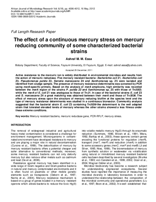
Figure 2. Gel electrophoresis of RFLP pattern of merA amplicons digested with Eco01091, AccI and
PshAI. Xanthomonas sp. Z5 containing Tn5053-like determinant is represented in lanes (2 to 4),
S.marcescens Z4 containing Tn5036-like determinant is represented in lanes (5 to 7) and E. coli Z1
containing Tn21-like determinant is represented in lanes (8 to 10). Digestion by Eco01091 is
represented in lanes (4, 7 and 10), AccI in lanes (3, 6 and 9) and PshAI in lanes (2, 5 and 8). Lane 1
contains Hyperladder I marker.
Z1 and Z3 were identified as Escherichia coli (99.6 and
99.9% identity, respectively), isolate Z2 was identified as
Pseudomonas putida (96.7% identity), isolate Z4 was
identified as Serratia marcescens (99.7% identity), and
isolate Z5 was identified as Xanthomonas sp. (97.8%
identity).
The purified plasmid DNA of each mercury resistant
strain was screened by PCR for merA genes. Results in
Figure 1B demonstrated the presence of the merA gene
(approximately 1300 bp) in the mercury resistant isolates.
The purified PCR products were sequenced and
databank compared; the merA region of E. coli Z1 strain
showed a high similarity to those of Tn21 (99%, Liebert et
al., 1999). In case of P. putida Z2 and Xanthomonas sp.
Z5 the amplified merA region recorded the highest
identity to those from Tn5053 (96 and 98%, Kholodii et
al., 1995) while, the amplified merA region of E. coli Z3
and S. marcescens Z4, showed the highest identity to
those from Tn5036 (96 and 99%, Yurieva et al., 1997).
According to the DNA sequence and the restriction
map of the merA region of the different determinants
(Restriction Site Analyzer and Map Generator,
www.algosome.com), some restriction enzymes were
used to digest the merA amplicons (Table 2) resulting in
a specific RFLP pattern (Figure 2). PshAI digested the
merA of Tn21-like determinant into two fragments (760 to
480 bp), AccI digested the merA of Tn5053-like
determinant into two fragments (880 to 360 bp) whereas
Eco01091 digested the merA of Tn5036-like determinant
into two fragments (790 to 450 bp); moreover, PCR with
specific primers (Table 1) was used to confirm the type of
these determinants in the bacterial strains. Data in Figure
3 showed that PCR amplicons were obtained by using
primers E3/E4 with Tn21-like determinant (730 bp),
MI3/MI2 with Tn5053-like determinant (500 bp), and
KI/KII with Tn5036-like determinant (350 bp).
Effect of mercury stress on a mercury reducing
biofilm
The isolated mercury resistant strains: E. coli Z1 and Z3,
P. putida Z2, Xanthomonas sp. Z5 and S. marcescens
Z4, were grown together inside a continuous bioreactor
for 30 days under selective continuous mercury stress
and the total protein content was monitored as a
parameter for the bacterial growth (Figure 4A). At the
same time, the DNA extracted from the effluent of the
bioreactor upon the pilot plant operation was subjected to
PCR by using specific primers for the different mercury
resistance determinants. The obtained data (Figure 4B)
demonstrated that the Tn5053-like determinant
disappeared after 18 days (at 40 µg/ml of HgCl2), the
Tn21-like determinant vanished after 24 days (at 60
µg/ml of HgCl2) whereas the Tn5036-like determinant
recorded a high tolerance capability under these extreme
conditions.
At the end of the experiment (after 30 days), the
composition of the mercury resistant community at the
strain level was analyzed on the basis of the 16S
ribosomal DNA gene showing the presence of only E. coli
(Z3), meanwhile the other strains were completely gone.
The use of 16S rRNA gene as a marker to study the
composition and the dynamics of some bacterial
communities has been reported in previous studies
(Wagner-Dobler et al., 2000; Saikaly et al., 2005).
�Afr. J. Microbiol. Res.
1
2
3
4
1000 bp
800 bp
730 bp
600 bp
500 bp
400 bp
350 bp
Figure 3. Gel electrophoresis of PCR products from plasmid DNA of E. coli Z1
containing Tn21-like determinant by using primers E3/E4 (lane 2), Xanthomonas sp.
Z5 containing Tn5053-like determinant by using primers MI3/MI2 (lane 3) and S.
marcescens Z4 containing Tn5036-like determinant by using primers KI/KII (lane 4).
Lane 1 contains the hyperladder I marker.
fE
400
A
Protein content (mg/L)
4010
300
200
100
0
0
3
6
9
12
15
18
21
24
27
30
Time (days)
B
1
2
3
4
5
6
Tn21-like determinant
Tn5053-like determinant
Tn5036-like determinant
Figure 4. (A) The growth of a mercury resistance bacterial population (expressed as mg/mL
d
protein) consisting of E. coli Z1, E.Z3, P. putida Z2, Xanthomonas sp. Z5 and S. marcescens Z4 in
a continuous aerobic bioreactor for 30 days on LB broth supplemented with different HgCl 2
concentrations: 20µg/mL up to the 3rd day, 30 µg/ml up to the 9th day, 40 µg/ml up to the 15th day,
50 µg/ml up to the 21st day, 60 µg/ml up to the end of the operation.(B) The gel electrophoresis of
PCR products from plasmid DNA of the bioreactor effluent obtained at the start of the exper iment
(lane 1), at 6 days (lane 2), at 12 days (lane 3), at 18 days (lane 4), at 24 days (lane 5), at 30 days
(lane 6) by using primers KI/KII for Tn5036-like determinant, MI3/MI2 for Tn5053-like determinant
and E3/E4 for Tn21-like determinant. Lane 1 contains Hyperladder I marker.
�Essa
1
2
3
4
1
2
3
4011
4
800 bp
600 bp
1000 bp
800 bp
600 bp
400 bp
790 bp
400 bp
A
350 bp
450 bp 200 bp
B
Figure 5. The gel electrophoresis of: A) RFLP pattern of merA amplicons obtained from plasmid DNA of the
bioreactor effluent at the end of the experiment (30 days) digested with Eco01091 (lane 2), AccI (lane 3) and
PshAI (lane 4), B) the PCR products from plasmid DNA of the bioreactor effluent obtained after 30 days by using
primers KI/KII (lane 2), MI3/MI2 (lane 3) and E3/E4. Lane 1 contains Hyperladder I marker.
Actually, the alteration of a bacterial community in
laboratory bioreactors under the influence of the heavy
metals stress by targeting of some marker genes especially those responsible for the resistance mechanism will
produce more accurate image for the biodiversity of the
bacterial population; so the mercury resistant community
was analysed to profile the type of their determinant
through using RFLP analysis of the obtained merA
amplicons which showed the presence of Tn5036determinant while the other determinants were not found
(Figure 5A). These results were confirmed via subjecting
the DNA extracted from the effluent of the bioreactor after
30 days to PCR by using some specific primers for the
different mer determinants (Figure 5B). A clear DNA band
(350 bp) was obtained with primers MR3/MR2 that are
specific to Tn5036-like determinant whereas no PCR
products were obtained by using the primers of the other
determinants. These results are compatible with those
who used the merA gene as a molecular marker to follow
the assortment of mercury resistant bacterial population
under the pressure of mercury toxicity in aerobic and
anaerobic environments (Felske et al., 2003; Simbahan
et al., 2005; Sotero-Martins et al., 2008).
This study clarified the presence of a strong selective
pressure on the microbial community inside the
bioreactor due to the mercury toxicity which led to the
predomination of E. coli (Z3) containing Tn5036-like
determinant. These results are in accordance with other
studies which showed that the continuous exposure to
elevated levels of mercury altered the microbial
community and exclusively select bacteria that can cope
with such levels (Osborn et al., 1993; Muller et al., 2001;
Ramaiah and De, 2003).
The domination of E. coli Z3 containing Tn5036-like
determinant over the other strains under the strong
selective pressure exerted by mercury toxicity was
attributed to the well adaptation of this strain which might
be linked with the type of mercury resistance determinant. This assumption is consistent with the finding of
previous studies that correlated the functional diversity
and the adaptation ability of some bacterial communities
under the influence of mercury with the frequency and the
type of mercury resistance operon (Smalla et al., 2006;
Chien et al., 2010); moreover, the capability of E. coli Z3
to tolerate an elevate level of mercury could be based on
the presence of some additional mechanisms beside
mercury volatilization that give this organism an extratolerance capability to cope with such mercury stress.
Agreeing with this hypothesis, Haferburg and Kothe
(2007) reported that the adaptation to heavy metal rich
environments resulted in microorganisms which show
activities for biosorption, bioprecipitation, extracellular
sequestration
and
chelation.
Such
resistance
mechanisms may play a role in transforming the toxic
metals into other forms that are not biologically available
to the cells. One of these mechanisms is the precipitation
of the soluble metal ions away from the cells via its
complexation into insoluble metal precipitates via the
production of some metabolites (Essa et al., 2006).
Finally, an environment with a raised concentration of
heavy metals constitutes a prospective stimulus for toxic
metal tolerant bacteria; such polluted environments
encourage adaptation for heavy metal resistance and
markedly affect on the composition of a bacterial biofilm.
The use of merA gene, the key enzyme of mercury
volatilization, as molecular markers in order to follow the
diversity of mercury resistant bacterial population under
the pressure of mercury toxicity can provide significant
information about the functional alterations of these
communities especially in contaminated environments.
Despite of the intrinsic role of the different mer operons in
mercury detoxification process, work is still necessary to
illustrate the distribution and diversity of these genetic
determinants in the bacterial communities under heavy
metals stress in order to employ them for the
bioremediation of these toxic pollutants.
REFERENCES
Barkay T, Miller SM, Summers AO (2003). Bacterial mercury resistance
from atoms to ecosystems. FEMS Microbiol. Rev., 27: 355–384.
Boon N, Windt WD, Verstraete W, Top EM (2002). Evaluation of nested
�4012
Afr. J. Microbiol. Res.
PCR-DGGE (denaturing gradient gel electrophoresis) with group
specific 16S rRNA primers for the analysis of bacterial communities
from different wastewater treatment plants. FEMS Microbiol. Ecol.,
39: 101–112.
Brown NL, Camakaris J, Lee BT, Williams T, Morby AP, Parkhill J,
Rouch DA (1991). Bacterial resistances to mercury and copper. J.
Cell Biochem., 46: 106-114.
Brunke M, Deckwer WD, Frischmuth A, Horn JM, Lunsdorf H, Rhode M,
Rohricht M, Timmis KN, Weppen P (1993). Microbial retention of
mercury from waste streams in a laboratory column containing merA
gene bacteria. FEMS Microbiol. Rev., 11: 145–152.
Chien M, Lin MK, Chang J, Huang C, Endo G, Suzuki S (2010).
Distribution of mercury resistance determinants in a highly mercury
polluted area in Taiwan. Interdisciplinary Studies on Environmental
Chemistry - Biological Responses to Contaminants, Eds., pp. 31–36.
Cursino L, Oberda SM, Cecilio RV, Moreira RM, Chartone-Souza E,
Nascimento AMA (1999). Mercury concentration in the sediment at
different gold prospecting sites along the Carmo stream, Minas
Gerias, Brazil & frequency of resistant bacteria in the respective
aquatic communities. Hydrobiologia, 394: 5-12.
De J, Ramaiah N, Vardanyan L (2008). Detoxification of toxic heavy
metals by marine bacteria highly resistant to mercury. Mar.
Biotechnol., 10(4): 471-477.
Dzair FZ, Zeroual Y, Moutaouakkil A, Taoufik J, Talbi M, Loutfi M, Lee
K, Blaghen M (2004). Bacterial volatilization of mercury by
immobilized bacteria in fixed and fluidized bed bioreactor. Ann.
Microbiol., 54: 353-364.
Essa AM, Julian DJ, Kidd SP, Brown NL, Hobman JL (2003). Mercury
resistance determinants related to Tn21, Tn1696, and Tn5053 in
enterobacteria from the preantibiotic era. Antimicrob. Agents
Chemother., 47: 1115-1119.
Essa AMM, Creamer NJ, Brown NL, Macaskie LE (2006). A new
approach to the remediation of heavy metal liquid wastes via offgases produced by Klebsiella pneumoniae M426. Biotechnol.
Bioeng., 95(6): 576–583.
Felske ADM, Fehr W, Pauling BV, von Canstein H, Wagner-Döbler I
(2003). Functional profiling of mercuric reductase (merA) genes in
biofilm communities of a technical scale biocatalyzer. BMC Microbiol.,
3: 22-27.
Fulladosa E, Murat JC, Villaescusa I (2005). Study on the toxicity of
binary equitoxic mixtures of metals using the luminescent bacteria
Vibrio fischeri as a biological target. Chemosphere, 58: 551-557.
Glendinning KG (2000). Studies on mercuric reductase and haemophilic
mercury resistance. Ph.D. thesis, the University of Birmingham, UK.
Goldberg JB, Ohman DE (1984). Cloning and expression in
Pseudomonas aeruginosa of a gene involved in production of
alginate. J. Bacteriol., 158: 1115–1121.
Haferburg G, Kothe E (2007). Microbes and metals: interactions in the
environment. J. Basic Microbiol., 47: 453-467.
Ji G, Silver S (1995). Bacterial resistance mechanisms for heavy metals
of environmental concern. J. Ind. Microbiol., 14: 61-75.
Kholodii GY, Mindlin SZ, Bass IA, Yurieva OV, Minakhina SV, Nikiforov
VG (1995). Four genes, two ends, and a res region are involved in
the transposition of Tn5053: a paradigm for a novel family of
transposons carrying either a mer operon or an integron. Mol.
Microbiol., 17: 1189-1200.
Lane DJ, Pace B, Olsen GL, Stahl DA, Pace NR (1985). Rapid
determination of 16S rRNA sequence for phylogenetic analysis. Proc.
Natl. Acad. Sci. USA, 82: 6955-6959.
Le Gouill C, Parent JL, Rola-Pleszczynski M, Stankova J (1994).
Analysis of recombinant plasmids by a modified alkaline lysis
method. Anal. Biochem., 219: 164-169.
Liu WT, Marsh TL, Cheng H, Forney LJ (1997). Characterization of
microbial diversity by determining terminal restriction fragment length
polymorphisms of genes encoding 16S rRNA. Appl. Environ.
Microbiol., 63: 4516–4522.
Liebert CA, Hall RM, Summers AO (1999). Transposon Tn21, flagship
of the floating genome. Microbiol. Mol. Biol. Rev., 63: 507-522.
Lowry OH, Rosebrough AL, Farr AL, Randall RJ (1951). Protein
measurement with the Folin phenol reagent. J. Biol. Chem., 193: 265275.
Misra TK (1992). Bacterial resistances to inorganic mercury salts and
organo-mercurials. Plasmid, 27: 4-16.
Muller AK, Rasmussen LD, Sorensen SJ (2001). Adaptation of the
bacterial community to mercury contamination. FEMS Microbiol.
Lett.,16: 49–53.
Narita M, Matsui K, Huang CC, Kawabata Z, Endo G (2004).
Dissemination of TnMERI1-like mercury resistance transposons
among Bacillus isolated from worldwide environmental samples.
FEMS Microbiol. Ecol., 48: 47-55.
Nies NH (1999). Microbial heavy-metal resistance. Appl. Microbiol.
Biotechnol., 51: 730-750.
Osborn AM, Bruce KD, Strike P, Ritchie DA (1993). Polymerase Chain
Reaction-restriction fragment length polymorphism analysis shows
divergence among mer determinants from Gram-negative soil
bacteria indistinguishable by DNA-DNA hybridization. Appl. Environ.
Microbiol., 59: 4024-4030.
Osborn AM, Bruce KD, Strike P, Ritchie DA (1997). Distribution,
diversity and evolution of the bacterial mercury resistance (mer)
operon. FEMS Microbiol. Rev., 19: 239-262.
Ramaiah N, De J (2003). Unusual rise in mercury-resistant bacteria in
coastal environs. Microbial Ecol., 45: 444-454.
Saikaly PE, Stroot PG, Oerther DB (2005). Use of 16S rRNA gene
terminal restriction fragment analysis to assess the impact of solids
retention time on the bacterial diversity of activated sludge. Appl.
Environ. Microbiol., 71(10): 5814-5822.
Simbahan J, Kurth E, Schelert J, Dillman A, Moriyama E, Jovanovich S,
Blum P (2005). Community analysis of a mercury hot spring supports
occurrence of domain-specific forms of mercuric reductase. Appl.
Environ. Microbiol., 71(12): 8836-8845.
Smalla K, Haines AS, Jones K, Krogerrecklenfort E, Heuer H, Schloter
M, Thomas CM (2006). Increased abundance of IncP-1 plasmids and
mercury resistance genes in mercury-polluted river sediments: First
discovery of IncP-1 plasmids with a complex mer transposon as the
sole accessory element. Appl. Environ. Microbiol., 72(11): 72537259.
Sobolev D, Begonia MFT (2008). Effects of heavy metal contamination
upon soil microbes: lead-induced changes in general and denitrifying
microbial communities as evidenced by molecular markers. Int. J.
Environ. Res. Pub. Heal., 5: 450-456.
Sotero-Martins A, de Jesus MS, Lacerda M, Moreira JC, Lauria
Filgueiras AL, Guimarães Barrocas PR (2008). A conservative region
of the mercury reductase gene (merA) as a molecular marker of
bacterial mercury resistance. Braz. J. Microbiol., 39: 307-310.
Summers AO (1986). Organization, expression and evolution of genes
for mercury resistance. Annu. Rev. Microbiol., 40: 607-634.
von Canstein H, Kelly S, Li Y, Wagner-Döbler I (2002). Species diversity
improves the efficiency of mercury-reducing biofilms under changing
environmental conditions. Appl. Environ. Microbiol., 68(6): 28292837.
von Canstein H, Li Y, Timmis KN, Deckwen WD, Wagner-Dobler I
(1999). Removal of mercury from chloralkali electrolysis wastewater
by a mercury-resistant Pseudomonas putida strain. Appl. Environ.
Microbiol., 65(12): 5279-5284.
Wagner-Döbler I, von Canstein H, Li Y, Timmis KN, Deckwer WD.
(2000). Removal of mercury from chemical wastewater by microorganisms in technical scale. Environ. Sci. Technol., 34: 4628-4634.
Yurieva O, Kholodii G, Minakhin L, Gorlenko Z, Kalyaeva E, Mindlin S,
Nikiforov V (1997). Intercontinental spread of promiscuous mercury
resistance transposons in environmental bacteria. Mol. Microbiol., 24:
321-329.
�
 Ashraf Essa