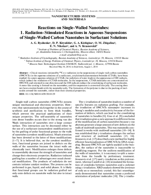
ISSN 0018-1439, High Energy Chemistry, 2015, Vol. 49, No. 1, pp. 48–52. © Pleiades Publishing, Ltd., 2015.
Original Russian Text © A.G. Ryabenko, D.P. Kiryukhin, G.A. Kichigina, O.M. Zhigalina, E.N. Nikolaev, A.N. Krasnovskii, 2015, published in Khimiya Vysokikh Energii, 2015,
Vol. 49, No. 1, pp. 51–55.
NANOSTRUCTURED SYSTEMS
AND MATERIALS
Reactions on Single-Walled Nanotubes:
1. Radiation-Stimulated Reactions in Aqueous Suspensions
of Single-Walled Carbon Nanotubes in Surfactant Solutions
A. G. Ryabenkoa, D. P. Kiryukhina, G. A. Kichiginaa, O. M. Zhigalinab,
E. N. Nikolaevc, and A. N. Krasnovskiid
a
Institute of Problems of Chemical Physics, Russian Academy of Sciences,
pr. Akademika Semenova 1, Chernogolovka, Moscow oblast, 142432 Russia
e-mail: aryabenk@icp.ac.ru
b Shubnikov Institute of Crystallography, Russian Academy of Sciences, Leninskii pr. 59, Moscow, 119333 Russia
c Talrose Institute of Energy Problems of Chemical Physics, Leninskii pr. 38, Moscow, 119334 Russia
d Moscow State Technological Institute STANKIN, Vadkovskii per. 1, Moscow, 127994 Russia
Received May 30, 2014; in final form, August 1, 2014
Abstract—Chical reactions initiated by 60Co γ-radiation in the suspensions of single-wall carbon nanotubes
(SWCNTs) in the aqueous solutions of a surfactant, cetyltrimethylammonium bromide (CTAB), have been
studied. In a pure aqueous solution of CTAB, the radiolysis of water leads to the appearance of OH radicals,
which induce the oxidation of CTAB molecules. In the suspensions, CTAB molecules are concentrated on
SWCNTs and oriented in one direction and the OH radicals induce the crosslinking of surfactant molecules.
A moss-like coating is formed on the nanotubes, and the suspension is converted into jelly. The coating does
not have covalent bonds with the nanotube walls. The formation of the polymer is due to the packing of molecules around the nanotube, rather than their chemical properties.
DOI: 10.1134/S0018143915010129
The γ-irradiation of nanotubes leads to a number of
specific features on radiation grafting. For example,
the irradiation of SWCNTs nanopaper increases its
Young’s modulus and conductivity because of the formation of defects, which can lead to the crosslinking
of nanotubes in bundles [8]. Guo et al. [9] concluded
that irradiation gives a new approach to different forms
of the modification of carbon nanotubes because γ-ray
photons generate a large amount of new defects on the
walls of multiwall nanotubes. This was repeatedly confirmed in works with multiwall nanotubes [10–14]. It
was established that γ-irradiation changes the surface
and structure of multiwall nanotubes [15, 16].
Jovanovic et al. [17] described a new method for highly
efficient functionalization of SWCNTs by DNA wrapping. Because SWCNTs are tightly packed in the bundles, the surface of the nanotubes is inaccessible to
modifying reagents. For the uniform modification of
the walls of nanotubes, it is necessary to split a nanotube from the bundle by preliminary treatment.
Jovanovic et al. [17] used γ-irradiation as this pretreatment, whereas Castell et al. [18] stimulated the formation of crosslinks between nanotubes and a polymer
using γ-irradiation. XRD analysis showed the presence
of only an α-monoclinic crystal; this fact unambiguously demonstrated that nanotubes create a preferred
orientation of polymer molecules [18]. The γ-irradia-
Single-wall carbon nanotubes (SWCNTs) possess
unique mechanical and electronic properties. However, they spontaneously stick together in the course of
synthesis and purification to form thick bundles,
which prevent the practical applications of their
unique properties. The self-assembly of nanotubes
into dense bundles occurs due to the strong van der
Waals interaction of nanotubes over a large contact
surface. This interaction can be decreased either by
the use of a surfactant (noncovalent modification) or
by the grafting of polar functional groups to the walls
of nanotubes (covalent modification). Much attention
has been devoted to the latter of these modifications
[1–7]. It is believed that, on the covalent modification, functional groups are joined to defects on the
walls of the nanotubes because the intact walls are
chemically inert. Modification enlarges these defects
to unavoidably decrease the strength of nanotubes and
change their electronic properties. Radiation-induced
grafting has a number of advantages over usual chemical modification. The products of radiolysis do not
contain initiator catalyst residues. The process can be
performed in gas, liquid, and solid phases. It is likely
that functional groups can be radiation grafted not
only onto defects on nanotube walls but also to intact
walls.
48
�REACTIONS ON SINGLE-WALLED NANOTUBES
As noted above, the surface of SWCNTs in bundles
is inaccessible to modifying agents. The ultrasonic
treatment of SWCNTs in the solutions of surfactants is
a standard method for splitting the bundles. In such
suspensions, the average thickness of the bundles
noticeably decreases; a portion of the tubes occurs in
an individual state, and the surfactant molecules are
concentrated on the surface of the nanotubes. In our
opinion, γ-irradiation will lead to the grafting of these
molecules to the walls of the nanotubes. In this work,
we studied the products of the radiolysis of the aqueous
suspensions of SWCNTs in the solutions of CTAB in
comparison with the radiolysis of the pure solutions
and the radiolysis of SWCNTs in water.
EXPERIMENTAL
The nanotubes were prepared by an electric arc
method with the use of a Ni/Y catalyst. The tubes
were purified to a 95% concentration by gas-phase
oxidation [22]. The suspensions were prepared by the
20-min ultrasonication of a 0.25% solution of CTAB,
which contained 1 mg of SWCNTs in 6 mL of the solution [22]. Nanopaper, which was used for determining
the radiation stability of the nanotubes, was prepared
by the filtration of this solution through a filter with
0.1-µm pores. All of the samples were irradiated with
60Co γ-rays on a Gammatok-100 source (dose rate,
0.15 Gy/s). Electron microscopy was performed on a
HIGH ENERGY CHEMISTRY
Vol. 49
No. 1
2015
284.331
CTAB
3500
N+ (CH3)3 = C19H42N + O
290
310
342.269
344.284
346.301
500
C19H42N + 3O–6H
C19H42N + 2O–4H + O
C19H42N + O–2H + 2O
C19H42N + 3O
326.274
328.288
330.304
332.321
1000
115 kGy
312.292
314.308
316.324
1500
H
C
H
C19H42N + 2O–4H
C19H42N + O–2H + O
C19H42N + 2O
2500
2000
CH3
H
C N+ CH3 = C19H42N
H
CH3
H
+
C N (CH3)3 = C19H42N + O–2H
H
H H
H O C C
H H
75 kGy
3000
H
HC
H
+
O
H
C C
H
35 kGy
4000
298.312
300.328
301.333
However, the reactions of the covalent modification of nanotubes can also be accompanied by reactions between the molecules of a grafted reagent. It is
reasonable to assume that some molecules, especially
long ones, can temporarily adhere and thus become
fixed on the walls of nanotubes due to van der Waals
interaction. Reactions between the adhered molecules
are limited by a specific orientation to the axis of a thin
nanotube on a molecular scale. This can produce
products that are strongly different from those formed
upon free reactions in a liquid and accelerate reactions
between the adhered molecules. This can be of special
importance for understanding the processes of the
production of a SWCNT/polymer composite by in situ
polymerization. As far as we know, this problem was
not studied in detail. Let us note two publications
where it was noted that the presence of nanotubes in a
reaction medium changes the structure of the resulting
polymer [18, 20]. Rance et al. [21] found an effect of
SWCNTs on chiral chemical reactions. That is, a
chiral nanosized carbon surface can control the chirality of the products of organic reactions.
C16
285.335
tion of threads woven from carbon nanotubes in an
atmosphere of air increases their tensile strength and
Young’s modulus, probably, because of an increase in
interactions between individual nanotubes [19]. All of
the cited publications were dedicated to reactions
between nanotubes or between a grafted reagent and a
nanotube.
49
330
350 m/z
Fig. 1. Electrospray mass spectra of a 0.1% CTAB aqueous
solution after γ-irradiation at doses of 35, 75, and 115 kGy.
Philips EM430ST instrument with an operating voltage of 200 kV.
RESULTS AND DISCUSSION
γ-Irradiation of SWCNT in Water
The irradiation of the nanopaper in water to a dose
of 7000 kGy did not change the UV–Vis–NIR spectrum of the nanotubes. The Raman spectrum did not
reveal changes with reference to the initial sample.
The IR spectrum of the nanopaper after irradiation
exhibited a considerable decrease in the absorption of
CTAB residues without changes in the spectrum of the
nanotubes. All the above suggests the high radiation
stability of the nanotubes in water; the OH radicals
resulting from the radiolysis of water did not interact
with the nanotubes. Consequently, unlike published
data [8, 10–17, 20, 21], radiation had no effect on the
structure of nanotube walls in water under the conditions of our experiments. At the same time, the irradiation of the nanopaper in air impaired the UV–Vis–
NIR spectrum; this is consistent with conclusions
made by Skakalova et al. [8]. We assume that this was
caused by ozone formed upon irradiation.
γ-Irradiation of CTAB Solutions
Figure 1 shows the electrospray mass spectra of a
0.1% solution of CTAB after γ-irradiation at doses of
35, 75, and 115 kGy. The degree of oxidation of CTAB
molecules increased with dose. From Fig. 1, it is evident that double bonds were formed on irradiation to
cause the appearance of additional absorption in the
UV range (Fig. 2a). Figure 2b shows the dependence
of this absorption on radiation dose and on the initial
�50
RYABENKO et al.
(а)
500 kGy
3.0
320 kGy
250 kGy
2.5
15
2.0
1.5
Total absorption, arb. units
Absorption, arb. units
(b)
120 kGy
1.0
40 kGy
0.5
0
0
237
262
Wavelength, nm
212
287
312
0.3%
0.2%
10
0.1%
5
0
100
200
300 400 500
Dose, kGy
600 700
Fig. 2. (a) Changes in the spectrum of a 0.3% CTAB solution with γ-irradiation dose. (b) The dose dependence of the accumulation of products with double bonds in solutions with different CTAB concentrations.
concentration of CTAB. It is evident that, at the initial
stage, the product was linearly accumulated with dose
regardless of the initial concentration. Consequently,
possible micelle formation (critical micelle concentration (CMC) = 0.25%) does not influence the rate of
oxidation. The CTAB content of the solution rapidly
decreased with γ-irradiation dose (see Fig. 1), and the
concentration of products also began to fall after the
initial linear increase (Fig. 2b). At high doses, CTAB
was completely oxidized.
7.5 Mrad
CTAB + SWCNTs
250
615.304
500
750
996.524
800.450
687.363
284.331
7.5 Mrad
11.5 Mrad
543.244
504.370
11.5 Mrad
127.123
45
40
35
30
25
20
15
10
5
C20H39O6NH2 ?
391.285
1000 m/z
Fig. 3. Electrospray mass spectra of the soluble fraction of
suspensions after γ-irradiation at doses of 75 and 115 kGy
and settling for eight months.
γ-Irradiation of SWCNT Suspensions
Other results were obtained upon the irradiation of
the suspensions of SWCNTs. Doses of 75 and 115 kGy
led to the loss of the long-time stability of the suspensions, and the nanotubes settled down after several
months. Figure 3 shows the mass spectrum of transparent liquid above these sediments. A comparison
between Figs. 1 and 3 indicates that, in the presence of
the nanotubes, free CTAB in solution was consumed
more rapidly, and the peaks of products in the solution
with the nanotubes were several times higher than the
peak of the parent substance (a peak at 284.331); the
products were very different, and their molecular
weights were noticeably higher; the fraction of heavy
masses increased with dose. Several peaks with distances of ~110 dalton between them stand out; that is,
a molecule of ~110 amu was formed in the process of
radiolysis in water, and these molecules were consecutively added to the CTAB molecule. At a dose of 200–
300 kGy, the suspension was converted into jelly,
which was insoluble in water and organic solvents.
Figure 4 shows a micrograph of a film of this jelly from
a transmission electron microscope (TEM). The
insert shows that the nanotubes were covered with a
moss-like coating, which was nonuniform in thickness
but no more than 1.5 nm thick (the thickness of the
coating was comparable with or smaller than the
thickness of a nanotube). In the suspension, the concentrations of water, CTAB, and SWCNTs were 99.73,
0.25, and 0.02 wt %, respectively. The only difference
of the suspension from pure solution was that CTAB
molecules in the suspension were concentrated at the
HIGH ENERGY CHEMISTRY
Vol. 49
No. 1
2015
�REACTIONS ON SINGLE-WALLED NANOTUBES
51
the latter version because the CTAB molecule is too
long to be arranged in the visible outgrowths according
to the former two versions. Figure 5 shows a hypothetical diagram of the formation of a coating. Note that,
in the course of irradiation (at the stages of 10, 20, 30,
and 50 kGy when the suspension remained stable), the
UV–Vis–NIR spectrum of the suspension did not
deteriorate; therefore, the SWCNTs did not undergo
chemical alteration in the course of the growth of the
coating. Nanotubes served as a catalyst for the formation of a polymer. Our preliminary experiments with
other surfactants (such as Triton-100 and sodium
dodecylbenzenesulfonate) showed that jelly was also
formed in the suspensions prepared based on these
surfactants under the action of γ-radiation. This fact
suggests that the formation of polymers was caused by
the packing of molecules around the nanotube rather
than their chemical properties.
25 nm
150 nm
CONCLUSIONS
Fig. 4. TEM image of a film of the jelly formed after the
irradiation of a suspension of SWCNTs at a dose of
250 kGy. Insert: a thin bundles and a separate nanotube
(marked with an arrow).
walls of SWCNTs. It is likely that, for this reason, the
OH radicals crosslinked the molecules; thus, a polymer was formed.
There is no general agreement on the packing of
surfactant molecules around a nanotube. O’Connell
et al. [23] and Shin et al. [24] assumed surfactant organization as a cylindrical micelle or micellar hemispheres, respectively, whereas Wallace and Sansom
[25] proposed the possibility of the stacking of surfactant molecules along the nanotube. Our data confirm
CTAB molecule
The presence of SWCNTs radically changes the
rate and direction of the reactions of OH radicals with
CTAB molecules; however, the addition of these latter
to the nanotube walls does not occur. In the presence
of the nanotubes, the OH radicals crosslink the molecules of CTAB with the formation of a polymer coating around the nanotubes and their bundles. The rate
of CTAB consumption increases.
ACKNOWLEDGMENTS
This work was supported by the Ministry of Education and Science of the Russian Federation within the
framework of a scientific state contract.
E.N. Nikolaev acknowledges the support of the
Russian Foundation for Basic Research, project
nos. 13-04-40110-N and 13-08-01445, and the Rus-
2.5 nm
2.5 nm
N
O
N
C
C
N
O
Carbon nanotube
Fig. 5. Illustration of polymer formation around a nanotube. Insert: TEM image of polymer outgrowths on the nanotube, which
shows that CTAB molecules concentrated at the nanotubes are arranged along the nanotube.
HIGH ENERGY CHEMISTRY
Vol. 49
No. 1
2015
�52
RYABENKO et al.
sian Science Foundation, grant no. 14-24-00114) for
the development of mass spectrometry methods.
REFERENCES
1. Mickelson, E.T., Chiang, I.W., Zimmerman, J.L.,
Boul, P.J., Lozano, J., Liu, J., Smalley, R.E.,
Hauge, R.H., and Margrave, J.L., J. Phys. Chem. B,
1999, vol. 103, no. 24, p. 4318.
2. Dyke, C.A. and Toiur, J.M., Nano-Lett., 2003, vol. 3,
no. 9, p. 1215.
3. Paloniemi, H., Aaritalo, T., Laiho, T., Liuke, H.,
Kocharova, N., Haapakka, K., Terzi, F., Seeber, R.,
and Lukkari, J., J. Phys. Chem. B, 2005, vol. 109,
no. 18, p. 8634.
4. Burghard, M., Surf. Sci. Rep., 2005, vol. 58, nos. 1–4,
p. 1.
5. Ciraci, S., Dag, S., Yildirim, T., Gulseren, O., and Senger, R.T., J. Phys.: Condens. Matter, 2004, vol. 16,
no. 29, p. Ð.901.
6. Tasis, D., Tagmatarchis, N., Bianco, A., and Prato, M.,
Chem. Rev., 2006, vol. 106, no. 3, p. 1105.
7. Kim, S.W., Kim, T., Kim, Y.S., Choi, H.S., Lim, H.J.,
Yang, S.J., and Park, C.R., Carbon, 2012, vol. 50, no. 1,
p. 3.
8. Skakalova, V., Dettlaff-Weglikowska, U., and Roth, S.,
Diamond Relat. Mater., 2004, vol. 13, no. 2, p. 296.
9. Guo, J., Li, Y., Wu, S., and Li, W., Nanotechnology,
2005, vol. 16, no. 10, p. 2385.
10. Xu, H.X., Wang, X.B., Zhang, Y.F., and Liu, S.Y.,
Chem. Mater., 2006, vol. 18, no. 13, p. 2929.
11. Chen, S.M., Wu, G.Z., Liu, Y.D., and Long, D.W.,
Macromolecules, 2006, vol. 39, no. 1, p. 330.
12. Wu, W.-T., Shi, L., Wang, Y., Pang, W., and Zhu, Q.,
Nanotechnology, 2008, vol. 19, no. 12, p. 125607.
13. Karim, M.R., Yeum, J.H., Lee, M.S., and Lim, K.T.,
Mater. Chem. Phys., 2008, vol. 112, no. 3, p. 779.
14. Jung, C.H., Kim, D.K., and Choi, J.H., Curr. Appl.
Phys., 2009, vol. 9, p. 85.
15. Xu, Z., Chen, L., Liu, L., Wu, X., and Chen, L., Carbon, 2011, vol. T.49, p. C.339.
16. Safibonab, B., Reyhani, A., Nozadgolikand, A., Mortazavi, S.Z., Mirershadi, S., and Ghoranneviss, M.,
Appl. Surf. Sci., 2011, vol. 258, no. 2, p. 766.
17. Jovanovic, S.P., Markovic, Z.M., Kleut, D.N., Romcevic, N.Z., Trajkovic, V.S., and Dramicanin, M.D.,
and Todorovic Markovic, B.M., Nanotechnology, 2009,
vol. 20, no. 44, p. 4456021.
18. Castell, P., Medel, F., Martinez, M., and Puertolas, J.,
J. Nanosci. Nanotechnol., 2009, vol. 9, no. 10, p. 6055.
19. Miao, M., Hawkins, S.C., Cai, J.Y., Gengenbach, T.R.,
Knott, R., and Huynh, C.P., Carbon, 2011, vol. 49,
no. 14, p. 4940.
20. Estrin, Ya.I., Badamshina, E.R., Grishchuk, A.A.,
Kulagina, G.S., Lesnichaya, Yu.A., Ol’khov, V.A., Ryabenko, A.G., and Sul’yanov, S.N., Polym. Sci., Ser. A,
2012, vol. 54, no. 4, p. 290.
21. Rance, G.A., Miners, S.A., Chamberlain, T.W., and
Khlobystov, A.N., Chem. Phys. Lett., 2013, vol. 557,
p. 10.
22. Ryabenko, A.G., Dorofeeva, T.V., and Zvereva, G.I.,
Carbon, 2004, vol. 42, no. 8/9, p. 1523.
23. O’Connell, M.J., Bachilo, S.M., Huffman, C.B.,
Moore, V.C., Strano, M.S., Haroz, E.H., Rialon, K.L.,
Boul, P.J., Noon, W.H., Kittrell, C., Ma, J.,
Hauge, R.H., Weisman, R.B., and Smalley, R.E., Science, 2002, vol. 297, p. 593.
24. Shin, Ji-Y., Premkumar, T., and Geckeler, K.E., Chem.
Eur. J., 2008, vol. 14, p. 6044.
25. Wallace, E.J. and Sansom, M.S.P., Nanotechnology,
2009, vol. 20, no. 4, p. 045101.
Translated by V. Makhlyarchuk
HIGH ENERGY CHEMISTRY
Vol. 49
No. 1
2015
�
 Evgeny N Nikolaev