579
J Physiol 578.2 (2007) pp 579–593
Suppression of testosterone does not blunt mRNA
expression of myoD, myogenin, IGF, myostatin or
androgen receptor post strength training in humans
Thue Kvorning1 , Marianne Andersen2 , Kim Brixen2 , Peter Schjerling3,4 , Charlotte Suetta5
and Klavs Madsen1
1
Institute of Sport Science and Clinical Biomechanics, University of Southern Denmark, Denmark
Department of Endocrinology, Odense University Hospital, Denmark
3
Department of Molecular Muscle Biology, CMRC, Rigshospitalet, Denmark
4
Department of Medical Biochemistry and Genetics, University of Copenhagen, Denmark
5
Institute of Sports Medicine, Copenhagen, Bispebjerg University Hospital, Denmark
2
We hypothesized that suppression of endogenous testosterone blunts mRNA expression post
strength training (ST). Twenty-two young men were randomized for treatment with the
GnRH analogue goserelin (3.6 mg every 4 weeks) or placebo for a period of 12 weeks. The
ST period of 8 weeks started at week 4. Strength test, blood sampling, muscle biopsies, and
whole-body dual-energy X-ray absorptiometry (DXA) scan were performed at weeks 4 and 12.
Muscle biopsies were taken during the final ST session (pre, post 4 h, and post 24 h). Resting
serum testosterone decreased significantly (P < 0.01) in the goserelin group from 22.6 ± 1.6
(mean ± S.E.M.) to 2.0 ± 0.1 nmol l−1 (week 4), whereas it remained unchanged in the placebo
group. An acute increase of serum testosterone was observed during the final ST session in
the placebo group (P < 0.05), whereas a decreased response was observed in the goserelin
group (P < 0.05). mRNA expression of IGF-IE(bc) and myogenin increased, while expression of
myostatin decreased (P < 0.01); however, no differences were observed between the groups.
Muscle strength and muscle mass showed a tendency to increase more in the placebo group than
in the goserelin group (P = 0.05). In conclusion, despite blocked acute responses of testosterone
and 10- to 20-fold lower resting levels in the goserelin group, ST resulted in a similar mRNA
expression of myoD, myogenin, IGF-IE(abc), myostatin and androgen receptor as observed in
the placebo group. Therefore, in the present study, the molecular events were the same, despite
divergent muscle hypertrophy and strength gains.
(Resubmitted 11 October 2006; accepted after revision 2 November 2006; first published online 9 November 2006)
Corresponding author T. Kvorning: Institute of Sports Science and Clinical Biomechanics, University of Southern
Denmark, Campusvej 55, DK-5230 Odense M, Denmark. Email: tkvorning@health.sdu.dk
Skeletal muscle has an incredible potential for adaptation
in response to strength training. The adaptation induced
by strength training and producing muscle hypertrophy
involves the orchestration of several anabolic mechanisms.
Deeper insight into this process is needed to fully
understand the signalling pathways that coordinate and
regulate this adaptation. As pointed out by Haddad &
Adams (2002), the majority of studies aimed at elucidating
the adaptive processes to strength training have been
dealing with end-point measurements on muscle strength
and muscle hypertrophy. It is difficult, however, to
precisely identify the signalling pathways and regulatory
mechanisms which operate prior to the gain in muscle
mass and muscle strength. A number of hormonal,
cellular and molecular mechanisms involved in the
�
C 2007 The Physiological Society
C 2007 The Authors. Journal compilation �
anabolic process have been characterized, but their specific
interactions are not understood. It appears that the first
anabolic response is accumulation of specific proteins
involved in the enlargement of muscle fibres. The second
step seems to be proliferation and differentiation of satellite
cells providing additional nuclei to the enlarging muscle
fibres (Kadi & Thornell, 2000; Charge & Rudnicki, 2004;
Ishido et al. 2004).
Different genes are expressed following strength
training, and they might be important for the observed
muscle hypertrophy (Cameron-Smith, 2002; Psilander
et al. 2003; Fluck & Hoppeler, 2003; Kim et al. 2005).
Myogenin and myoD, also called myogenic regulating
factors (MRF), are expressed in satellite cells and muscle
fibres, and they have been implicated in mediating the
DOI: 10.1113/jphysiol.2006.122671
�580
T. Kvorning and others
processes of cell proliferation and differentiation, as well
as defining muscle phenotype (Charge & Rudnicki, 2004;
Ishido et al. 2004). The expression of myoD and myogenin has been reported to increase after strength training
in humans (Hespel et al. 2001; Willoughby & Nelson,
2002; Psilander et al. 2003; Willoughby & Rosene, 2003;
Coffey et al. 2006). Unchanged expression of myoD
(Hespel et al. 2001; Hameed et al. 2003; Bamman et al.
2004) and myogenin (Bamman et al. 2004), however, has
also been reported. IGF-IEa, IGF-IEb and IGF-IEc are
isoforms of IGF-I (insulin-like growth factor I) (Hameed
et al. 2004). The IGF-IEc isoform, also called
mechano growth factor (MGF), is thought to stimulate
myofibrillar protein synthesis and satellite cell activation
and proliferation (Adams, 1998; Goldspink, 1999; Yang &
Goldspink, 2002; Hameed et al. 2004), whereas IGF-IEa
promotes differentiation into muscle fibres (Hameed
et al. 2003). Both increased (Hameed et al. 2003, 2004)
and unchanged (Hameed et al. 2003; Psilander et al.
2003) expression of IGF-IEa and IGF-IEc have been
reported in response to strength training. Myostatin is a
transforming growth factor defined as a negative regulator
of muscle mass (Doumit et al. 1996; McPherron &
Lee, 1997; Reisz-Porszasz et al. 2003). Most of the
previous studies have shown decreased myostatin
expression following strength training (Roth et al. 2003;
Kim et al. 2005; Coffey et al. 2006), although a single study
showed increased expression (Willoughby, 2004).
Endogenous testosterone increases acutely in response
to strength training (Kraemer et al. 1990, 1991,
1993, 1995, 1998, 1999; Hakkinen & Pakarinen, 1993;
Hansen et al. 2001). The importance of testosterone
in strength-training-induced muscle hypertrophy seems
clear (Inoue et al. 1994; Hickson et al. 1994; Bhasin et al.
1996, 2001; Bamman et al. 2001; Hansen et al. 2001;
Storer et al. 2003; Willoughby & Taylor, 2004; Kraemer &
Ratamess, 2005). Moreover, previously published results
from this study demonstrated that suppression of serum
testosterone below 10% of normal levels attenuated the
increase in lean mass and muscle strength during strength
training (Kvorning et al. 2006).
However, the observation that muscle hypertrophy
seems to occur only in the trained muscle, and not
in the untrained muscle, tells us two things. First,
this observation excludes a solely systemic mechanism.
Secondly, the muscle ability to interact with the circulating
levels of endogenous testosterone seems to be very
important (Harridge, 2003). This suggests that the
challenged muscles increase the sensitivity to this specific
circulating anabolic hormone. Androgen receptor (AR)
are expressed in myonuclei (Dorlochter et al. 1994)
and satellite cells (Doumit et al. 1996). The importance
of AR for the adaptation to electrical stimulation in
rats has been investigated, and the increase in muscle
mass was effectively suppressed by AR blockade (Inoue
J Physiol 578.2
et al. 1994). In addition, Bamman et al. (2001) observed
increased AR mRNA concentrations 48 h after a bout of
leg training. Furthermore, Willoughby & Taylor (2004)
reported that the mRNA expression for AR correlated
to serum testosterone concentrations. It is not known if
endogenous testosterone regulates the transcription of the
above mentioned genes.
Therefore, the aim of the present study was to
elucidate whether endogenous testosterone is involved
in the regulation of genes proposed to be involved
in strength-training-induced muscle hypertrophy in
a randomized, placebo-controlled, and blinded
intervention study. The endogenous production
of testosterone was suppressed by the use of a
GnRH analogue during the intervention period. We
hypothesized that low testosterone levels blunt mRNA
expression of myoD, myogenin, AR, IGF-IEa, IGF-IEb
and IGF-IEc, and blunt the decrease in mRNA expression
of myostatin, resulting in attenuation of the gain in
muscle mass and muscle strength in response to strength
training.
Methods
Subjects and study design
Details of this study design have been reported elsewhere
(Kvorning et al. 2006). Briefly, 26 subjects volunteered
to participate in the study. The subjects participated in
leisure sport only once or twice per week, and previous
experience with strength training did not exceed
1 h week−1 . The study conformed to the guidelines in
the Declaration of Helsinki and was approved by the
local ethical committee (VF 20040173). All subjects were
informed of the risks and purposes of the study before their
written consent was obtained. The subjects were carefully
matched in pairs with regard to isometric knee extension
strength, body–mass index and age. Within each pair, the
subjects were randomized to placebo (saline) or goserelin
3.6 mg (GnRH analogue) injections once every fourth
week, three times in total. Clinical examination of the
subjects was performed before the experiment and
two subjects were disqualified due to exclusion criteria
(metabolic disorders, low testosterone levels, angina
pectoris, lower back disorders, prescription medication
for heart or lung diseases, or any recent physical trauma).
Moreover, two subjects did not complete the study due to
an injury unrelated to the study and due to side-effects of
the GnRH analogue treatment (hot flushes), respectively.
Therefore, 22 young men completed the study (Table 1).
The subjects and investigators involved in training and
testing were blinded regarding the allocation of the subjects
while two investigators (M.A. and K.B.) administering the
study drugs and monitoring safety parameters were aware
�
C 2007 The Physiological Society
C 2007 The Authors. Journal compilation �
�Gene expression, suppressed testosterone and strength training
J Physiol 578.2
Table 1. Anthropometric measurements of the subjects before
the strength training period
581
Group
Age
(years)
Height
(cm)
Body mass
(kg)
BMI
(kg m−2 )
subject was carefully corrected until proper technique
was achieved. Subsequently, a 10 repetition maximum
(RM) load was measured for all exercises in the training
programme, to determine the initial training load.
Goserelin (n = 12)
Placebo (n = 10)
25 ± 1
23 ± 1
179.5 ± 1.6
185.0 ± 1.4
80.7 ± 3.7
83.4 ± 3.9
25.3 ± 1.1
24.5 ± 1.1
Treatment with goserelin
BMI, body–mass index. Values are means ± S.E.M. None of the
parameters differed significantly between the groups.
of the allocation. The schedule of study procedures are
shown in Fig. 1.
Testing procedures
The subjects underwent three test procedures during
the study. Tests 1, 2, and 3 included measurements of
hormonal resting levels, isometric strength testing, and
measurements of acute hormonal responses to a strength
training session. These measurements were completed
in succession and on a separate day. Muscle biopsies
and whole body dual-energy X-ray absorptiometry (DXA
scan) were performed on separate days in relation to Test 2
and Test 3. In addition, at Tests 2 and 3, 2–3 days separated
biopsies from the measurements of hormonal resting
levels, isometric strength testing, etc. (Fig. 1). The subjects
were familiarized with the study procedures approximately
2 weeks before entering Test 1. This included measuring of
anthropometrics of the subjects and a careful introduction
to the testing procedures. Furthermore, each subject
completed the entire strength testing protocol and was
introduced to the strength training exercises, where the
Muscle biopsy taken pre the strength
training period
Goserelin (Zoladex; AstraZeneca) 3.6 mg depot was
injected subcutaneously in the abdomen once every
fourth week, in order to reduce and maintain endogenous testosterone concentrations within castrate range.
Goserelin prevents the reappearance of luteinising
hormone releasing hormone (LHRH) receptors and
consequently inhibits the secretion of luteinising hormone
(LH) from the pituitary gland and thus testicular
production of testosterone (Cockshott, 2000). All subjects
received three injections in total, starting immediately after
Test 1 (Fig. 1).
Training
A standardized warm-up was performed before training
consisting of four sets of squats with 20 repetitions without
load, with 1 min rest between sets. Subjects from both
groups trained the same progressive strength training
programme. The programme was designed in accordance
with Kraemer et al. (2002). Previous studies with
similar strength training programmes have demonstrated
significant acute increases in the level of testosterone
(Hakkinen & Pakarinen, 1993; Kraemer et al. 1998) and
significant increases for muscle strength and muscle mass
(Braith et al. 1989; Narici et al. 1996; Aagaard et al. 2002;
Glowacki et al. 2004; Moore et al. 2005). The programmes
Muscle biopsies pre, 4 and 24 hours
post a strength training session
Treatment with goserelin (3.6 mg)
Familiarization
Test 2
8 weeks strength training
Test 3
Test 2
8 weeks strength training
Test 3
Test 1
Treatment with placebo (saline)
Week
0
4
12
Figure 1. Overview of the study design
After completion of Test 1 the subjects were randomized in to a goserelin group and a placebo group. Tests 1, 2 and
3 included blood sampling (resting levels and acute hormonal response to a strength training session), isometric
strength testing and in addition whole-body dual-energy X-ray absorptiometry (DXA) scan and muscle biopsies
were performed at Tests 2 and 3.
�
C 2007 The Physiological Society
C 2007 The Authors. Journal compilation �
�582
T. Kvorning and others
were performed three times a week for 8 weeks and
consisted of leg press, knee extension, leg curl, bench press,
lat pull down, biceps curl and elbow extension. Subjects
did four sets of each exercise for the legs, and three sets
of each exercise for the upper body. The strength training
period consisted of 24 training sessions comprising three
periods of eight training sessions. In the first and third
period, subjects trained 10 repetitions with corresponding
10 RM loads in all exercises, with 2 min rests between sets.
In the second period, they trained six repetitions with
corresponding 6 RM loads in all exercises, with 3 min rests
between sets. The training loads were increased due to
RM tests at the start of each of the three periods. The
goserelin group increased the training load (10 RM) in the
exercises leg press and bench press, measured before and
after the training period, from 242 ± 10 to 320 ± 7 kg and
48 ± 3 to 56 ± 3 kg, respectively. The same measurements
for the placebo group were 258 ± 17 to 327 ± 12 kg and
47 ± 2 to 55 ± 3 kg, respectively (n.s. between groups).
Mean training volume (calculated as load multiplied by
repetitions) for the leg press was 194 752 ± 8119 kg for the
goserelin group and 205 913 ± 13 023 kg for the placebo
group, and for bench press it was 26 443 ± 1335 kg for
the goserelin group and 28 202 ± 1732 kg for the placebo
group (n.s. between groups). All training sessions were
supervised and both groups carried out the same number
of training sessions (except for one training session);
therefore, subjects in the goserelin group completed 23.7
training sessions on average, and the placebo group
completed 23.6 training sessions on average. All subjects
participated in a minimum of 22 training sessions.
Blood sampling (hormonal resting levels)
Subjects reported to the laboratory between 07.00 and
09.00 h, and were fasting from 24.00 h the day before, and
refrained from strenuous physical activity for 48 h. Blood
samples were drawn at the same time of the day for each
subject during Tests 1, 2, and 3 after 30 min of supine rest
from an antecubital vein for determination of serum endogenous total testosterone, free testosterone, sex hormone
binding globulin (SHBG), growth hormone (GH) and
cortisol. Blood (30 ml) was drawn for serum samples and
immediately chilled on ice, and centrifuged at 3000 r.p.m.
(1300 g) for 10 min at 20◦ C. All serum samples were then
distributed to appropriate tubes and stored at −80◦ C
until analysed. After blood sampling, a standardized
breakfast was served for the subjects, followed by a 1 h
rest before proceeding to isometric strength testing. The
amount of food was adjusted in relation to body weight.
Subjects were divided in three groups (e.g. light, medium
and heavy body mass group) receiving different sizes
of breakfast, containing in total 6.46 ± 0.10 kcal kg−1 ,
consisting of 0.21 ± 0.01 (g protein) kg−1 , 1.25 ± 0.02
(g carbohydrate) kg−1 and 0.10 ± 0.00 (g fat) kg−1 .
J Physiol 578.2
Blood sampling (acute hormonal response to a
strength training session)
Concurrent with the first (Test 2) and final (Test 3) strength
training session, three blood samples were taken-before
the strength training session (pre), immediately after
the training session (post 0 min) and subsequently after
15 min of rest following the training session (post
15 min). Blood samples were drawn at the same time
of the day for each subject during Tests 2 and 3. For
analysis of testosterone, GH, SHBG and cortisol, 10 ml
of blood was collected in pre-cooled tubes containing
ethylendiaminetetraacetic acid (EDTA). The samples were
immediately chilled on ice, centrifuged at 3000 r.p.m.
(1300 g) for 10 min at 20◦ C, and plasma was stored at
−80◦ C until assayed.
Analysis of hormones
Serum total testosterone was measured using an
in-house assay based on extraction, chromatography, and
radioimmunoassay (RIA), as described in Lykkesfeldt
et al. (1985). Free testosterone (non protein bound) was
calculated as described by Bartsch (1980). Serum GH,
cortisol, and SHBG were measured by a time-resolved
fluoroimmunoassay by AutoDelfia (Turku, Finland).
Muscle biopsies
In a resting condition, muscle biopsy samples (∼100 mg)
from the middle portion of the vastus lateralis were
obtained by using the Bergström needle technique
(Bergström, 1962). Incisions were made through the skin
and muscle fascia following the administration of local
anaesthesia (2–3 ml 1% lidocaine (lignocaine)). Pre- and
post-training biopsy samples were taken from the same
region and depth of the muscle. The tissue was immediately
freed from blood and visible connective tissue, rapidly
frozen in liquid N2 , and stored at −80◦ C for mRNA
isolation. Biopsy samples were obtained at four time points
to measure the mRNA expression of myoD, myogenin,
myostatin, IGF-IEa, IGF-IEb, IGF-IEc and AR. The first
biopsy was taken in the right leg (Test 2) and served as a
pre-training period biopsy. In connection with the second,
though last strength training session (Test 3), three biopsy
samples were taken. One biopsy was taken in the right
leg before the start of the training session and served
both as a post-training period biopsy and a pre-training
biopsy. This biopsy was taken 48 h after the previous
strength training session. The subjects then completed
the exercises, and another biopsy was taken in the left
leg 4 h after completion of the strength training session.
The final biopsy was taken in the right leg 24 h after
the pre-training biopsy. Time points for all subjects were
standardized and equal from day to day. The subjects had
been fasting from 24.00 h the day before and had refrained
�
C 2007 The Physiological Society
C 2007 The Authors. Journal compilation �
�Gene expression, suppressed testosterone and strength training
J Physiol 578.2
583
Table 2. Primers for real-time RT-PCR and Northern probes
mRNA
Sense primer
Anti-sense primer
RT-PCR
IGF-IEa
IGF-IEb
IGF-IEc
Myostatin
AR
RPLP0
GAPDHa
GACATGCCCAAGACCCAGAAGGA
GCCCCCATCTACCAACAAGAACAC
GCCCCCATCTACCAACAAGAACAC
TGCTGTAACCTTCCCAGGACCA
CAAGACGCTTCTACCAGCTCACCA
GGAAACTCTGCATTCTCGCTTCCT
CCTCCTGCACCACCAACTGCTT
CGGTGGCATGTCACTCTTCACTC
CAGACTTGCTTCTGTCCCCTCCTTC
CGGTGGCATGTCACTCTTCACTC
GCTCATCACAGTCAAGACCAAAATCC
CGGAAAGTCCACGCTCACCA
CCAGGACTCGTTTGTACCCGTTG
GAGGGGCCATCCACAGTCTTCT
Northern
Myogenin
MyoD
GAPDHb
GCAGGCTCAAGAAGGTGAAT
GCTCCGACGGCATGATGG
GAACATCATCCCTGCCTCTACT
ATGGATGAGGAAGGGGATAG
TAAAGCGCTGTTGGGAGG
GTCTACATGGCAACTGTGAGGA
AR, androgen receptor; GAPDH, gylceraldehyde-3-phosphate dehydrogenase; a GAPDH primers
for RT-PCR, b GAPDH primers for Northern probes.
from strenuous physical activity for 48 h. Two hours
prior to all biopsies, subjects were served a standardized
meal. Subjects were divided in three groups (e.g. light,
medium and heavy body mass group) receiving different
sizes of meals, containing in total 8.01 ± 0.14 kcal kg−1 ,
consisting of 0.45 ± 0.01 (g protein) kg−1 , 1.31 ± 0.03
(g carbohydrate) kg−1 and 0.10 ± 0.00 (g fat) kg−1 .
RNA purification
Total RNA was isolated from muscle biopsy samples
by phenol extraction (TriReagent; Molecular Research
Center, OH, USA) as previously described (Kadi et al.
2004). Intact RNA was confirmed by denaturing agarose
gel electrophoresis.
Real-time RT-PCR
mRNA expression of IGF-IEa, IGF-IEb, IGF-IEc, myostatin, AR and RPLP0 was analysed by real-time RT-PCR.
Total RNA (500 ng) was converted into cDNA in 20 µl
using the OmniScript reverse transcriptase (Qiagen, CA,
USA) according to the manufacturer’s protocol. For each
target mRNA, 0.25 µl cDNA was amplified in a 25 µl
SYBR Green PCR reaction containing 1× Quantitect SYBR
Green Master Mix (Qiagen) and 100 nm of each primer
(Table 2). The amplification was monitored real-time
using the MX3000P real-time PCR machine (Stratagene,
CA, USA). The threshold cycle (Ct) values were related
to a standard curve made with the cloned PCR products
and specificity ensured by melting curve analysis. The
quantities were normalized to the GAPDH mRNA (Kadi
et al. 2004).
Northern blotting
mRNA expression of myoD and myogenin was analysed
by Northern Blotting. Northern analysis was performed
�
C 2007 The Physiological Society
C 2007 The Authors. Journal compilation �
as previously described (Kadi et al. 2004). Briefly, 350 ng
total RNA was separated on a 1% denaturing formaldehyde
agarose gel and blotted to a positively charged nylon
membrane using alkaline transfer. Samples from the same
subject were loaded together. The membrane was then
hybridized with the specific single-stranded DNA probe
(below) at 50◦ C (42◦ C for 28S) overnight in UltraHyb
(Ambion, Austin, USA) followed by washing in 0.1× SSPE
and 0.1% SDS at 60◦ C (42◦ C) to remove excess probe. The
32
P-labelled probes were made from cloned PCR products
(primers in Table 2) as previously described (Kadi et al.
2004). The 28S probe was made by 5′ phosphorylation of an
oligonucleotide complementary to 28S rRNA (TCG CCG
TTA CTG AGG GAA TCC TGG TTA GTT TCT TT) using
T4 polynucleotide kinase and [γ -32 P]ATP. The signals
were detected and quantified on a PhosphorImager. The
membranes were stripped for probe and hybridized with
gylceraldehyde-3-phosphate dehydrogenase (GAPDH) for
normalization succeeded by hybridization with the 28S
rRNA oligo.
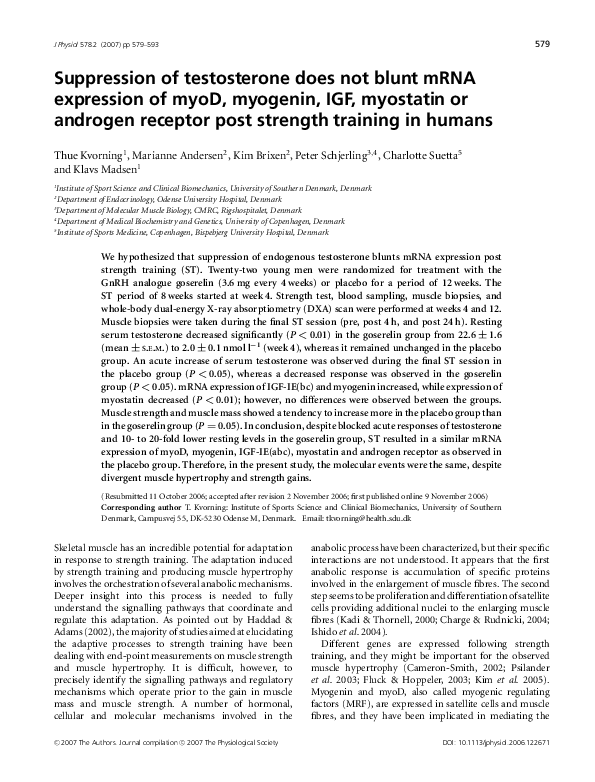
Changes in ‘housekeeping’ gene expression
To test the stability of the ‘housekeeping’ gene, GAPDH,
used for normalization of mRNA data, two other
‘housekeeping’ genes, 28S rRNA and RPLP0, were
measured and normalized to GAPDH. There was a slight
increase in 28S rRNA after the training period in the
goserelin group, and RPLP0 mRNA increased slightly 24 h
after the last training session in both groups (P < 0.05)
(Fig. 2). If either of the apparent changes in 28S rRNA
or RPLP0 mRNA expression was in reality due to a
change in the normalization gene (encoding GAPDH) this
would mean that GAPDH mRNA level would decrease.
However, from a biological point of view, an increase in
protein synthesis components (28S and RPLP0) is more
likely than a decrease in a glycolytic enzyme following
�584
T. Kvorning and others
strength training. Furthermore, the changes seen in the
other mRNAs were larger and can therefore not simply
be an artefact of the chosen normalizing gene encoding
GAPDH.
Whole-body DXA scan
Subjects were DXA scanned (Hologic 4500 A, Waltham,
MA, USA) before and after the training period (Tests 2
and 3). The DXA scan was conducted between 08.00 and
16.00 h and at least 24 h after training sessions (in order
to avoid any impact of changes in hydration). Regional
lean body mass was measured. The coefficient of variation
(CV) for lean body mass is 0.5–1%.
Isometric strength testing
Subjects were strength tested at Tests 1, 2 and 3. After
a 5 min standardized warm-up procedure on a bicycle
ergometer, the dominant leg was tested in a KinCom
dynamometer (KinCom 500H, software version 4.03;
Chattecx Corp., USA). The protocol implicated isometric
knee extensions performed at a locked position of 70◦ knee
flexion (0◦ = full extension). Subjects were instructed to
extend the knee as explosively and forcefully as possible and
three attempts were performed with maximal contraction
held for 3 s. A period of 45 s of recovery between trials
was given and the highest absolute value for isometric
10
measurements was used for further analysis. The isometric
measurements were sampled on an external computer with
a sampling rate of 1000 Hz and corrected for the influence
of gravity (Aagaard et al. 1995). All measurements were
filtered by a fourth-order zero-lag Butterworth low-pass
filter (10 Hz cut off frequency) and analysed for peak
torque. The isometric strength measurements at Test 1
were obtained to serve as control comparisons; however,
for the ease of illustration, only Tests 2 and 3 are
depicted.
Statistics
Differences in mean (pre and post the strength training
period) within or between groups were tested using
paired and unpaired t tests for mRNA expression
(following logaritmic transformation) and for strength
and DXA measurements and for training load and volume.
Measurements of acute hormonal responses were analysed
by two-way ANOVA repeated measurements. mRNA
expression measured pre, 4th and 24th post the strength
training session was analysed by two-way ANOVA repeated
measurements on logarithmic values. mRNA expression
values are presented as geometric means ± back
transformed s.e.m. in figures. All other data are presented
as means ± s.e.m. A significance level of P < 0.05 was
chosen. Statistical analyses were performed using Stat
View, SAS Institute 1998.
10
RPLP0
Goserelin
Placebo
RPLP0
Goserelin
Placebo
*
1
1
0.1
0.1
10
J Physiol 578.2
10
28S rRNA
Goserelin
Placebo
*
28S rRNA
Goserelin
Placebo
1
*
1
0.1
Pre
Post
Pre
Post 4 h
Post 24 h
Figure 2. Changes in 28S rRNA and RPLP0 mRNA
Changes in 28S rRNA and RPLP0 mRNA measured pre and post the strength training period and pre, 4 and 24 h
post the strength training session, respectively (geomean ± S.E.M.) (n = 10 in the goserelin group, and n = 7 in the
placebo group). ∗ Significant difference compared with the corresponding pre value (P < 0.05).
�
C 2007 The Physiological Society
C 2007 The Authors. Journal compilation �
�Gene expression, suppressed testosterone and strength training
J Physiol 578.2
585
Table 3. Resting levels of hormones
Hormone
Group
Test 1
Test 2
0.1∗
Test 3
Testosterone
(nmol l−1 )
Goserelin†
Placebo
22.6 ± 1.6
22.2 ± 1.4
2.0 ±
24.7 ± 1.7
1.1 ± 0.2∗
22.0 ± 1.5
Free testosterone
(nmol l−1 )
Goserelin†
Placebo
0.62 ± 0.03
0.60 ± 0.03
0.05 ± 0.00∗
0.69 ± 0.05
0.02 ± 0.00∗
0.57 ± 0.03
GH
(mU l−1 )
Goserelin
Placebo
0.33 ± 0.17 (n = 11)
0.30 ± 0.12
0.14 ± 0.02
0.27 ± 0.13
0.37 ± 0.11 (n = 11)
0.17 ± 0.09
GH, growth hormone. Values are means ± S.E.M. Test 1, before treatment; Test 2, after 3 weeks of
treatment with either goserelin or placebo and before strength training; Test 3, after the strength
training period. ∗ Significant different from Test 1 (P < 0.01). †Significant treatment effect compared with
placebo (P < 0.01).
Results
Baseline values
No significant differences were observed between the
groups regarding baseline values before the intervention
period (Test 1) or before the strength training period
(Test 2) in any of the variables measured.
Resting levels of serum testosterone, free
testosterone, GH, SHBG and cortisol
As previously published (Kvorning et al. 2006), the
change in serum endogenous testosterone levels differed
significantly between the groups (P < 0.01). Testosterone
remained constant in the placebo group throughout
the intervention period, but decreased significantly in
the goserelin group (P < 0.01). A similar difference
between the groups was observed for the endogenous
free testosterone levels (P < 0.01), with a decrease in the
goserelin group from Test 1 to Tests 2 and 3 (P < 0.01),
whereas it remained unchanged in the placebo group
(Table 3). There were no changes observed in the resting
levels of serum GH during the study (Table 3). The resting
levels of cortisol and SHBG remained also unchanged
throughout the intervention period (data not shown).
Acute hormonal response to strength training
sessions
The acute response of testosterone, free testosterone
and SHBG was similar at Tests 2 and 3. Only data
from Test 3 are shown, since they corresponded to the
measurements of acute mRNA expression. The placebo
group responded to the final strength training session with
a significant larger acute response in serum testosterone
compared with the goserelin group (P < 0.01). The level
of testosterone increased ∼15% immediately after the
strength training session in the placebo group (P < 0.05).
The goserelin group showed a decrease in testosterone
�
C 2007 The Physiological Society
C 2007 The Authors. Journal compilation �
and free testosterone 15 min post the strength training
session (P < 0.05). In addition, the level trended to be
below rest immediately after strength training (P = 0.05)
(Fig. 3). A significant acute increase from rest in the level
of SHBG was observed in the placebo group immediately
after strength training (P < 0.05). The increase in serum
SHBG in the placebo group, however, was not significantly
different from the goserelin group. No changes were
seen in serum SHBG in the goserelin group (Fig. 4).
There was no significant difference between the groups
regarding the acute response in serum GH at Test 3. Thus,
a significant acute increase from rest in the level of GH
was observed in the goserelin group immediately after
strength training and 15 min post training (P < 0.05).
The same picture was seen in the placebo group but the
change was only significant immediately after the training
session (P < 0.05) (Fig. 4). However, the goserelin group
showed significantly lower GH response during Test 2
compared with Test 3 (P < 0.01). Conversely, the placebo
group showed significantly higher GH response during
Test 2 compared with Test 3 (P < 0.05) (Fig. 5). Serum
cortisol showed no acute response at Test 2 or Test 3 for
any of the two groups (data not shown).
Resting mRNA expression measured pre and post the
strength training period
No differences were observed between the groups for
the resting mRNA expression measured pre and post
the strength training period. However, a significantly
increased expression of IGF-IEa, IGF-IEb and IGF-IEc
was seen in the goserelin group (P < 0.05). The placebo
group showed a significant increase for IGF-IEa (P < 0.05)
while IGF-IEb tended to increase (P = 0.07). There was a
significant increased expression of myogenin following the
strength training period in the goserelin group (P < 0.05),
whereas a trend toward decreased mRNA expression of
myostatin was observed (P = 0.06). Finally, no changes in
the mRNA expression of AR or myoD were seen after the
strength training period (Fig. 6).
�586
T. Kvorning and others
J Physiol 578.2
Figure 3. Acute responses of testosterone and free testosterone measured during the final strength
training session (Test 3)
Values are means ± S.E.M. ∗ Significant difference from the corresponding pre value (P < 0.05). #Significant
treatment effect compared with placebo (P < 0.01).
Acute mRNA expression measured pre and post the
strength training session
No differences were observed between the groups for
the acute mRNA expression measured pre and post the
strength training session. However, a significant increase
was seen in the goserelin group 4 h post strength training
regarding IGF-IEb and 24 h post training for IGF-IEc
(P < 0.05). The placebo group showed a trend to increase
24 h post training for IGF-IEb and IGF-IEc, with P values
of 0.07 and 0.05, respectively. Myostatin mRNA expression
decreased in both groups 4 h post strength training
(P < 0.05), and was still significantly reduced 24 h post
training in the goserelin group (P < 0.05). Both groups
showed increased mRNA expression for myogenin 4 h and
24 h post the strength training session (P < 0.05). There
were no changes in the mRNA expression of AR and myoD
after the strength training session (Fig. 7).
Figure 4. Acute responses of growth hormome (GH) and sex hormone binding globulin (SHBG) measured
during the final strength training session (Test 3)
Values are means ± S.E.M. (GH, Test 3, n = 11 in the goserelin group). ∗ Significant difference compared with the
corresponding pre value (P < 0.05).
�
C 2007 The Physiological Society
C 2007 The Authors. Journal compilation �
�Gene expression, suppressed testosterone and strength training
J Physiol 578.2
Isometric strength and lean leg mass
As previously published (Kvorning et al. 2006), only
the placebo group showed a significant increase in
isometric strength after 8 weeks of training (P < 0.05) and
the change trended to be higher in the placebo group
compared with goserelin group (P = 0.05). Lean leg mass
increased significant in both groups (P < 0.05). However,
the increase in the placebo group showed a trend to be
larger than the increase in the goserelin group (P = 0.05)
(Fig. 8).
Discussion
In the present study the use of a GnRH analogue effectively
suppressed the resting levels and blocked the acute increase
in serum testosterone in response to strength training. The
absence of the acute increase of testosterone, however, had
no influence on the acute mRNA expression of myoD,
myogenin, myostatin, IGF-IEa, IGF-IEb, IGF-IEc and AR
after the strength training session. Similarly, the lower
resting level of testosterone had no effect on the resting
mRNA expression before or after the strength training
period. Therefore, endogenous testosterone does not seem
to be involved in the transcriptional regulation of these
particular genes, which are supposed to be involved in
the adaptation to strength training. On the other hand,
suppression of the level of testosterone attenuates the
increase in lean mass and muscle strength. Therefore,
the important news in the present study is that the
molecular events were the same in spite of divergent muscle
hypertrophy and strength gains.
The acute changes in mRNA expression seen in our
study within the 24 h window are supported by previous
studies (Willoughby & Nelson, 2002; Hameed et al. 2003,
2004; Psilander et al. 2003; Kim et al. 2005; Coffey
et al. 2006). We found no changes, however, in the
expression of IGF-IEa and myoD in agreement with
previous observations (Hameed et al. 2003). The resting
mRNA expression of IGF-IEa, IGF-IEb, IGF-IEc and
myogenin increased after the strength training period and
the mRNA expression of myostatin trended to decrease.
Similar results have been obtained earlier (Roth et al. 2003;
Willoughby & Rosene, 2003; Hameed et al. 2004; Bickel
et al. 2005). The mRNA expression of myoD showed no
changes as previously observed by Bamman et al. (2004).
In accordance with earlier studies with similar strength
training programmes, the strength training session
induced significant acute increases in the level of
testosterone in the placebo group (Hakkinen & Pakarinen,
1993, Kraemer et al. 1998). The acute response of
testosterone was parallel by and acute increase in the
serum level of SHBG. On the other hand, the acute
response in the goserelin group, showed a decreased level
of testosterone. We can therefore relate the attenuated
response to the strength training period (e.g. less gain in
muscle mass and no gain in isometric muscle strength)
seen in the goserelin group to endogenous testosterone.
This implies that testosterone may regulate intracellular
factors downstream from myoD, myogenin, myostatin,
IGF-IEa, IGF-IEb and IGF-IEc mRNA transcription.
In addition, testosterone could alter post-translational
processes such as protein breakdown or efficiency of
intracellular amino acid utilization. In support of
this, a study without training intervention but with
suppression of endogenous testosterone showed that the
gene expression of actin and myosin were not altered;
however, both lean mass and muscle strength decreased
(Mauras et al. 1998). In addition, 4 weeks of functional
overload in rats was shown to have no effect on myoD
and myogenin expression even though lean mass was
increased (Mozdziak et al. 1998). On the other hand,
Mauras et al. (1998) reported decreases in IGF-I mRNA
expression with suppression of endogenous testosterone
Figure 5. Acute response of growth hormone (GH) measured during the first (Test 2) and final strength
training session (Test 3)
Values are means ± S.E.M. (Test 3, n = 11 in the goserelin group). ∗ Test 2 significantly different from Test 3
(P < 0.05). #Significant treatment effect compared with placebo (P < 0.05).
�
C 2007 The Physiological Society
C 2007 The Authors. Journal compilation �
587
�588
T. Kvorning and others
and argued that androgens are necessary for local IGF-I
production. The finding by Mauras et al. (1998) fits
well with the observation of Urban et al. (1995) and
Ferrando et al. (2002) where increasing testosterone levels
by supplementation in elderly men were associated with
increased IGF-I mRNA expression in skeletal muscle.
It was surprising to observe in the present study that
no changes took place in the expression of AR either at
rest or acute as a reaction to the dramatic changes of
endogenous testosterone. Bamman et al. (2001) registered
an increase in the expression of AR 48 h after a single
strength training session. Furthermore, Willoughby &
Taylor (2004) measured an increased mRNA expression
of AR 48 h after two sequential strength training sessions.
10
J Physiol 578.2
In both of the above-mentioned studies, biopsies were
performed 48 h post the training session, whereas in the
present study biopsies were taken 4 and 24 h post the
training session. Therefore, the expression of AR seems
to peak later than 24 h and timing of the biopsies may
explain the divergent results. Finally, when comparing
studies on gene expression, one must bear in mind
that the impact on expression of exercise performed
without prior familiarization or training is likely to
differ markedly from the response to repeated exercise
bouts or the trained response (Cameron-Smith, 2002;
Coffey et al. 2006). The pre training session biopsy
in the present study was taken 48 h after the previous
training session in the present study, but we cannot be
Myogenin
Goserelin
Placebo
*
1
10
10
AR
Goserelin
Placebo
MyoD
Goserelin
Placebo
1
1
0.1
0.1
10
10
IGF-IEa
Goserelin
Placebo
*
*
1
10
IGF-IEb
Goserelin
Placebo
*
1
1
IGF-IEc
Goserelin
Placebo
Myostatin
Goserelin
Placebo
*
1
0.1
Pre
Post
Pre
Post
Figure. 6. Changes in resting mRNA expression
Changes in resting mRNA expression measured pre and post the 8 weeks strength training period
(geomean ± S.E.M.) (n = 10 in the goserelin group, and n = 7 in the placebo group). ∗ Significant difference
compared with the corresponding pre value (P < 0.05). No treatment effect (goserelin versus placebo) was observed
in any of the genes.
�
C 2007 The Physiological Society
C 2007 The Authors. Journal compilation �
�Gene expression, suppressed testosterone and strength training
J Physiol 578.2
certain that this is a true baseline, since an elevated mRNA
expression may be present in response to the preceding
training session. We did not test whether the expression
of myoD, myogenin, IGF-IEa, IGF-IEb, IGF-IEc,
myostatin and AR was back to baseline 48 h post strength
training. Previous studies have shown divergent results
on this matter. Thus, studies demonstrate that expression
of the respective genes seems to peak in a 24 h window post
training (Psilander et al. 2003; Yang et al. 2005), whereas
other studies show that the genes may continue to be
upregulated 48 h post strength training (Roth et al. 2003;
Bickel et al. 2005).
An important observation in the present study was
that suppression of testosterone influenced the acute
10
589
response of GH in the goserelin group. In addition,
there seemed to be a trend towards a lower resting level
of GH after 3 weeks of GnRH analogue treatment, but
the level re-established after 8 weeks strength training.
These findings are congruent with the previous finding
by Mauras et al. (1987) where testosterone was shown
to influence GH secretion. However, suppression of
endogenous testosterone production had no significant
influence on the resting level or acute response of serum
cortisol. The trend towards a lower resting level and the
lower acute response of GH was only present in the initial
part of the strength training period, since the placebo and
goserelin group showed identical resting levels and acute
responses of GH at Test 3. In contrast, Mauras et al. (1998)
Myogenin
Goserelin
Placebo
*
*
*
*
1
10
10
AR
Goserelin
Placebo
1
1
0.1
0.1
10
10
IGF-IEa
Goserelin
Placebo
MyoD
Goserelin
Placebo
IGF-IEb
Goserelin
Placebo
1
*
0.1
10
1
1
IGF-IEc
Goserelin
Placebo
*
Myostatin
Goserelin
Placebo
1
0.1
*
*
*
Post 4 h
Post 24 h
0.1
Pre
Post 4 h
Post 24 h
Pre
Figure. 7. Changes in acute mRNA expression
Changes in acute mRNA expression measured pre, 4 and 24 h post the strength training session (geomean ± S.E.M.)
(n = 10 in the goserelin group, and n = 7 in the placebo group). ∗ Significant difference compared with the
corresponding pre value (P < 0.05). No treatment effect (goserelin versus placebo) was observed in any of the
genes.
�
C 2007 The Physiological Society
C 2007 The Authors. Journal compilation �
�590
T. Kvorning and others
found that suppression of testosterone by GnRH analogues
was not accompanied by decreases in GH concentration.
Instead the GH secretion increased after 10 weeks of
hypogonadism. Ultimately, these findings are interesting
since it has been postulated that GH and cortisol are
involved in the regulation of the mRNA expression of
IGF-I and myostatin (Rennie et al. 2004). In support of
this, Hameed et al. (2004) reported that GH treatment
increased IGF-IEa and IGF-IEc expression in the elderly
and myostatin expression has been shown to increase in
response to elevations in serum glucocorticoids (Lang et al.
2001; Ma et al. 2003). With these observations in mind, it
could be speculated that the lower acute increase in the
concentration of GH seen in the goserelin group during
the first strength training session may have affected the
acute IGF-IEa and IGF-IEc expression during the first
part of the strength training period, thus leading to a
lesser pronounced expression compared with the placebo
group where a larger acute increase in the level of GH was
present. However, similar serum cortisol levels in the
#
Isometric strength (Nm)
280
*
260
240
220
Test 2
Test 3
Test 2
Test 3
200
12
#
*
Lean leg mass (kg)
11
*
10
9
Test 2
Test 3
Test 2
Test 3
J Physiol 578.2
placebo and goserelin groups may help to explain why there
was no difference in the mRNA expression of myostatin
between the groups. In contrast to the goserelin group, the
placebo group showed a reduced acute response of GH
at Test 3 compared with Test 2. This is in accordance with
an earlier study (Ahtiainen et al. 2003), whereas Kraemer
et al. (1998) reported unchanged acute response to training
sessions after a training period.
Finally, it is important to stress that the relative
contribution of transcriptional versus translational
adaptations to strength training induced increase in
muscle hypertrophy is not well understood (CameronSmith, 2002). Thus, increased protein synthesis could
result from more mRNA molecules being translated or
from an increased rate of translation of each molecule
of mRNA. Chesley et al. (1992) demonstrated an
increased protein synthesis after strength training without
simultaneous increases in RNA content. In addition,
Welle et al. (1999) concluded that the stimulation of
protein synthesis by resistance exercise was mediated by
more efficient translation of mRNA. Consequently, a
translational mechanism may explain increased protein
synthesis without increases in mRNA expression (Bolster
et al. 2003). Therefore, caution must be applied to the
analysis of adaptive changes in both mRNA responses to
exercise and the impact of transcriptional compared with
translational events (Cameron-Smith, 2002). If hormonal
factors regulate the genes involved in the adaptation
process through translational events or post-translational
events, they were not detected in the present study. It may
be speculated that a decreased translation was present
in the goserelin group compared with the placebo
group, induced by the lack of acute response of
testosterone or/and by the low resting level of testosterone.
Furthermore, the effect of testosterone on transcription
may have occurred early in the training period and
not been detected, since muscle biopsies were taken
in relation to the final strength training session.
Transcriptional events may have occurred after the first
few training sessions and were attenuated later on when a
new steady-state level of protein was attained. On the other
hand, the present study does not exclude the possibility
that endogenous testosterone may regulate other hypertrophic signalling genes besides the one measured, or affect
other mechanisms responsible for gain in muscle mass
and muscle strength. These speculations are supported by
the observation that the goserelin group adapted to the
strength training period by attenuated increases in both
lean leg mass and isometric knee extension strength.
8
Goserelin
Placebo
Figure 8. Isometric strength and lean leg mass measured before
(Test 2) and after (Test 3) the strength training period
Values are means ± S.E.M.∗ Significant increase (P < 0.05). #P = 0.05
between groups.
Conclusions
In spite of both blocked acute responses and very
low resting levels of endogenous testosterone in the
GnRH-analogue-treated group, strength training resulted
�
C 2007 The Physiological Society
C 2007 The Authors. Journal compilation �
�J Physiol 578.2
Gene expression, suppressed testosterone and strength training
in a similar mRNA expression of myoD, myogenin,
IGF-IEa, IGF-IEb, IGF-IEc, myostatin and AR, as observed
in a placebo group showing acute responses of testosterone
to strength training and 10–20 times higher resting
levels of testosterone. Therefore, endogenous testosterone
does not seem to be involved in the regulation of the
expression of these previously established signalling genes
in the processes of strength-training-induced muscle
hypertrophy. On the other hand, suppression of the level
of endogenous testosterone attenuates the increase in
lean mass and muscle strength. Therefore, the important
finding in the present study is that the molecular events
were the same despite divergent muscle hypertrophy and
strength gains.
References
Aagaard P, Simonsen EB, Andersen JL, Magnusson P &
Dyhre-Poulsen P (2002). Increased rate of force development
and neural drive of human skeletal muscle following
resistance training. J Appl Physiol 93, 1318–1326.
Aagaard P, Simonsen EB, Trolle M, Bangsbo J & Klausen K
(1995). Isokinetic hamstring/quadriceps strength ratio:
influence from joint angular velocity, gravity correction
and contraction mode. Acta Physiol Scand 4, 421–427.
Adams GR (1998). Role of insulin-like growth factor-I in the
regulation of skeletal muscle adaptation to increased loading.
Exerc Sport Sci Rev 26, 31–60.
Ahtiainen JP, Pakarinen A, Alen M, Kraemer WJ & Hakkinen K
(2003). Muscle hypertrophy, hormonal adaptations and
strength development during strength training in
strength-trained and untrained men. Eur J Appl Physiol 89,
555–563.
Bamman MM, Ragan RC, Kim JS, Cross JM, Hill VJ, Tuggle SC
& Allman RM (2004). Myogenic protein expression before
and after resistance loading in 26 and 64-yr-old men and
women. J Appl Physiol 97, 1329–1337.
Bamman MM, Shipp JR, Jiang J, Gower BA, Hunter GR,
Goodman A, McLafferty CL Jr & Urban RJ (2001).
Mechanical load increases muscle IGF-I and androgen
receptor mRNA concentrations in humans. Am J Physiol
Endocrinol Metab 280, 383–390.
Bartsch W (1980). Interrelationships between sex hormonebinding globulin and testosterone, 5 alphadihydrotestosterone and oestradiol-17 beta in blood of
normal men. Maturitas 2, 109–118.
Bergström J (1962). Muscle electrolytes in man. Scand J Clin
Lab Invest 68, 1–110.
Bhasin S, Storer TW, Berman N, Callegari C, Clevenger B,
Phillips J, Bunnell TJ, Tricker R, Shirazi A & Casaburi R
(1996). The effects of supraphysiologic doses of testosterone
on muscle size and strength in normal men. Engl J Med 335,
1–7.
Bhasin S, Woodhouse L, Casaburi R, Singh AB, Bhasin D,
Berman N, Chen X, Yarasheski KE, Magliano L, Dzekov C,
Dzekov J, Bross R, Phillips J, Sinha-Hikim I, Shen R & Storer
TW (2001). Testosterone dose–response relationships in
healthy young men. Am J Physiol Endocrinol Metab 281,
1172–1181.
�
C 2007 The Physiological Society
C 2007 The Authors. Journal compilation �
591
Bickel CS, Slade J, Mahoney E, Haddad F, Dudley GA & Adams
GR (2005). Time course of molecular responses of human
skeletal muscle to acute bouts of resistance exercise. J Appl
Physiol 98, 482–488.
Bolster DR, Kimball SR & Jefferson LS (2003). Translational
control mechanisms modulate skeletal muscle gene expression during hypertrophy. Exerc Sport Sci Rev 31, 111–116.
Braith RW, Graves JE, Pollock ML, Leggett SL, Carpenter DM
& Colvin AB (1989). Comparison of 2 vs 3 days/week of
variable resistance training during 10- and 18-week
programs. Int J Sports Med 10, 450–454.
Cameron-Smith D (2002). Exercise and skeletal muscle gene
expression. Clin Exp Pharmacol Physiol 29, 209–213.
Charge SB & Rudnicki MA (2004). Cellular and molecular
regulation of muscle regeneration. Physiol Rev 84, 209–238.
Chesley A, MacDougall JD, Tarnapolsky MA, Atkinson SA &
Smith K (1992). Changes in human muscle protein synthesis
after exercise. J Appl Physiol 73, 1383–1388.
Cockshott ID (2000). Clinical pharmacokinetics of goserelin.
Clin Pharmacokinet 39, 27–48.
Coffey VG, Shield A, Canny BJ, Carey KA, Cameron-Smith D &
Hawley JA (2006). Interaction of contractile activity and
training history on mRNA abundance in skeletal muscle
from trained athletes. Am J Physiol Endocrinol Metab 290,
849–855.
Dorlochter M, Astrow SH & Herrera AA (1994). Effects of
testosterone on a sexually dimorphic frog muscle: repeated in
vivo observations and androgen receptor distribution.
J Neurobiol 25, 897–916.
Doumit ME, Cook DR & Merkel RA (1996). Testosterone
up-regulates androgen receptors and decreases
differentiation of porcine myogenic satellite cells in vitro.
Endocrinology 137, 1385–1394.
Ferrando AA, Sheffield-Moore M, Yeckel CW, Gilkison C, Jiang
J, Achacosa A, Lieberman SA, Tipton K, Wolfe RR & Urban
RJ (2002). Testosterone administration to older men
improves muscle function: molecular and physiological
mechanisms. Am J Physiol Endocrinol Metab 282, 601–607.
Fluck M & Hoppeler H (2003). Molecular basis of skeletal
muscle plasticity – from gene to form and function. Rev
Physiol Biochem Pharmacol 146, 159–216.
Glowacki SP, Martin SE, Maurer A, Baek W, Green JS & Crouse
SF (2004). Effects of resistance, endurance, and concurrent
exercise on training outcomes in men. Med Sci Sports Exerc
36, 2119–2127.
Goldspink G (1999). Changes in muscle mass and phenotype
and the expression of autocrine and systemic growth factors
by muscle in response to stretch and overload. J Anat 194,
323–334.
Haddad F & Adams GR (2002). Selected contribution: acute
cellular and molecular responses to resistance exercise. J Appl
Physiol 93, 394–403.
Hakkinen K & Pakarinen A (1993). Acute hormonal responses
to two different fatiguing heavy-resistance protocols in male
athletes. J Appl Physiol 74, 882–887.
Hameed M, Lange KH, Andersen JL, Schjerling P, Kjaer M,
Harridge SD & Goldspink G (2004). The effect of
recombinant human growth hormone and resistance
training on IGF-I mRNA expression in the muscles of elderly
men. J Physiol 555, 231–240.
�592
T. Kvorning and others
Hameed M, Orrell RW, Cobbold M, Goldspink G & Harridge
SD (2003). Expression of IGF-I splice variants in young and
old human skeletal muscle after high resistance exercise.
J Physiol 547, 247–254.
Hansen S, Kvorning T, Kjaer M & Sjoegaard G (2001). The
effect of short-term strength training on human skeletal
muscle: the importance of physiologically elevated hormone
levels. Scand J Med Sci Sports 11, 347–354.
Harridge SD (2003). Ageing and local growth factors in muscle.
Scand J Med Sci Sports 13, 34–39.
Hespel P, Op’t Eijnde B, Van Leemputte M, Urso B, Greenhaff
PL, Labarque V, Dymarkowski S, Van Hecke P & Richter EA
(2001). Oral creatine supplementation facilitates the
rehabilitation of disuse atrophy and alters the expression of
muscle myogenic factors in humans. J Physiol 536, 625–633.
Hickson RC, Hidaka K, Foster C, Falduto MT, Chatterton RT &
JR (1994). Successive time courses of strength development
and steroid hormone responses to heavy-resistance training.
J Appl Physiol 76, 663–670.
Inoue K, Yamasaki S, Fushiki T, Okada Y & Sugimoto E (1994).
Androgen receptor antagonist suppresses exercise-induced
hypertrophy of skeletal muscle. Eur J Appl Physiol 69, 88–91.
Ishido M, Kami K & Masuhara M (2004). Localization of
MyoD, myogenin and cell cycle regulatory factors in
hypertrophying rat skeletal muscles. Acta Physiol Scand 180,
281–289.
Kadi F, Schjerling P, Andersen LL, Charifi N, Madsen JL,
Christensen LR & Andersen JL (2004). The effects of heavy
resistance training and detraining on satellite cells in human
skeletal muscles. J Physiol 558, 1005–1012.
Kadi F & Thornell LE (2000). Concomitant increases in
myonuclear and satellite cell content in female trapezius
muscle following strength training. Histochem Cell Biol 113,
99–103.
Kim JS, Cross JM & Bamman MM (2005). Impact of resistance
loading on myostatin expression and cell cycle regulation in
young and older men and women. Am J Physiol Endocrinol
Metab 288, 1110–1119.
Kraemer WJ, Adams K, Cafarelli E, Dudley GA, Dooly C,
Feigenbaum MS, Fleck SJ, Franklin B, Fry AC, Hoffman JR,
Newton RU, Potteiger J, Stone MH, Ratamess NA &
Triplett-McBride T (2002). American College of Sports
Medicine. American College of Sports Medicine position
stand. Progression models in resistance training for healthy
adults. Med Sci Sports Exerc 34, 364–380.
Kraemer WJ, Häkkinen K, Newton RU, Nindl BC, Volek JS,
McCormick M, Gotshalk LA, Gordon SE, Fleck SJ, Campbell
WW, Putukian M & Evans WJ (1999). Effects of
heavy-resistance training on hormonal response patterns in
younger vs. older men. J Appl Physiol 87, 982–992.
Kraemer WJ, Marchitelli LJ, Gordon SE, Harman E, Dziados
JE, Friedl K, Maresh C, Fry AC, Mello P & Fleck SJ (1991).
Endogenous anabolic hormonal and growth factor responses
to heavy resistance exercise in males and females. Int J Sports
Med 12, 228–235.
Kraemer WJ, Marchitelli LJ, Gordon SE, Harman E, Dziados
JE, Frykman P, McCurry D & Fleck SJ (1990). Hormonal
and growth factor responses to heavy resistance exercise
protocols. J Appl Physiol 69, 1442–1450.
J Physiol 578.2
Kraemer WJ, Marchitelli LJ, Gordon SE, Harman E, Dziados
JE, Frykman P, Mello P, Koziris LP, Triplett NT & Fleck SJ
(1993). Changes in hormonal concentrations after different
heavy-resistance exercise protocols in women. J Appl Physiol
75, 594–604.
Kraemer WJ, Patton JF, Gordon SE, Harman EA, Deschenes
MR, Reynolds K, Newton RU, Triplett NT & Dziados JE
(1995). Compatibility of high-intensity strength and
endurance training on hormonal and skeletal muscle
adaptations. J Appl Physiol 78, 976–989.
Kraemer WJ & Ratamess NA (2005). Hormonal responses and
adaptations to resistance exercise and training. Sports Med
35, 339–361.
Kraemer WJ, Staron RS, Hagerman FC, Hikida RS,
Fry AC, Gordon SE, Nindl BC, Gothshalk LA, Volek JS,
Marx JO, Newton RU & Häkkinen K (1998). The effects
of short-term resistance training on endocrine function
in men and women. Eur J Appl Physiol 78,
69–76.
Kvorning T, Andersen M, Brixen K & Madsen K (2006).
Suppression of endogenous testosterone production
attenuates the response to strength training; a randomized,
placebo-controlled and blinded intervention study.
Am J Physiol Endocrinol Metab 291, 1325–1332.
Lang CH, Silvis C, Nystrom G & Frost RA (2001). Regulation of
myostatin by glucocorticoids after thermal injury. FASEB J
15, 1807–1809.
Lykkesfeldt G, Bennett P, Lykkesfeldt AE, Micic S, Moller S &
Svenstrup B (1985). Abnormal androgen and oestrogen
metabolism in men with steroid sulphatase deficiency
and recessive X-linked ichthyosis. Clin Endocrinol 4,
385–393.
Ma K, Mallidis C, Bhasin S, Mahabadi V, Artaza J,
Gonzalez-Cadavid N, Arias J & Salehian B (2003).
Glucocorticoid-induced skeletal muscle atrophy is associated
with upregulation of myostatin gene expression. Am J Physiol
Endocrinol Metab 285, 363–371.
Mauras N, Blizzard RM, Link K, Johnson ML, Rogol AD &
Veldhuis JD (1987). Augmentation of growth hormone
secretion during puberty: evidence for a pulse
amplitude-modulated phenomenon. J Clin Endocrinol Metab
64, 596–601.
Mauras N, Hayes V, Welch S, Rini A, Helgeson K, Dokler M,
Veldhuis JD & Urban RJ (1998). Testosterone deficiency in
young men: Marked alterations in whole body protein
kinetics, strength and adiposity. J Clin Endocrinol Metab 83,
1886–1892.
McPherron AC & Lee SJ (1997). Double muscling in cattle due
to mutations in the myostatin gene. Proc Natl Acad Sci U S A
94, 12457–12461.
Moore DR, Phillips SM, Babraj JA, Smith K & Rennie MJ
(2005). Myofibrillar and collagen protein synthesis in human
skeletal muscle in young men after maximal shortening and
lengthening contractions. Am J Physiol Endocrinol Metab 6,
1153–1159.
Mozdziak PE, Greaser ML & Schultz E (1998). Myogenin,
MyoD, and myosin expression after pharmacologically and
surgically induced hypertrophy. J Appl Physiol 84,
1359–1364.
�
C 2007 The Physiological Society
C 2007 The Authors. Journal compilation �
�J Physiol 578.2
Gene expression, suppressed testosterone and strength training
Narici MV, Hoppeler H, Kayser B, Landoni L, Claassen H,
Gavardi C, Conti M & Cerretelli P (1996). Human
quadriceps cross-sectional area, torque and neural activation
during 6 months strength training. Acta Physiol Scand 157,
175–186.
Psilander N, Damsgaard R & Pilegaard H (2003). Resistance
exercise alters MRF and IGF-I mRNA content in human
skeletal muscle. J Appl Physiol 95, 1038–1044.
Reisz-Porszasz S, Bhasin S, Artaza JN, Shen R, Sinha-Hikim I,
Hogue A, Fielder TJ & Gonzalez-Cadavid NF (2003). Lower
skeletal muscle mass in male transgenic mice with
muscle-specific overexpression of myostatin. Am J Physiol
Endocrinol Metab 285, 876–888.
Rennie MJ, Wackerhage H, Spangenburg EE & Booth FW
(2004). Control of the size of the human muscle mass. Annu
Rev Physiol 66, 799–828.
Roth SM, Martel GF, Ferrell RE, Metter EJ, Hurley BF & Rogers
MA (2003). Myostatin gene expression is reduced in humans
with heavy-resistance strength training: a brief
communication. Exp Biol Med 228, 706–709.
Storer TW, Magliano L, Woodhouse L, Lee ML, Dzekov C,
Dzekov J, Casaburi R & Bhasin S (2003). Testosterone
dose-dependently increases maximal voluntary strength and
leg power, but does not affect fatigability or specific tension.
J Clin Endocrinol Metab 88, 1478–1485.
Urban RJ, Bodenburg YH, Gilkison C, Foxworth J, Coggan AR,
Wolfe RR & Ferrando A (1995). Testosterone administration
to elderly men increases skeletal muscle strength and protein
synthesis. Am J Physiol Endocrinol Metab 269, 820–826.
Welle S, Bhatt K & Thornton CA (1999). Stimulation of
myofibrillar synthesis by exercise is mediated by more
efficient translation of mRNA. J Appl Physiol 86,
1220–1225.
�
C 2007 The Physiological Society
C 2007 The Authors. Journal compilation �
593
Willoughby DS (2004). Effects of heavy resistance training on
myostatin mRNA and protein expression. Med Sci Sports
Exerc 36, 574–582.
Willoughby DS & Nelson MJ (2002). Myosin heavy-chain
mRNA expression after a single session of heavy-resistance
exercise. Med Sci Sports Exerc 34, 1262–1269.
Willoughby DS & Rosene JM (2003). Effects of oral creatine
and resistance training on myogenic regulatory factor
expression. Med Sci Sports Exerc 35, 923–929.
Willoughby DS & Taylor L (2004). Effects of sequential bouts of
resistance exercise on androgen receptor expression. Med Sci
Sports Exerc 36, 1499–1506.
Yang Y, Creer A, Jemiolo B & Trappe S (2005). Time course of
myogenic and metabolic gene expression in response to
acute exercise in human skeletal muscle. J Appl Physiol 5,
1745–1752.
Yang SY & Goldspink G (2002). Different roles of the IGF-I Ec
peptide (MGF) and mature IGF-I in myoblast proliferation
and differentiation. FEBS Lett 522, 156–160.
Acknowledgements
First of all we would like to thank the subjects who participated
in the study. Secondly, we would like to thank the laboratory
technicians Gitte Scheel Klemmensen, Bente Tøt, Donna
Arbuckle-Lund, Kirsten Westermann and Anette Riis Madsen.,
engineer Cuno Rasmussen, Professor Per Aagaard, PhD student
Anders Holsgaard Larsen, and the students Emil Pedersen and
Jacob Søndergaard, for their helpful cooperation during the
study. We would like to thank Anti Doping Denmark and the
Team Denmark Foundation for their financial support.
�
 Charlotte Suetta