Hindawi
Journal of Ophthalmology
Volume 2018, Article ID 6390706, 5 pages
https://doi.org/10.1155/2018/6390706
Clinical Study
Effect of Reformation of the Anterior Chamber by Air or by a
Balanced Salt Solution (BSS) on Corneal Endothelium after
Phacoemulsification: A Comparative Study
Alahmady Hamad Alsmman ,1 Mohammed Ezzeldawla,1 Amr Mounir ,1
Ashraf Mostafa Elhawary,1 Osama Ali Mohammed,1 Mahmoud Farouk ,1
and Ahmed Mohamed Sherif 2
1
2
Department of Ophthalmology, Sohag Faculty of Medicine, Sohag University, Sohag, Egypt
Department of Ophthalmology, Cairo Faculty of Medicine, Cairo University, Cairo, Egypt
Correspondence should be addressed to Alahmady Hamad Alsmman; alahmady20@yahoo.com
Received 5 November 2017; Accepted 18 March 2018; Published 8 April 2018
Academic Editor: Van C. Lansingh
Copyright © 2018 Alahmady Hamad Alsmman et al. This is an open access article distributed under the Creative Commons
Attribution License, which permits unrestricted use, distribution, and reproduction in any medium, provided the original work
is properly cited.
Aim. To study the effect of reformation of the anterior chamber by air or by a balanced salt solution, after smooth
phacoemulsification on the corneal endothelial count and morphology. Methods. A prospective interventional nonrandomized
comparative study included 500 eyes of 500 patients with age range between 50 and 60 years, prepared for cataract surgery and
presented to the Ophthalmology department of Sohag University Hospital in the period from October 2016 to May 2017.
Corneal endothelial morphology and count were examined, and the results were recorded for all cases before the surgery.
Patients were divided into two groups, and both groups were diagnosed with grade 2 cataract and underwent uncomplicated
phacoemulsification performed by well-trained surgeons. At the end of the surgery, group 1 was subjected to a reformation of
the anterior chamber via a balanced salt solution (BSS) injection while group 2 was subjected to a reformation of the anterior
chamber via air injection. Corneal endothelial morphology and count were evaluated in the first and 3rd month postoperatively.
Results. The study included 500 patients (250 in each group), 220 males (44%) and 280 females (56%) with no significant
statistical age differences. Both preoperative and postoperative (3 months after the operation) recorded parameters of the
corneal endothelium did not show any significant statistical differences. The cumulative dissipated energy was recorded, for all
cases of both groups, during phacoemulsification with no significant statistical differences (P = 0 7). Conclusion. There is no
difference between the effect of reformation of the anterior chamber after phacoemulsification, using air or using a BSS
injection, on the corneal endothelial count and morphology.
1. Introduction
Cataract surgery is one of the most frequently performed surgeries worldwide. It is well established that this surgery has a
negative effect on corneal endothelium, as it decreases the
endothelial cell count. The severity of the affection depends
on many variables, as phacoemulsification time and energy,
surgical technique, anterior chamber depth, and the use of
ophthalmic viscoelastic devices (OVDs) [1].
The corneal endothelium functions as an active pump
and also as a barrier against the aqueous humor of the anterior chamber; thus, it holds an important role in the process
�2
of corneal tissue hydration. It has no ability of regeneration,
so any decrease in its density is irreversible and can lead to
permanent blurring of vision and pain [2].
The corneal injury caused by phacoemulsification usually
leads to corneal edema, and if it is severe enough, it might
result in irreversible bullous keratopathy, making corneal
tissue transplantation the only effective treatment [2].
Corneal endothelial injury associated with phacoemulsification is assessed by specular microscopy through measuring
changes of the cell density (CD) and the cell morphology [3].
Air injection has become widely used in many anterior
segment surgeries [4], for example, restoration of normal
intraocular pressure and reformation of the anterior chamber
and many other surgical procedures [5].
Direct contact between gases and the corneal endothelial
layer is not natural, and many experimental and clinical studies have proved the occurrence of corneal injury as a result of
air injection into the anterior chamber [6].
Nowadays, most surgeons prefer to use a balanced salt
solution to avoid the harms of air injection, even though
there were no reported complications of air injection over
the long term [7].
This study aims at comparing the effect of reformation of
the anterior chamber after phacoemulsification, using air and
BSS injection, on corneal endothelial count and morphology.
2. Patient and Methods
This is a prospective interventional nonrandomized comparative study, which included 500 eyes of 500 patients with age
range between 50 and 60 years, prepared for cataract surgery
and presented to the Ophthalmology department of Sohag
University Hospital in the period from October 2016 to
May 2017. The study only included cases diagnosed with
grade 2 cataract according to the Lens Opacities Classification System III (LOCS III) [8].
Exclusion criteria included the following: patients aged
less than 50 years or more than 60 years; patients with a
history of previous corneal pathology, pseudoexfoliation,
ocular trauma or intraocular surgery, or intraocular inflammation; and patients having preoperative endothelial cell
count < 1500 cells per square millimeter, preoperative
anterior chamber depth < 2.5 mm, or short axial length
eyes < 21 mm and long axial length > 25 mm.
The ethical committee of Sohag University approved this
study protocol, which was carried out according to the
Declaration of Helsinki. A written informed consent was
obtained from each included case.
All patients were divided into two groups; group 1 was
subjected to phacoemulsification with a reformation of the
anterior chamber using a balanced salt solution (BSS) injection at the end of the surgery, while group 2 was subjected
to phacoemulsification with a reformation of the anterior
chamber by air injection at the end of the surgery.
Patients who fit the criteria were allocated to each group
in turn.
All patients were subjected to a full ophthalmological
evaluation before the operation The evaluation included a
slit-lamp examination, measurement of the best corrected
Journal of Ophthalmology
visual acuity, measurement of the intraocular pressure
(IOP), and specular microscopic examination (using Specular Microscope, SP-3000P, Topcon, Tokyo, Japan, with the
IMAGEnet system (version 2.1, Topcon)). Also, central corneal endothelium morphology assessment was conducted,
which includes central corneal thickness (CCT), endothelial
cell density (ECD), corneal endothelial cell size variations
as the percentage of the abnormal sizes (corneal polymegathism), and corneal cell shape variations as the percentage of
the hexagonal cells (corneal pleomorphism). Postoperative
follow-up occurred on the first day (after 6 hours), second
day, one week, and one month and after three months.
Specular microscopic examination postoperatively occurred
twice after one month and three months.
All patients received the same regimen of 1 drop of
cyclopentolate 1%, 1 drop of phenylephrine 10%, and 1
drop of diclofenac 0.1% 20 minutes before surgery. Also,
a dose of 5-6 ml lidocaine hydrochloride 2% with adrenaline
1 : 200,000 was used for the peribulbar anesthesia.
All operations were performed by three well-experienced
surgeons (first three authors) using the standard divide and
conquer technique and the same phacoemulsification equipment (INFINITI Vision System; Alcon Laboratories Inc.,
Fort Worth, TX, USA) at similar settings. Any cases that
developed intraoperative complications were excluded from
the study.
The patients were allocated to different surgeons in turn,
the first surgeon 167 eyes, second surgeon 167 eyes, and third
surgeon 166 eyes.
All cases were operated on using the same standardized
surgical technique, which includes the use of a sterile drape
with speculum, application of the corneal topical anesthesia
(lidocaine 2% in gel suspension), performance of a 2.75 mm
self-sealing corneal incision, injection of a viscoelastic agent
(Healon; Advanced Medical Optics Inc., Santa Ana, CA),
application of capsulorrhexis, application of hydrodissection,
application of the conventional phacoemulsification (longitudinal ultrasound) (divide and conquer), irrigation and
aspiration of the cortical material, introduction of viscoelastic in a bag to implant a foldable acrylic lens (AcrySof
SA60AT; Alcon Laboratories), implantation of the lens
through a 2.8 mm incision with the use of injector, and aspiration of the viscoelastic. Closure of the incision was done by
hydration using a 30-gauge cannula with no sutures. The
cumulative dissipated energy (CDE; phaco energy) was documented for all patients.
In the postoperative period, all patients received the same
treatment including topical antibiotic (moxifloxacin) at a rate
of 5 times per day for one week and prednisolone acetate at a
rate of 5 times per day for one week with a gradual decrease
in the second week, then replaced by nonsteroidal antiinflammatory drops at a rate of three times per day for the
following two weeks.
2.1. Statistical Analysis. The data were analyzed using SPSS
for Windows version 18.0 software (SPSS Inc., Chicago, IL,
USA). The data are shown as the mean and standard deviation. The results were analyzed using Student’s t-test to compare the mean values of both groups. The qualitative data was
�Journal of Ophthalmology
3
Table 1: Preoperative demographics and corneal endothelial parameters.
Sample size (male : female)
Age (year)
CDE (joules)
Cell density (cells/mm2)
Coefficient of variance % (polymegathism)
Hexagonality % (pleomorphism)
Central corneal thickness (micron)
Group 1 (BSS)
Mean ± SD
Range
Mean ± SD
Group 2 (air)
Range
P value
250 (120 : 130)
56 ± 2.3
52 to 60
7.19 ± 1.1
4.96 to 9.14
2646 ± 284
1858 to 2890
38.2 ± 6
27 to 49
49 ± 8
39 to 64
508 ± 22
474 to 571
57.9 ± 3.9
6.51 ± 1
2604 ± 367
40.3 ± 8.4
51 ± 10
509 ± 34
250 (100 : 150)
53 to 60
4.97 to 8.52
1998 to 3209
31 to 61
34 to 73
450 to 571
0.1
0.7
0.16
0.8
0.3
0.28
Table 2: Pre- and postoperative corneal endothelial parameters.
Group 1 (BSS)
Mean ± SD
Post 1
Preoperative
month
Cell density (cells/mm2)
Endothelial cell loss
Coefficient of variance %
(polymegathism)
Hexagonality % (pleomorphism)
Central corneal thickness, micron
2646 ± 284.1
Group 2 (Air)
Mean ± SD
Post 1
Preoperative
month
Post 3
month
2479 ± 303
189 ± 132
7.1%
2605 ± 367
—
2523 ±311
146 ± 84
5.5%
38.2 ± 6
1644±
49 ± 8
508 ± 22
39 ± 15
518 ± 23
expressed in the form of numbers and percentages and compared using the chi-square test. The multivariable regression
analysis was done to identify the different corneal endothelial
parameters. P value was considered significant if it was less
than 0.05.
Post 3
month
P value
2424 ± 336
172 ± 95
6.6%
0.16
—
2465 ± 351
164 ± 125
6.3%
39.8 ± 10
40.3 ± 8.4
35.8 ± 11
34.4 ± 11
0.8
45 ± 10
508 ± 20
51 ± 10
509 ± 34
42 ± 8
530 ± 40
43 ± 9
510 ± 41
0.3
0.28
0.1
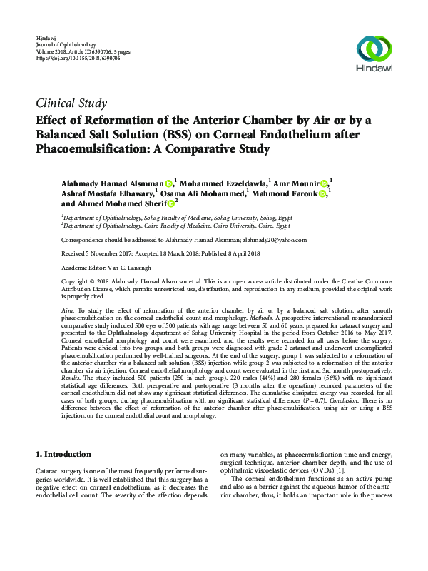
2700.
2625.
2550.
2475.
3. Results
2400.
The study included 500 patients (250 on each group), 220
males (44%) and 280 females (56%); group 1 included 120
males and 130 females with age range between 52 and 60
years, while group 2 included 100 males and 150 females with
age range between 53 and 60 years. No significant statistical
differences were recorded in the preoperative data about the
age and the corneal parameters which include endothelial cell
density, the coefficient of variance, hexagonality, and central
corneal thickness (Table 1).
The cumulative dissipated energy was recorded for all
cases during the phacoemulsification; the mean CDE in
group 1 was 7.19 with SD 1.1, while the mean CDE in group
2 was 6.51 with SD 1. There was no significant statistical
difference between both groups with P value = 0.7 (Table 1).
In group 1, the mean endothelial cell loss was 146 with
SD 84 in the first month and 189 with SD 132 in the third
month, while in group 2, the mean endothelial cell loss was
164 with SD 125 in the first month and 172 with SD 95 in
the third month, with P value = 0.1 for both groups at the
3rd month.
2325.
2250.
M3
M1
Preop
Endothelial cell density (air)
Endothelial cell density (BSS)
Figure 1: Pre- and post mean endothelial cell density.
There were no operative complications reported for all
cases. There was no significant statistical difference between
both groups regarding the corneal endothelial data in the
3rd month postoperatively. The postoperative corneal
parameters are summarized in Table 2 and Figures 1–3.
4. Discussion
Normally the cornea is transparent. This state is maintained
by the corneal endothelium, which keeps the corneal stroma
�4
Journal of Ophthalmology
55.
44.
33.
22.
11.
0.
M3
M1
Preop
Corneal endothelial variation (air)
Corneal endothelial variation (BSS)
Figure 2: Pre- and post mean corneal endothelial variation.
60
45
30
15
0
M3
M1
Preop
Corneal endothelial hexagonality (air)
Corneal endothelial hexagonality (BSS)
Figure 3: Pre- and post mean corneal endothelial hexagonality.
continuously dehydrated by acting as a barrier and an active
fluid pump. This essential function is easily compromised by
any damage that could happen during any eye surgery, especially phacoemulsification surgeries. This has prompted
many studies to compare the severity of the damage that
results from different cataract operation techniques [9].
Although the safety of the phacoemulsification has been
markedly improved, the prevention of the corneal endothelial damage during phacoemulsification is still an important
interest for all cataract surgeons [10].
In this study, we investigated the effect of reformation of
the anterior chamber by air injection and by BSS injection on
different corneal endothelial cell parameters, which were
evaluated using the specular microscope to detect any
damage resulting from the injection.
All preoperative data as regards age, the degree of
cataract, and corneal endothelial cell parameters showed no
statistical significance between both groups. Patients with
other factors which might affect the corneal endothelial
count were excluded, for example, patients older than 60
years and patients with advanced grades of cataract [11].
Sutureless corneal wounds have become the standard
technique in cataract surgery, based on the fact that a watertight wound is an airtight wound and not vice versa. Hence,
air injection is used for the reformation of the anterior chamber after phacoemulsification [12]. However, some studies
reported air leakiness in 1/3 of the included cases [13].
In this study, we proved that there is no statistically
significant difference between air and BSS injection in the
anterior chamber reformation.
Our results agree with the study of Galin et al. [14]
which were performed on rabbits’ eyes. The authors examined the effect of air injection in the anterior chamber on
the corneal endothelium. They used a light microscope and
an electronic microscope for their study. They reported that
the presence of air in the anterior chamber in contact with
the corneal endothelium has no toxic effect on the corneal
endothelium but even stimulates the proliferation of the
corneal endothelial cells.
Also, our results are similar to the results of the Ventura
et al. study [15], who confirmed that the air has no damaging
effect on the corneal endothelium of the cat. Our results
coincide with the results of the Norn study [5]. Norn studied
the effect of reformation of the anterior chamber, after cataract extraction, using air injection on the corneal endothelium of humans. His study included an examination of the
patients before and after the surgery. He proved that the corneal thickness was thinner in patients injected with air with
no other adverse effects over a six-month period following
the operation.
This study disagreed with the study of Olson et al. [16],
who compared the effect of air and balanced saline solution
injection into the anterior chamber on the corneal endothelium of cats. They reported a significant decrease in the endothelial cell density after air injection into the anterior
chamber. He also noted significant endothelial damage
during corneal perfusion studies.
Corneal pachymetry is assessed as the occurrence of
edema is an indirect tool to evaluate corneal endothelial
changes. It is important in cases of surgically induced endothelial cell loss [17].
Although, in this study, an initial increase in the corneal thickness caused by postoperative edema was reported.
But the difference between both groups was not of statistically significance.
A significant positive correlation was found in many
studies between the endothelial cell loss and the nuclear
sclerosis grade, also between the endothelial cell loss and
phacoemulsification power and time [18]. So only cases with
grade 2 cataract were included in this study. Also, the power
used in the surgery did not show a significant difference
between both groups. So the endothelial cell loss reported
in our study was not affected by the previous factors, and as
a result, the effect of air on the corneal endothelium had
not been masked by any factor.
Corneal endothelial parameters as regards cell density,
endothelial cell loss, hexagonality, and coefficient of variance
did not show any significant difference between both groups,
which means that the reformation of the anterior chamber by
air injection has no toxic effect on the corneal endothelium.
�Journal of Ophthalmology
This was expected as thousands of phacoemulsification
surgeries with air use in the anterior chamber reformation
have been performed in our society “South Egypt” with
satisfactory results.
5. Conclusions
There is no difference between the effect of reformation
of the anterior chamber, after phacoemulsification, using
air or using BSS injection on the corneal endothelial
count and morphology. Also, there is no reported toxic
effect of air on corneal endothelial parameters evaluated
by specular microscope.
Conflicts of Interest
The authors declare that they have no conflicts of interest.
References
[1] I. Conrad-Hengerer, F. H. Hengerer, T. Schultz, and H. B.
Dick, “Effect of femtosecond laser fragmentation on effective
phacoemulsification time in cataract surgery,” Journal of
Refractive Surgery, vol. 28, no. 12, pp. 879–884, 2012.
[2] T. Igarashi, I. Ohsawa, M. Kobayashi et al., “Hydrogen prevents corneal endothelial damage in phacoemulsification cataract surgery,” Scientific Reports, vol. 6, no. 1, article 31190,
2016.
[3] G. O. Waring III, W. M. Bourne, H. F. Edelhauser, and K. R.
Kenyon, “The corneal endothelium. Normal and pathologic
structure and function,” Ophthalmology, vol. 89, no. 6,
pp. 531–590, 1982.
[4] H. Landry, A. Aminian, L. Hoffart et al., “Corneal endothelial
toxicity of air and SF6,” Investigative Ophthalmology & Visual
Science, vol. 52, no. 5, pp. 2279–2286, 2011.
[5] M. S. Norn, “Corneal thickness after cataract extraction with
air in the anterior chamber,” Acta Ophthalmologica, vol. 53,
no. 5, pp. 747–750, 1975.
[6] D. A. Sim, R. Wong, and M. F. P. Griffiths, “Injecting an air
bubble at the end of sutureless cataract surgery to prevent
inflow of ocular surface fluid,” Eye, vol. 21, no. 11, pp. 14441445, 2007.
[7] D. A. Lee, M. R. Wilson, M. O. Yoshizumi, and M. Hall, “The
ocular effects of gases when injected into the anterior chamber
of rabbit eyes,” Archives of Ophthalmology, vol. 109, no. 4,
pp. 571–575, 1991.
[8] G. Bencić, M. Zorić-Geber, D. Sarić, M. Čorak, and Z. Mandić,
“Clinical importance of the lens opacities classification system
III (LOCS III) in phacoemulsification,” Collegium Antropologicum, vol. 29, Supplement 1, pp. 91–94, 2005.
[9] P. K. Sahu, G. K. Das, S. Agrawal, and S. Kumar, “Comparative
evaluation of corneal endothelium in patients with diabetes
undergoing phacoemulsification,” Middle East African Journal
of Ophthalmology, vol. 24, no. 2, pp. 74–80, 2017.
[10] H. Takahashi, “Corneal endothelium and phacoemulsification,” Cornea, vol. 35, pp. S3–S7, 2016.
[11] M. Orski, A. Synder, D. Pałenga-Pydyn, W. Omulecki, and
M. Wilczyński, “The effect of the selected factors on corneal
endothelial cell loss following phacoemulsification,” Klinika
Oczna, vol. 116, no. 2, pp. 94–99, 2014.
5
[12] D. Calladine and R. Packard, “Clear corneal incision architecture in the immediate postoperative period evaluated using
optical coherence tomography,” Journal of Cataract & Refractive Surgery, vol. 33, no. 8, pp. 1429–1435, 2007.
[13] C. Matossian, S. Makari, and R. Potvin, “Cataract surgery and
methods of wound closure: a review,” Clinical Ophthalmology,
vol. 9, pp. 921–928, 2015.
[14] M. A. Galin, E. Fetherolf, L. Lin, and D. L. Van Horn, “Experimental cataract surgeryelectron microscopy,” Ophthalmology,
vol. 86, no. 4, pp. 608–620, 1979.
[15] A. S. Ventura, R. Walti, and M. Bohnke, “Corneal thickness
and endothelial density before and after cataract surgery,”
British Journal of Ophthalmology, vol. 85, no. 1, pp. 18–20,
2001.
[16] L. E. Olson, J. Marshall, N. S. Rice, and R. Andrews, “Effects of
ultrasound on the corneal endothelium: I. The acute lesion,”
British Journal of Ophthalmology, vol. 62, no. 3, pp. 134–144,
1978.
[17] A. Assaf and M. Roshdy, “Comparative analysis of corneal
morphological changes after transversal and torsional phacoemulsification through 2.2 mm corneal incision,” Clinical
Ophthalmology, vol. 7, pp. 55–61, 2013.
[18] M. Mahdy, Eid, Bhatia, Mohammed, and Hafez, “Relationship
between endothelial cell loss and microcoaxial phacoemulsification parameters in noncomplicated cataract surgery,”
Clinical Ophthalmology, vol. 6, pp. 503–510, 2012.
�MEDIATORS
of
INFLAMMATION
The Scientific
World Journal
Hindawi Publishing Corporation
http://www.hindawi.com
www.hindawi.com
2013
Volume 2018
Gastroenterology
Research and Practice
Hindawi
www.hindawi.com
Journal of
Diabetes Research
Hindawi
www.hindawi.com
Volume 2018
Volume 2018
Hindawi
www.hindawi.com
Volume 2018
Hindawi
www.hindawi.com
Volume 2018
International Journal of
Journal of
Endocrinology
Immunology Research
Hindawi
www.hindawi.com
Disease Markers
Hindawi
www.hindawi.com
Volume 2018
Volume 2018
Submit your manuscripts at
www.hindawi.com
BioMed
Research International
PPAR Research
Hindawi
www.hindawi.com
Hindawi
www.hindawi.com
Volume 2018
Volume 2018
Journal of
Obesity
Journal of
Ophthalmology
Hindawi
www.hindawi.com
Volume 2018
Evidence-Based
Complementary and
Alternative Medicine
Stem Cells
International
Hindawi
www.hindawi.com
Volume 2018
Hindawi
www.hindawi.com
Volume 2018
Journal of
Oncology
Hindawi
www.hindawi.com
Volume 2018
Hindawi
www.hindawi.com
Volume 2013
Parkinson’s
Disease
Computational and
Mathematical Methods
in Medicine
Hindawi
www.hindawi.com
Volume 2018
AIDS
Behavioural
Neurology
Hindawi
www.hindawi.com
Research and Treatment
Volume 2018
Hindawi
www.hindawi.com
Volume 2018
Hindawi
www.hindawi.com
Volume 2018
Oxidative Medicine and
Cellular Longevity
Hindawi
www.hindawi.com
Volume 2018
�
 mahmoud farouk