Location via proxy:   [ UP ]  
[Report a bug]   [Manage cookies]                
Previous Issue
Volume 11, June


Bioengineering, Volume 11, Issue 7 (July 2024) – 97 articles

Cover Story (view full-size image): 
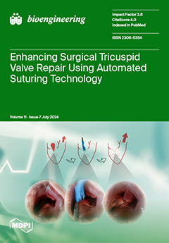
Valvular heart disease is a significant cause of cardiovascular morbidity and mortality. Minimal invasive cardiac surgery aims to restore health while decreasing the burden of intervention for affected patients. In the present study, we describe the use of automated suturing technology to facilitate valve repair in the setting of tricuspid regurgitation. Furthermore, we compare this modified approach to the conventional surgical technique in a passive beating heart model. The isolated and combined procedure proved to be effective in our experimental setup, offering a promising solution for the future treatment of tricuspid valve disease. View this paper
  • Issues are regarded as officially published after their release is announced to the table of contents alert mailing list.
  • You may sign up for e-mail alerts to receive table of contents of newly released issues.
  • PDF is the official format for papers published in both, html and pdf forms. To view the papers in pdf format, click on the "PDF Full-text" link, and use the free Adobe Reader to open them.
Order results
Result details
Section
Select all
Export citation of selected articles as:
20 pages, 8525 KiB  
Article
Uncertainty Quantification in SAR Induced by Ultra-High-Field MRI RF Coil via High-Dimensional Model Representation
by Xi Wang, Shao Ying Huang and Abdulkadir C. Yucel
Bioengineering 2024, 11(7), 730; https://doi.org/10.3390/bioengineering11070730 (registering DOI) - 18 Jul 2024
Abstract
As magnetic field strength in Magnetic Resonance Imaging (MRI) technology increases, maintaining the specific absorption rate (SAR) within safe limits across human head tissues becomes challenging due to the formation of standing waves at a shortened wavelength. Compounding this challenge is the uncertainty [...] Read more.
As magnetic field strength in Magnetic Resonance Imaging (MRI) technology increases, maintaining the specific absorption rate (SAR) within safe limits across human head tissues becomes challenging due to the formation of standing waves at a shortened wavelength. Compounding this challenge is the uncertainty in the dielectric properties of head tissues, which notably affects the SAR induced by the radiofrequency (RF) coils in an ultra-high-field (UHF) MRI system. To this end, this study introduces a computational framework to quantify the impacts of uncertainties in head tissues’ dielectric properties on the induced SAR. The framework employs a surrogate model-assisted Monte Carlo (MC) technique, efficiently generating surrogate models of MRI observables (electric fields and SAR) and utilizing them to compute SAR statistics. Particularly, the framework leverages a high-dimensional model representation technique, which constructs the surrogate models of the MRI observables via univariate and bivariate component functions, approximated through generalized polynomial chaos expansions. The numerical results demonstrate the efficiency of the proposed technique, requiring significantly fewer deterministic simulations compared with traditional MC methods and other surrogate model-assisted MC techniques utilizing machine learning algorithms, all while maintaining high accuracy in SAR statistics. Specifically, the proposed framework constructs surrogate models of a local SAR with an average relative error of 0.28% using 289 simulations, outperforming the machine learning-based surrogate modeling techniques considered in this study. Furthermore, the SAR statistics obtained by the proposed framework reveal fluctuations of up to 30% in SAR values within specific head regions. These findings highlight the critical importance of considering dielectric property uncertainties to ensure MRI safety, particularly in 7 T MRI systems. Full article
21 pages, 3747 KiB  
Article
ViT-PSO-SVM: Cervical Cancer Predication Based on Integrating Vision Transformer with Particle Swarm Optimization and Support Vector Machine
by Abdulaziz AlMohimeed, Mohamed Shehata, Nora El-Rashidy, Sherif Mostafa, Amira Samy Talaat and Hager Saleh
Bioengineering 2024, 11(7), 729; https://doi.org/10.3390/bioengineering11070729 (registering DOI) - 18 Jul 2024
Abstract
Cervical cancer (CCa) is the fourth most prevalent and common cancer affecting women worldwide, with increasing incidence and mortality rates. Hence, early detection of CCa plays a crucial role in improving outcomes. Non-invasive imaging procedures with good diagnostic performance are desirable and have [...] Read more.
Cervical cancer (CCa) is the fourth most prevalent and common cancer affecting women worldwide, with increasing incidence and mortality rates. Hence, early detection of CCa plays a crucial role in improving outcomes. Non-invasive imaging procedures with good diagnostic performance are desirable and have the potential to lessen the degree of intervention associated with the gold standard, biopsy. Recently, artificial intelligence-based diagnostic models such as Vision Transformers (ViT) have shown promising performance in image classification tasks, rivaling or surpassing traditional convolutional neural networks (CNNs). This paper studies the effect of applying a ViT to predict CCa using different image benchmark datasets. A newly developed approach (ViT-PSO-SVM) was presented for boosting the results of the ViT based on integrating the ViT with particle swarm optimization (PSO), and support vector machine (SVM). First, the proposed framework extracts features from the Vision Transformer. Then, PSO is used to reduce the complexity of extracted features and optimize feature representation. Finally, a softmax classification layer is replaced with an SVM classification model to precisely predict CCa. The models are evaluated using two benchmark cervical cell image datasets, namely SipakMed and Herlev, with different classification scenarios: two, three, and five classes. The proposed approach achieved 99.112% accuracy and 99.113% F1-score for SipakMed with two classes and achieved 97.778% accuracy and 97.805% F1-score for Herlev with two classes outperforming other Vision Transformers, CNN models, and pre-trained models. Finally, GradCAM is used as an explainable artificial intelligence (XAI) tool to visualize and understand the regions of a given image that are important for a model’s prediction. The obtained experimental results demonstrate the feasibility and efficacy of the developed ViT-PSO-SVM approach and hold the promise of providing a robust, reliable, accurate, and non-invasive diagnostic tool that will lead to improved healthcare outcomes worldwide. Full article
Show Figures

Graphical abstract

12 pages, 2466 KiB  
Article
Association between Elastic Modulus of Foot Soft Tissues and Gait Characteristics in Young Individuals with Flatfoot
by Xin Jiao, Tianyi Hu, Yongjin Li, Binbin Wang, Mirabel Ewura Esi Acquah, Zengguang Wang, Qianqian Chen, Yaokai Gan and Dongyun Gu
Bioengineering 2024, 11(7), 728; https://doi.org/10.3390/bioengineering11070728 (registering DOI) - 18 Jul 2024
Abstract
Flatfoot is a common foot deformity, causing foot pain, osteoarthritis of the midfoot, and even knee and hip dysfunction. The elastic modulus of foot soft tissues and its association with gait biomechanics still remain unclear. For this study, we recruited 20 young individuals [...] Read more.
Flatfoot is a common foot deformity, causing foot pain, osteoarthritis of the midfoot, and even knee and hip dysfunction. The elastic modulus of foot soft tissues and its association with gait biomechanics still remain unclear. For this study, we recruited 20 young individuals with flatfoot and 22 age-matched individuals with normal foot arches. The elastic modulus of foot soft tissues (posterior tibial tendon, flexor digitorum brevis, plantar fascia, heel fat pad) was obtained via ultrasound elastography. Gait data were acquired using an optical motion capture system. The association between elastic modulus and gait data was analyzed via correlation analysis. The elastic modulus of the plantar fascia (PF) in individuals with flatfoot was higher than that in individuals with normal foot arches. There was no significant difference in the elastic modulus of the posterior tibial tendon (PTT), the flexor digitorum brevis (FDB), or the heel fat pad (HFD), or the thickness of the PF, PTT, FDB, and HFD. Individuals with flatfoot showed greater motion of the hip and pelvis in the coronal plane, longer double-support phase time, and greater maximum hip adduction moment during walking. The elastic modulus of the PF in individuals with flatfoot was positively correlated with the maximum hip extension angle (r = 0.352, p = 0.033) and the maximum hip adduction moment (r = 0.429, p = 0.039). The plantar fascia is an important plantar structure in flatfoot. The alteration of the plantar fascia’s elastic modulus is likely a significant contributing factor to gait abnormalities in people with flatfoot. More attention should be given to the plantar fascia in the young population with flatfoot. Full article
(This article belongs to the Section Biomechanics and Sports Medicine)
Show Figures

Graphical abstract

16 pages, 5685 KiB  
Article
Production of Reverse Transcriptase and DNA Polymerase in Bacterial Expression Systems
by Kristína Hriňová, Johana Dlapová, Bohuš Kubala, Ľubica Kormanová, Zdenko Levarski, Eva Struhárňanská, Ján Turňa and Stanislav Stuchlík
Bioengineering 2024, 11(7), 727; https://doi.org/10.3390/bioengineering11070727 (registering DOI) - 18 Jul 2024
Abstract
DNA amplification and reverse transcription enzymes have proven to be invaluable in fast and reliable diagnostics and research applications because of their processivity, specificity, and robustness. Our study focused on the production of mutant Taq DNA polymerase and mutant M-MLV reverse transcriptase in [...] Read more.
DNA amplification and reverse transcription enzymes have proven to be invaluable in fast and reliable diagnostics and research applications because of their processivity, specificity, and robustness. Our study focused on the production of mutant Taq DNA polymerase and mutant M-MLV reverse transcriptase in the expression hosts Vibrio natriegens and Escherichia coli under various expression conditions. We also examined nonspecific extracellular production in V. natriegens. Intracellularly, M-MLV was produced in V. natriegens at the level of 11% of the total cell proteins (TCPs) compared with 16% of TCPs in E. coli. We obtained a soluble protein that accounted for 11% of the enzyme produced in V. natriegens and 22% of the enzyme produced in E. coli. Taq pol was produced intracellularly in V. natriegens at the level of 30% of TCPs compared with 26% of TCPs in E. coli. However, Taq pol was almost non-soluble in E. coli, whereas in V. natriegens, we obtained a soluble protein that accounted for 23% of the produced enzyme. We detected substantial extracellular production of Taq pol. Thus, V. natriegens is a suitable alternative host with the potential for production of recombinant proteins. Full article
(This article belongs to the Section Cellular and Molecular Bioengineering)
Show Figures

Figure 1

4 pages, 167 KiB  
Editorial
Editorial Topical Collection: “Biomedical Imaging and Data Analytics for Disease Diagnosis and Treatment”
by Cosimo Ieracitano and Xuejun Zhang
Bioengineering 2024, 11(7), 726; https://doi.org/10.3390/bioengineering11070726 (registering DOI) - 18 Jul 2024
Abstract
The integration of biomedical imaging techniques with advanced data analytics is at the forefront of a transformative era in healthcare [...] Full article
29 pages, 18319 KiB  
Review
Pulsing Addition to Modulated Electro-Hyperthermia
by Andras Szasz
Bioengineering 2024, 11(7), 725; https://doi.org/10.3390/bioengineering11070725 - 17 Jul 2024
Viewed by 52
Abstract
Numerous preclinical results have been verified, and clinical results have validated the advantages of modulated electro-hyperthermia (mEHT). This method uses the nonthermal effects of the electric field in addition to thermal energy absorption. Modulation helps with precisely targeting and immunogenically destroying malignant cells, [...] Read more.
Numerous preclinical results have been verified, and clinical results have validated the advantages of modulated electro-hyperthermia (mEHT). This method uses the nonthermal effects of the electric field in addition to thermal energy absorption. Modulation helps with precisely targeting and immunogenically destroying malignant cells, which could have a vaccination-like abscopal effect. A new additional modulation (high-power pulsing) further develops the abilities of the mEHT. My objective is to present the advantages of pulsed treatment and how it fits into the mEHT therapy. Pulsed treatment increases the efficacy of destroying the selected tumor cells; it is active deeper in the body, at least tripling the penetration of the energy delivery. Due to the constant pulse amplitude, the dosing of the absorbed energy is more controllable. The induced blood flow for reoxygenation and drug delivery is high enough but not as high as increasing the risk of the dissemination of malignant cells. The short pulses have reduced surface absorption, making the treatment safer, and the increased power in the pulses allows the reduction of the treatment time needed to provide the necessary dose. Full article
Show Figures

Figure 1

16 pages, 14753 KiB  
Article
Fabrication and Dielectric Validation of an Arm Phantom for Electromyostimulation
by Katja Uhrhan, Esther Schwindt and Hartmut Witte
Bioengineering 2024, 11(7), 724; https://doi.org/10.3390/bioengineering11070724 - 17 Jul 2024
Viewed by 123
Abstract
Electromyostimulation (EMS) is an up-and-coming training method that demands further fundamental research regarding its safety and efficacy. To investigate the influence of different stimulation parameters, electrode positions and electrode sizes on the resulting voltage in the tissue, a tissue mimicking phantom is needed. [...] Read more.
Electromyostimulation (EMS) is an up-and-coming training method that demands further fundamental research regarding its safety and efficacy. To investigate the influence of different stimulation parameters, electrode positions and electrode sizes on the resulting voltage in the tissue, a tissue mimicking phantom is needed. Therefore, this study describes the fabrication of a hydrogel arm phantom for EMS applications with the tissue layers of skin, fat, blood and muscle. The phantom was dielectrically validated in the frequency range of 20 Hz to 100 Hz. We also conducted electromyography (EMG) recordings during EMS on the phantom and compared them with the same measurements on a human arm. The phantom reproduces the dielectric properties of the tissues with deviations ranging from 0.8% to more than 100%. Although we found it difficult to find a compromise between mimicking the permittivity and electrical conductivity at the same time, the EMS–EMG measurements showed similar waveforms (1.9–9.5% deviation) in the phantom and human. Our research contributes to the field of dielectric tissue phantoms, as it proposes a multilayer arm phantom for EMS applications. Consequently, the phantom can be used for initial EMS investigations, but future research should focus on further improving the dielectric properties. Full article
Show Figures

Graphical abstract

12 pages, 11945 KiB  
Article
Evaluation of Denoising Performance of ResNet Deep Learning Model for Ultrasound Images Corresponding to Two Frequency Parameters
by Hyekyoung Kang, Chanrok Park and Hyungjin Yang
Bioengineering 2024, 11(7), 723; https://doi.org/10.3390/bioengineering11070723 - 16 Jul 2024
Viewed by 171
Abstract
Ultrasound imaging is widely used for accurate diagnosis due to its noninvasive nature and the absence of radiation exposure, which is achieved by controlling the scan frequency. In addition, Gaussian and speckle noises degrade image quality. To address this issue, filtering techniques are [...] Read more.
Ultrasound imaging is widely used for accurate diagnosis due to its noninvasive nature and the absence of radiation exposure, which is achieved by controlling the scan frequency. In addition, Gaussian and speckle noises degrade image quality. To address this issue, filtering techniques are typically used in the spatial domain. Recently, deep learning models have been increasingly applied in the field of medical imaging. In this study, we evaluated the effectiveness of a convolutional neural network-based residual network (ResNet) deep learning model for noise reduction when Gaussian and speckle noises were present. We compared the results with those obtained from conventional filtering techniques. A dataset of 500 images was prepared, and Gaussian and speckle noises were added to create noisy input images. The dataset was divided into training, validation, and test sets in an 8:1:1 ratio. The ResNet deep learning model, comprising 16 residual blocks, was trained using optimized hyperparameters, including the learning rate, optimization function, and loss function. For quantitative analysis, we calculated the normalized noise power spectrum, peak signal-to-noise ratio, and root mean square error. Our findings showed that the ResNet deep learning model exhibited superior noise reduction performance to median, Wiener, and median-modified Wiener filter algorithms. Full article
(This article belongs to the Special Issue Radiological Imaging and Its Applications)
Show Figures

Figure 1

14 pages, 1146 KiB  
Article
Surfactant-Mediated Microalgal Flocculation: Process Efficiency and Kinetic Modelling
by Carolina Maia, Vânia Pôjo, Tânia Tavares, José C. M. Pires and Francisco Xavier Malcata
Bioengineering 2024, 11(7), 722; https://doi.org/10.3390/bioengineering11070722 - 16 Jul 2024
Viewed by 315
Abstract
Microalgae are a valuable source of lipids, proteins, and pigments, but there are challenges in large-scale production, especially in harvesting. Existing methods lack proven efficacy and cost-effectiveness. However, flocculation, an energy-efficient technique, is emerging as a promising solution. Integrating surfactants enhances microalgal harvesting [...] Read more.
Microalgae are a valuable source of lipids, proteins, and pigments, but there are challenges in large-scale production, especially in harvesting. Existing methods lack proven efficacy and cost-effectiveness. However, flocculation, an energy-efficient technique, is emerging as a promising solution. Integrating surfactants enhances microalgal harvesting and disruption simultaneously, reducing processing costs. This study investigated cetyltrimethylammonium bromide (CTAB), dodecyltrimethylammonium bromide (DTAB), and sodium dodecyl sulphate (SDS) for harvesting Tetraselmis sp. strains (75LG and 46NLG). CTAB exhibits superior results, with 88% harvesting efficiency at 1500 and 2000 mg L−1 for 75LG and 46NLG, respectively, for 60 min of sedimentation—thus being able to reduce the operating time. Beyond evaluating harvesting efficiency, our study explored the kinetics of the process; the modified Gompertz model led to the best fit. Furthermore, the largest kinetic constants were observed with CTAB, thus highlighting its efficacy in optimising the microalgal harvesting process. With the incorporation of the suggested enhancements, which should be addressed in future work, CTAB could hold the potential to optimise microalgal harvesting for cost-effective and sustainable large-scale production, eventually unlocking the commercial potential of microalgae for biodiesel production. Full article
(This article belongs to the Special Issue 10th Anniversary of Bioengineering: Biochemical Engineering)
Show Figures

Graphical abstract

18 pages, 4697 KiB  
Article
Developing a Swallow-State Monitoring System Using Nasal Airflow, Surface Electromyography, and Thyroid Cartilage Movement Detection
by Wann-Yun Shieh, Mohammad Anwar Khan and Ya-Cheng Shieh
Bioengineering 2024, 11(7), 721; https://doi.org/10.3390/bioengineering11070721 - 16 Jul 2024
Viewed by 273
Abstract
The safe ingestion of food and water requires appropriate coordination between the respiratory and swallowing pathways. This coordination can be disrupted because of aging or various diseases, thereby resulting in swallowing disorders. No comparative research has been conducted on methods for effectively screening [...] Read more.
The safe ingestion of food and water requires appropriate coordination between the respiratory and swallowing pathways. This coordination can be disrupted because of aging or various diseases, thereby resulting in swallowing disorders. No comparative research has been conducted on methods for effectively screening swallowing disorders in individuals and providing timely alerts to their caregivers. Therefore, the present study developed a monitoring and alert system for swallowing disorders by using three types of noninvasive sensors, namely those measuring nasal airflow, surface electromyography signals, and thyroid cartilage movement. Two groups of participants, one comprising healthy individuals (58 participants; mean age 49.4 years) and another consisting of individuals with a history of unilateral stroke (21 participants; mean age 54.4 years), were monitored when they swallowed five volumes of water. Through an analysis of the data from both groups, seven indicators of swallowing disorders were identified, and the proposed system characterized the individual’s swallowing state as having a green (safe), yellow (unsafe), or red (highly unsafe) status on the basis of these indicators. The results indicated that the symptoms of swallowing disorders are detectable. Healthcare professionals can then use these data to conduct assessments, perform screening, and provide nutrient intake suggestions. Full article
(This article belongs to the Section Biosignal Processing)
Show Figures

Figure 1

20 pages, 5195 KiB  
Article
Chitosan Scaffolds from Crustacean and Fungal Sources: A Comparative Study for Bone-Tissue-Engineering Applications
by Neelam Iqbal, Payal Ganguly, Lemiha Yildizbakan, El Mostafa Raif, Elena Jones, Peter V. Giannoudis and Animesh Jha
Bioengineering 2024, 11(7), 720; https://doi.org/10.3390/bioengineering11070720 - 16 Jul 2024
Viewed by 215
Abstract
Chitosan (CS), a biopolymer, holds significant potential in bone regeneration due to its biocompatibility and biodegradability attributes. While crustacean-derived CS is conventionally used in research, there is growing interest in fungal-derived CS for its equally potent properties in bone regenerative applications. Here, we [...] Read more.
Chitosan (CS), a biopolymer, holds significant potential in bone regeneration due to its biocompatibility and biodegradability attributes. While crustacean-derived CS is conventionally used in research, there is growing interest in fungal-derived CS for its equally potent properties in bone regenerative applications. Here, we investigated the physicochemical and biological characteristics of fungal (MDC) and crustacean (ADC)-derived CS scaffolds embedded with different concentrations of tricalcium phosphate minerals (TCP), i.e., 0(wt)%: ADC/MDC-1, 10(wt)%: ADC/MDC-2, 20(wt)%: ADC/MDC-3 and 30(wt)%: ADC/MDC-4. ADC-1 and MDC-1 lyophilised scaffolds lacking TCP minerals presented the highest zeta potentials of 47.3 ± 1.2 mV and 55.1 ± 1.6 mV, respectively. Scanning electron microscopy revealed prominent distinctions whereby MDC scaffolds exhibited striation-like structural microarchitecture in contrast to the porous morphology exhibited by ADC scaffold types. With regard to the 4-week scaffold mass reductions, MDC-1, MDC-2, MDC-3, and MDC-4 indicated declines of 55.98 ± 4.2%, 40.16 ± 3.6%, 27.05 ± 4.7%, and 19.16 ± 5.3%, respectively. Conversely, ADC-1, ADC-2, ADC-3, and ADC-4 presented mass reductions of 35.78 ± 5.1%, 25.19 ± 4.2%, 20.23 ± 6.3%, and 13.68 ± 5.4%, respectively. The biological performance of the scaffolds was assessed through in vitro bone marrow mesenchymal stromal cell (BMMSCs) attachment via indirect and direct cytotoxicity studies, where all scaffold types presented no cytotoxic behaviours. MDC scaffolds indicated results comparable to ADC, where both CS types exhibited similar physiochemical properties. Our data suggest that MDC scaffolds could be a potent alternative to ADC-derived scaffolds for bone regeneration applications, particularly for 10(wt)% TCP concentrations. Full article
(This article belongs to the Section Regenerative Engineering)
Show Figures

Figure 1

22 pages, 4120 KiB  
Article
Three-Dimensionally Printed Agarose Micromold Supports Scaffold-Free Mouse Ex Vivo Follicle Growth, Ovulation, and Luteinization
by Emily J. Zaniker, Prianka H. Hashim, Samuel Gauthier, James A. Ankrum, Hannes Campo and Francesca E. Duncan
Bioengineering 2024, 11(7), 719; https://doi.org/10.3390/bioengineering11070719 - 15 Jul 2024
Viewed by 485
Abstract
Ex vivo follicle growth is an essential tool, enabling interrogation of folliculogenesis, ovulation, and luteinization. Though significant advancements have been made, existing follicle culture strategies can be technically challenging and laborious. In this study, we advanced the field through development of a custom [...] Read more.
Ex vivo follicle growth is an essential tool, enabling interrogation of folliculogenesis, ovulation, and luteinization. Though significant advancements have been made, existing follicle culture strategies can be technically challenging and laborious. In this study, we advanced the field through development of a custom agarose micromold, which enables scaffold-free follicle culture. We established an accessible and economical manufacturing method using 3D printing and silicone molding that generates biocompatible hydrogel molds without the risk of cytotoxicity from leachates. Each mold supports simultaneous culture of multiple multilayer secondary follicles in a single focal plane, allowing for constant timelapse monitoring and automated analysis. Mouse follicles cultured using this novel system exhibit significantly improved growth and ovulation outcomes with comparable survival, oocyte maturation, and hormone production profiles as established three-dimensional encapsulated in vitro follicle growth (eIVFG) systems. Additionally, follicles recapitulated aspects of in vivo ovulation physiology with respect to their architecture and spatial polarization, which has not been observed in eIVFG systems. This system offers simplicity, scalability, integration with morphokinetic analyses of follicle growth and ovulation, and compatibility with existing microphysiological platforms. This culture strategy has implications for fundamental follicle biology, fertility preservation strategies, reproductive toxicology, and contraceptive drug discovery. Full article
(This article belongs to the Special Issue Bioengineering Technologies to Advance Reproductive Health)
Show Figures

Graphical abstract

10 pages, 1861 KiB  
Article
CARING: Cannula for Alleviation of Retinal Injury Caused by Needle Fluidic Gashing
by Kaersti L. Rickels, Anthony L. Gunderman, Mattie S. McLellan, Muhammad M. Shamim, Joseph A. Sanford and Sami H. Uwaydat
Bioengineering 2024, 11(7), 718; https://doi.org/10.3390/bioengineering11070718 - 15 Jul 2024
Viewed by 293
Abstract
Infusion-related iatrogenic retinal breaks (IRBs) are a significant complication in vitrectomies, particularly when smaller-gauge cannulas are used during fluid infusion. Using two-dimensional finite element analysis (FEA), we analyzed forces exerted on the retina from different cannulas: traditional 25-gauge, 20-gauge, 23-gauge, and 27-gauge, then [...] Read more.
Infusion-related iatrogenic retinal breaks (IRBs) are a significant complication in vitrectomies, particularly when smaller-gauge cannulas are used during fluid infusion. Using two-dimensional finite element analysis (FEA), we analyzed forces exerted on the retina from different cannulas: traditional 25-gauge, 20-gauge, 23-gauge, and 27-gauge, then investigated four alternative new cannula designs: (A) oblique orifices, (B) external obstruction, (C) side ports, and (D) perpendicular orifices. The analysis revealed that the standard 25-gauge cannula had a force of 0.546 milli-Newtons (mN). Optimized cannulas demonstrated decreased forces: 0.072 mN (A), 0.266 mN (B), 0.417 mN (C), and 0.117 mN (D). While all the designs decrease fluid jet force, each has unique challenges: Design A may complicate manufacturing, B requires unique attachment techniques, C could misdirect fluid toward the lens and peripheral retina, and D requires a sealed trocar/cannula design to prevent unwanted fluid ejection. These four innovative cannula designs, identified with detailed engineering simulations, provide promising strategies to reduce the risk of IRBs during vitrectomy, bridging the gap between engineering insights and clinical application. Full article
(This article belongs to the Special Issue Ophthalmic Engineering (2nd Edition))
Show Figures

Figure 1

18 pages, 2562 KiB  
Article
Electroencephalogram Alpha Oscillations in Stroke Recovery: Insights into Neural Mechanisms from Combined Transcranial Direct Current Stimulation and Mirror Therapy in Relation to Activities of Daily Life
by Chia-Lun Liu, Ya-Wen Tu, Ming-Wei Li, Ku-Chou Chang, Chih-Hung Chang, Chih-Kuang Chen and Ching-Yi Wu
Bioengineering 2024, 11(7), 717; https://doi.org/10.3390/bioengineering11070717 - 15 Jul 2024
Viewed by 277
Abstract
The goal of stroke rehabilitation is to establish a robust protocol for patients to live independently in community. Firstly, we examined the impact of 3 hybridized transcranial direct current stimulation (tDCS)-mirror therapy interventions on activities of daily life (ADL) in stroke patients. Secondly, [...] Read more.
The goal of stroke rehabilitation is to establish a robust protocol for patients to live independently in community. Firstly, we examined the impact of 3 hybridized transcranial direct current stimulation (tDCS)-mirror therapy interventions on activities of daily life (ADL) in stroke patients. Secondly, we explored the underlying therapeutic mechanisms with theory-driven electroencephalography (EEG) indexes in the alpha band. This was achieved by identifying the unique contributions of alpha power in motor production to ADL in relation to the premotor cortex (PMC), primary cortex (M1), and Sham tDCS with mirror therapy. The results showed that, although post-intervention ADL improvement was comparable among the three tDCS groups, one of the EEG indexes differentiated the interventions. Neural-behavioral correlation analyses revealed that different types of ADL improvements consistently corresponded with alpha power in the temporal lobe exclusively in the PMC tDCS group (all rs > 0.39). By contrast, alterations in alpha power in the central-frontal region were found to vary, with ADL primarily in the M1 tDCS group (r = −0.6 or 0.7), with the benefit depending on the complexity of the ADL. In conclusion, this research suggested two potential therapeutic mechanisms and demonstrated the additive benefits of introducing theory-driven neural indexes in explaining ADL. Full article
(This article belongs to the Special Issue Technologies for Monitoring and Rehabilitation of Motor Disabilities)
Show Figures

Graphical abstract

15 pages, 5811 KiB  
Project Report
The Effects of Khat Chewing among Djiboutians: Dental Chemical Studies, Gingival Histopathological Analyses and Bioinformatics Approaches
by Fatouma Mohamed Abdoul-Latif, Ayoub Ainane, Ali Merito, Ibrahim Houmed Aboubaker, Houda Mohamed, Sanaa Cherroud and Tarik Ainane
Bioengineering 2024, 11(7), 716; https://doi.org/10.3390/bioengineering11070716 - 15 Jul 2024
Viewed by 612
Abstract
This study examined the effects of khat chewing on oral gingival conditions by adopting a targeted process which combined physicochemical analyses of the teeth, histopathological examinations of the gums, and bioinformatics modeling. The physicochemical evaluation of teeth in khat consumers compared to non-consumers [...] Read more.
This study examined the effects of khat chewing on oral gingival conditions by adopting a targeted process which combined physicochemical analyses of the teeth, histopathological examinations of the gums, and bioinformatics modeling. The physicochemical evaluation of teeth in khat consumers compared to non-consumers was carried out using specific analytical techniques; hence, the results of this initial investigation revealed significant erosion of the tooth enamel due to khat chewing, as well as an alteration of the essential chemical composition of the teeth. Additionally, the histopathological analyses complemented preliminary studies by showing severe inflammation of the gums and oral mucosa in khat users. The understanding of these studies was enriched by bioinformatics analysis, where modeling was carried out via computational methods. This analytical phase examined molecular docking mechanisms, including the interaction between cathinone, the main alkaloid of khat, and the protein receptors involved in the protection of gingival tissues against infections. In summary, this multidisciplinary research provided an in-depth view of the oral health issues related to khat chewing, combining experimental studies with bioinformatics perspectives. Full article
(This article belongs to the Special Issue Biomaterials in Dental Applications)
Show Figures

Graphical abstract

21 pages, 4198 KiB  
Article
Discriminant Input Processing Scheme for Self-Assisted Intelligent Healthcare Systems
by Mohamed Medani, Shtwai Alsubai, Hong Min, Ashit Kumar Dutta and Mohd Anjum
Bioengineering 2024, 11(7), 715; https://doi.org/10.3390/bioengineering11070715 - 14 Jul 2024
Viewed by 300
Abstract
Modern technology and analysis of emotions play a crucial role in enabling intelligent healthcare systems to provide diagnostics and self-assistance services based on observation. However, precise data predictions and computational models are critical for these systems to perform their jobs effectively. Traditionally, healthcare [...] Read more.
Modern technology and analysis of emotions play a crucial role in enabling intelligent healthcare systems to provide diagnostics and self-assistance services based on observation. However, precise data predictions and computational models are critical for these systems to perform their jobs effectively. Traditionally, healthcare monitoring has been the primary emphasis. However, there were a couple of negatives, including the pattern feature generating the method’s scalability and reliability, which was tested with different data sources. This paper delves into the Discriminant Input Processing Scheme (DIPS), a crucial instrument for resolving challenges. Data-segmentation-based complex processing techniques allow DIPS to merge many emotion analysis streams. The DIPS recommendation engine uses segmented data characteristics to sift through inputs from the emotion stream for patterns. The recommendation is more accurate and flexible since DIPS uses transfer learning to identify similar data across different streams. With transfer learning, this study can be sure that the previous recommendations and data properties will be available in future data streams, making the most of them. Data utilization ratio, approximation, accuracy, and false rate are some of the metrics used to assess the effectiveness of the advised approach. Self-assisted intelligent healthcare systems that use emotion-based analysis and state-of-the-art technology are crucial when managing healthcare. This study improves healthcare management’s accuracy and efficiency using computational models like DIPS to guarantee accurate data forecasts and recommendations. Full article
Show Figures

Graphical abstract

13 pages, 714 KiB  
Review
Prostate-Specific Membrane Antigen Radioligand Therapy in Non-Prostate Cancers: Where Do We Stand?
by Francesco Dondi, Alberto Miceli, Guido Rovera, Vanessa Feudo, Claudia Battisti, Maria Rondini, Andrea Marongiu, Antonio Mura, Riccardo Camedda, Maria Silvia De Feo, Miriam Conte, Joana Gorica, Cristina Ferrari, Anna Giulia Nappi and Giulia Santo
Bioengineering 2024, 11(7), 714; https://doi.org/10.3390/bioengineering11070714 - 14 Jul 2024
Viewed by 291
Abstract
Introduction: The term theragnostic refers to the combination of a predictive imaging biomarker with a therapeutic agent. The promising application of prostate-specific membrane antigen (PSMA)-based radiopharmaceuticals in the imaging and treatment of prostate cancer (PCa) patients opens the way to investigate a possible [...] Read more.
Introduction: The term theragnostic refers to the combination of a predictive imaging biomarker with a therapeutic agent. The promising application of prostate-specific membrane antigen (PSMA)-based radiopharmaceuticals in the imaging and treatment of prostate cancer (PCa) patients opens the way to investigate a possible role of PSMA-based radiopharmaceuticals in cancers beyond the prostate. Therefore, the aim of this review was to evaluate the role of 177Lu-PSMA radioligand therapy (RLT) in malignancies other than prostate cancer by evaluating preclinical, clinical studies, and ongoing clinical trials. Methods: An extensive literature search was performed in three different databases using different combinations of the following terms: “Lu-PSMA”, “177Lu-PSMA”, “preclinical”, “mouse”, “salivary gland cancer”, “breast cancer”, “glioblastoma”, “solid tumour”, “renal cell carcinoma”, “HCC”, “thyroid”, “salivary”, “radioligand therapy”, and “lutetium-177”. The search had no beginning date limit and was updated to April 2024. Only articles written in English were included in this review. Results: A total of four preclinical studies were selected (breast cancer model n = 3/4). PSMA-RLT significantly reduced cell viability and had anti-angiogenic effects, especially under hypoxic conditions, which increase PSMA binding and uptake. Considering the clinical studies (n = 8), the complexity of evaluating PSMA-RLT in cancers other than prostate cancer was clearly revealed, since in most of the presented cases a sufficient tumour radiation dose was not achieved. However, encouraging results can be found in some types of diseases, such as thyroid cancer. Some clinical trials are still ongoing, and results from prospective larger cohorts of patients are awaited. Conclusions: The need for larger patient cohorts and more RLT cycles administered underscores the need for further comprehensive studies. Given the very preliminary results of both preclinical and clinical studies, ongoing clinical trials in the near future may provide stronger evidence of both the safety and therapeutic efficacy of PSMA-RLT in malignancies other than prostate cancer. Full article
(This article belongs to the Special Issue Applications of Radioimmunotherapy and Imaging in Nuclear Medicine)
Show Figures

Figure 1

12 pages, 2820 KiB  
Article
Effect of Sextant Fixating Angle of Spiral Clavicle Plate on Biomechanical Stability—A Preliminary Finite Element Study
by Ming-Hsien Hu, Po-Feng Su, Kun-Jhih Lin, Wen-Chuan Chen and Shun-Ping Wang
Bioengineering 2024, 11(7), 713; https://doi.org/10.3390/bioengineering11070713 - 13 Jul 2024
Viewed by 306
Abstract
Introduction: A spiral clavicle plate has been accepted for its superior multidirectional compatibility in the treatment of midshaft clavicle fractures from a biomechanical perspective. However, the influence of the sextant angle (spiral level) definition on biomechanical performance has not been clarified. A conceptual [...] Read more.
Introduction: A spiral clavicle plate has been accepted for its superior multidirectional compatibility in the treatment of midshaft clavicle fractures from a biomechanical perspective. However, the influence of the sextant angle (spiral level) definition on biomechanical performance has not been clarified. A conceptual finite element analysis was conducted to identify the advantages and drawbacks of spiral clavicle plates with various sextant angle definitions. Methods: Conventional superior and three different conceptual spiral plates with sextant angle definitions ranging from 45 to 135 degrees were constructed to restore an OTA 15-B1.3 midshaft clavicle fracture model. Three major loading scenarios (cantilever downward bending, axial compression, and axial torsion) were simulated to evaluate the reconstructed structural stiffness and the stress on the clavicle plate and bone screws. Results: The spiral clavicle plate demonstrated greater capability in resisting cantilever downward bending with an increase in sextant angle and showed comparable structural stiffness and implant stress compared to the superior clavicle plate. However, weakened resistance to axial compression load was noted for the spiral clavicle plate, with lowered stiffness and increased stress on the clavicle plate and screws as the spiral level increased. Conclusion: The spiral clavicle plate has been reported to offer multidirectional compatibility for the treatment of midshaft clavicle fractures, as well as geometric advantages in anatomical matching and reduced skin prominence after surgery. The current study supports that remarkable cantilever bending strength can be achieved with this plate. However, users must consider the potential drawback of lowered axial compression resistance in safety considerations. Full article
Show Figures

Figure 1

14 pages, 3053 KiB  
Article
Comparison of Transcranial Magnetic Stimulation Dosimetry between Structured and Unstructured Grids Using Different Solvers
by Francesca Camera, Caterina Merla and Valerio De Santis
Bioengineering 2024, 11(7), 712; https://doi.org/10.3390/bioengineering11070712 - 13 Jul 2024
Viewed by 285
Abstract
In recent years, the interest in transcranial magnetic stimulation (TMS) has surged, necessitating deeper understanding, development, and use of low-frequency (LF) numerical dosimetry for TMS studies. While various ad hoc dosimetric models exist, commercial software tools like SimNIBS v4.0 and Sim4Life v7.2.4 are [...] Read more.
In recent years, the interest in transcranial magnetic stimulation (TMS) has surged, necessitating deeper understanding, development, and use of low-frequency (LF) numerical dosimetry for TMS studies. While various ad hoc dosimetric models exist, commercial software tools like SimNIBS v4.0 and Sim4Life v7.2.4 are preferred for their user-friendliness and versatility. SimNIBS utilizes unstructured tetrahedral mesh models, while Sim4Life employs voxel-based models on a structured grid, both evaluating induced electric fields using the finite element method (FEM) with different numerical solvers. Past studies primarily focused on uniform exposures and voxelized models, lacking realism. Our study compares these LF solvers across simplified and realistic anatomical models to assess their accuracy in evaluating induced electric fields. We examined three scenarios: a single-shell sphere, a sphere with an orthogonal slab, and a MRI-derived head model. The comparison revealed small discrepancies in induced electric fields, mainly in regions of low field intensity. Overall, the differences were contained (below 2% for spherical models and below 12% for the head model), showcasing the potential of computational tools in advancing exposure assessment required for TMS protocols in different bio-medical applications. Full article
Show Figures

Figure 1

46 pages, 2893 KiB  
Review
A Comprehensive Review of AI Diagnosis Strategies for Age-Related Macular Degeneration (AMD)
by Aya A. Abd El-Khalek, Hossam Magdy Balaha, Ashraf Sewelam, Mohammed Ghazal, Abeer T. Khalil, Mohy Eldin A. Abo-Elsoud and Ayman El-Baz
Bioengineering 2024, 11(7), 711; https://doi.org/10.3390/bioengineering11070711 - 13 Jul 2024
Viewed by 234
Abstract
The rapid advancement of computational infrastructure has led to unprecedented growth in machine learning, deep learning, and computer vision, fundamentally transforming the analysis of retinal images. By utilizing a wide array of visual cues extracted from retinal fundus images, sophisticated artificial intelligence models [...] Read more.
The rapid advancement of computational infrastructure has led to unprecedented growth in machine learning, deep learning, and computer vision, fundamentally transforming the analysis of retinal images. By utilizing a wide array of visual cues extracted from retinal fundus images, sophisticated artificial intelligence models have been developed to diagnose various retinal disorders. This paper concentrates on the detection of Age-Related Macular Degeneration (AMD), a significant retinal condition, by offering an exhaustive examination of recent machine learning and deep learning methodologies. Additionally, it discusses potential obstacles and constraints associated with implementing this technology in the field of ophthalmology. Through a systematic review, this research aims to assess the efficacy of machine learning and deep learning techniques in discerning AMD from different modalities as they have shown promise in the field of AMD and retinal disorders diagnosis. Organized around prevalent datasets and imaging techniques, the paper initially outlines assessment criteria, image preprocessing methodologies, and learning frameworks before conducting a thorough investigation of diverse approaches for AMD detection. Drawing insights from the analysis of more than 30 selected studies, the conclusion underscores current research trajectories, major challenges, and future prospects in AMD diagnosis, providing a valuable resource for both scholars and practitioners in the domain. Full article
(This article belongs to the Section Biosignal Processing)
Show Figures

Figure 1

16 pages, 4186 KiB  
Article
Mathematical Modeling of Vedolizumab Treatment’s Effect on Microbiota and Intestinal Permeability in Inflammatory Bowel Disease Patients
by Antonio D’Ambrosio, Annamaria Altomare, Tamara Boscarino, Manuele Gori, Paola Balestrieri, Lorenza Putignani, Federica Del Chierico, Simone Carotti, Michele Cicala, Michele Pier Luca Guarino and Vincenzo Piemonte
Bioengineering 2024, 11(7), 710; https://doi.org/10.3390/bioengineering11070710 - 12 Jul 2024
Viewed by 442
Abstract
Growing evidence suggests that impaired gut permeability and gut microbiota alterations are involved in the pathogenesis of Inflammatory Bowel Diseases (IBDs), which include Ulcerative Colitis (UC) and Crohn’s Disease (CD). Vedolizumab is an anti-α4β7 antibody approved for IBD treatment, used as the first [...] Read more.
Growing evidence suggests that impaired gut permeability and gut microbiota alterations are involved in the pathogenesis of Inflammatory Bowel Diseases (IBDs), which include Ulcerative Colitis (UC) and Crohn’s Disease (CD). Vedolizumab is an anti-α4β7 antibody approved for IBD treatment, used as the first treatment or second-line therapy when the first line results in inadequate effectiveness. The aim of this study is to develop a mathematical model capable of describing the pathophysiological mechanisms of Vedolizumab treatment in IBD patients. In particular, the relationship between drug concentration in the blood, colonic mucosal permeability and fecal microbiota composition was investigated and modeled to detect and predict trends in order to support and tailor Vedolizumab therapies. To pursue this aim, clinical data from a pilot study on a cluster of 11 IBD patients were analyzed. Enrolled patients underwent colonoscopy in three phases (before (t0), after 24 weeks of (t1) and after 52 weeks of (t2 ) Vedolizumab treatment) to collect mucosal biopsies for transepithelial electrical resistance (TEER) evaluation (permeability to ions), intestinal permeability measurement and histological analysis. Moreover, fecal samples were collected for the intestinal microbiota analysis at the three time points. The collected data were compared to those of 11 healthy subjects at t0, who underwent colonoscopy for screening surveillance, and used to implement a three-compartmental mathematical model (comprising central blood, peripheral blood and the intestine). The latter extends previous evidence from the literature, based on the regression of experimental data, to link drug concentration in the peripheral blood compartment with Roseburia abundance and intestinal permeability. The clinical data showed that Vedolizumab treatment leads to an increase in TEER and a reduction in intestinal permeability to a paracellular probe, improving tissue inflammation status. Microbiota analysis showed increasing values of Roseburia, albeit not statistically significant. This trend was adequately reproduced by the mathematical model, which offers a useful tool to describe the pathophysiological effects of Vedolizumab therapy on colonic mucosal permeability and fecal microbiota composition. The model’s satisfactory predictive capabilities and simplicity shed light on the relationship between the drug, the microbiota and permeability and allow for its straightforward extension to diverse therapeutic conditions. Full article
Show Figures

Graphical abstract

16 pages, 3031 KiB  
Article
Two-Stream Convolutional Neural Networks for Breathing Pattern Classification: Real-Time Monitoring of Respiratory Disease Patients
by Jinho Park, Thien Nguyen, Soongho Park, Brian Hill, Babak Shadgan and Amir Gandjbakhche
Bioengineering 2024, 11(7), 709; https://doi.org/10.3390/bioengineering11070709 - 12 Jul 2024
Viewed by 476
Abstract
A two-stream convolutional neural network (TCNN) for breathing pattern classification has been devised for the continuous monitoring of patients with infectious respiratory diseases. The TCNN consists of a convolutional neural network (CNN)-based autoencoder and classifier. The encoder of the autoencoder generates deep compressed [...] Read more.
A two-stream convolutional neural network (TCNN) for breathing pattern classification has been devised for the continuous monitoring of patients with infectious respiratory diseases. The TCNN consists of a convolutional neural network (CNN)-based autoencoder and classifier. The encoder of the autoencoder generates deep compressed feature maps, which contain the most important information constituting data. These maps are concatenated with feature maps generated by the classifier to classify breathing patterns. The TCNN, single-stream CNN (SCNN), and state-of-the-art classification models were applied to classify four breathing patterns: normal, slow, rapid, and breath holding. The input data consisted of chest tissue hemodynamic responses measured using a wearable near-infrared spectroscopy device on 14 healthy adult participants. Among the classification models evaluated, random forest had the lowest classification accuracy at 88.49%, while the TCNN achieved the highest classification accuracy at 94.63%. In addition, the proposed TCNN performed 2.6% better in terms of classification accuracy than an SCNN (without an autoencoder). Moreover, the TCNN mitigates the issue of declining learning performance with increasing network depth, as observed in the SCNN model. These results prove the robustness of the TCNN in classifying breathing patterns despite using a significantly smaller number of parameters and computations compared to state-of-the-art classification models. Full article
(This article belongs to the Special Issue Monitoring and Analysis of Human Biosignals, Volume II)
Show Figures

Graphical abstract

11 pages, 2682 KiB  
Article
Visualization of Cerebrospinal Fluid Outflow and Egress along the Nerve Roots of the Lumbar Spine
by Diana Vucevic, Vadim Malis, Won C. Bae, Hideki Ota, Koichi Oshio, Marin A. McDonald and Mitsue Miyazaki
Bioengineering 2024, 11(7), 708; https://doi.org/10.3390/bioengineering11070708 - 12 Jul 2024
Viewed by 304
Abstract
Intrinsic cerebrospinal fluid (CSF) dynamics in the brain have been extensively studied, particularly the egress sites of tagged intrinsic CSF in the meninges. Although spinal CSF recirculates within the central nervous system (CNS), we hypothesized that CSF outflows from the lumbar spinal canal. [...] Read more.
Intrinsic cerebrospinal fluid (CSF) dynamics in the brain have been extensively studied, particularly the egress sites of tagged intrinsic CSF in the meninges. Although spinal CSF recirculates within the central nervous system (CNS), we hypothesized that CSF outflows from the lumbar spinal canal. We aimed to visualize and semi-quantify the outflow using non-contrast MRI techniques. We utilized a 3 Tesla clinical MRI with a 16-channel spine coil, employing time–spatial labeling inversion (Time-SLIP) with tag-on and tag-off acquisitions, T2-weighted coronal 2D fluid-attenuated inversion recovery (FLAIR) and T2-weighted coronal 3D centric ky-kz single-shot FSE (cSSFSE). Images were acquired using time–spatial labeling inversion pulse (Time-SLIP) with tag-on and tag-off acquisitions with varying TI periods. Ten healthy volunteers with no known spinal diseases participated. Variations in tagged CSF outflow were observed across different thoracolumbar nerve root segments in all participants. We quantified CSF outflow at all lumbar levels and the psoas region. There was no significant difference among the ROIs for signal intensity. The tagged CSF outflow from the spinal canal is small but demonstrates egress to surrounding tissues. This finding may pave the way for exploring intrathecal drug delivery, understanding of CSF-related pathologies and its potential as a biomarker for peripheral neuropathy and radiculopathy. Full article
(This article belongs to the Special Issue Advancements in Medical Imaging Technology)
Show Figures

Graphical abstract

27 pages, 4322 KiB  
Article
Adaptive Filtering with Fitted Noise Estimate (AFFiNE): Blink Artifact Correction in Simulated and Real P300 Data
by Kevin E. Alexander, Justin R. Estepp and Sherif M. Elbasiouny
Bioengineering 2024, 11(7), 707; https://doi.org/10.3390/bioengineering11070707 - 12 Jul 2024
Viewed by 284
Abstract
(1) Background: The electroencephalogram (EEG) is frequently corrupted by ocular artifacts such as saccades and blinks. Methods for correcting these artifacts include independent component analysis (ICA) and recursive-least-squares (RLS) adaptive filtering (-AF). Here, we introduce a new method, AFFiNE, that applies Bayesian adaptive [...] Read more.
(1) Background: The electroencephalogram (EEG) is frequently corrupted by ocular artifacts such as saccades and blinks. Methods for correcting these artifacts include independent component analysis (ICA) and recursive-least-squares (RLS) adaptive filtering (-AF). Here, we introduce a new method, AFFiNE, that applies Bayesian adaptive regression spline (BARS) fitting to the adaptive filter’s reference noise input to address the known limitations of both ICA and RLS-AF, and then compare the performance of all three methods. (2) Methods: Artifact-corrected P300 morphologies, topographies, and measurements were compared between the three methods, and to known truth conditions, where possible, using real and simulated blink-corrupted event-related potential (ERP) datasets. (3) Results: In both simulated and real datasets, AFFiNE was successful at removing the blink artifact while preserving the underlying P300 signal in all situations where RLS-AF failed. Compared to ICA, AFFiNE resulted in either a practically or an observably comparable error. (4) Conclusions: AFFiNE is an ocular artifact correction technique that is implementable in online analyses; it can adapt to being non-stationarity and is independent of channel density and recording duration. AFFiNE can be utilized for the removal of blink artifacts in situations where ICA may not be practically or theoretically useful. Full article
Show Figures

Graphical abstract

10 pages, 1987 KiB  
Article
Ultrasound Elastography Assessment of Knee Intra-Articular Adhesions at Varying Knee Angles
by Jiling Ye, Linjing Peng, Angang Ding, Shijie Chen, Bin Cai and Yifei Yao
Bioengineering 2024, 11(7), 706; https://doi.org/10.3390/bioengineering11070706 - 12 Jul 2024
Viewed by 277
Abstract
We aimed to verify the feasibility of using shear wave elastography (SWE) to quantify knee scars and the elastic modulus of scar tissues. Overall, 16 participants underwent SWE assessments and range-of-motion measurement and completed the Knee Injury and Osteoarthritis Outcome Score. The inter-rater [...] Read more.
We aimed to verify the feasibility of using shear wave elastography (SWE) to quantify knee scars and the elastic modulus of scar tissues. Overall, 16 participants underwent SWE assessments and range-of-motion measurement and completed the Knee Injury and Osteoarthritis Outcome Score. The inter-rater reliability for SWE in the suprapatellar bursa, below the patellar tendon, and in the medial and lateral trochlear groove remained within 0.861–0.907. The SWE values in the four regions increased with increasing knee angle, and significant differences were observed between the values for below the patellar tendon and the suprapatellar bursa at knee flexion angles of 60° and 90°. The SWE values of the medial and lateral trochlear groove at 30°, 60°, and 90° knee flexion were higher on the affected side. A negative correlation was observed between the SWE values for the lateral trochlear groove at 0°, 30°, and 60° and those for below the patellar tendon at 0° and the suprapatellar bursa at 30° with both active and passive knee extension. The suprapatellar bursa value at 60° exhibited a positive correlation with both knee flexion and passive knee flexion, whereas that of the suprapatellar bursa at 90° exhibited a positive correlation with both the range of motion and passive range of motion. SWE is a replicable and effective method for detecting scar strength in the knee joint. Full article
(This article belongs to the Special Issue Recent Advances in Biomechanics of Soft Tissues)
Show Figures

Figure 1

24 pages, 4908 KiB  
Review
Emerging Biomedical and Clinical Applications of 3D-Printed Poly(Lactic Acid)-Based Devices and Delivery Systems
by Allan John R. Barcena, Prashanth Ravi, Suprateek Kundu and Karthik Tappa
Bioengineering 2024, 11(7), 705; https://doi.org/10.3390/bioengineering11070705 - 11 Jul 2024
Viewed by 325
Abstract
Poly(lactic acid) (PLA) is widely used in the field of medicine due to its biocompatibility, versatility, and cost-effectiveness. Three-dimensional (3D) printing or the systematic deposition of PLA in layers has enabled the fabrication of customized scaffolds for various biomedical and clinical applications. In [...] Read more.
Poly(lactic acid) (PLA) is widely used in the field of medicine due to its biocompatibility, versatility, and cost-effectiveness. Three-dimensional (3D) printing or the systematic deposition of PLA in layers has enabled the fabrication of customized scaffolds for various biomedical and clinical applications. In tissue engineering and regenerative medicine, 3D-printed PLA has been mostly used to generate bone tissue scaffolds, typically in combination with different polymers and ceramics. PLA’s versatility has also allowed the development of drug-eluting constructs for the controlled release of various agents, such as antibiotics, antivirals, anti-hypertensives, chemotherapeutics, hormones, and vitamins. Additionally, 3D-printed PLA has recently been used to develop diagnostic electrodes, prostheses, orthoses, surgical instruments, and radiotherapy devices. PLA has provided a cost-effective, accessible, and safer means of improving patient care through surgical and dosimetry guides, as well as enhancing medical education through training models and simulators. Overall, the widespread use of 3D-printed PLA in biomedical and clinical settings is expected to persistently stimulate biomedical innovation and revolutionize patient care and healthcare delivery. Full article
Show Figures

Figure 1

9 pages, 5297 KiB  
Article
Single versus Double Plate Fixation in Condylar Neck Fractures: Clinical Results and Biomechanics Simulation
by Chien-Chung Chen, Ting-Han Chiu, Cheng-Yu Yan, Ya-Pei Hou and Ting-Sheng Lin
Bioengineering 2024, 11(7), 704; https://doi.org/10.3390/bioengineering11070704 - 11 Jul 2024
Viewed by 218
Abstract
The open reduction of mandibular condyle neck fractures is difficult due to the limited surgical field and complex facial nerve structures. The most effective fixation method for narrow fractured segments is debated as standard double four-hole plate fixation is often not feasible. This [...] Read more.
The open reduction of mandibular condyle neck fractures is difficult due to the limited surgical field and complex facial nerve structures. The most effective fixation method for narrow fractured segments is debated as standard double four-hole plate fixation is often not feasible. This research compared bone stability and force resistance between single-long-plate and double-short-plate fixations using clinical outcomes, a Sawbones mandible model, and finite element analysis. In patients with condyle neck fractures, nine were fixed with single-long-plate and twelve with double-short-plate fixations, with no significant differences in malocclusion and facial palsy rates. In compression tests with a Sawbones model, displacements in the posterior part were similar in both fixation groups. In contrast, the anterior part had significantly higher displacements in the single-long-plate group. Finite element analysis showed higher displacements in both anterior and posterior parts in the single-plate group compared to the double-short-plate group. Maximum stresses were at the second screw hole in single-long-plate fixation and the turning point of the upper plate at the condyle neck in double-short-plate fixation. Double-short-plate fixations demonstrated better stability and force resistance than single-long-plate fixations. Full article
Show Figures

Figure 1

18 pages, 6011 KiB  
Article
Difficult Airway Assessment Based on Multi-View Metric Learning
by Jinze Wu, Yuan Yao, Guangchao Zhang, Xiaofan Li and Bo Peng
Bioengineering 2024, 11(7), 703; https://doi.org/10.3390/bioengineering11070703 - 11 Jul 2024
Viewed by 231
Abstract
The preoperative assessment of difficult airways is of great significance in the practice of anesthesia intubation. In recent years, although a large number of difficult airway recognition algorithms have been investigated, defects such as low recognition accuracy and poor recognition reliability still exist. [...] Read more.
The preoperative assessment of difficult airways is of great significance in the practice of anesthesia intubation. In recent years, although a large number of difficult airway recognition algorithms have been investigated, defects such as low recognition accuracy and poor recognition reliability still exist. In this paper, we propose a Dual-Path Multi-View Fusion Network (DMF-Net) based on multi-view metric learning, which aims to predict difficult airways through multi-view facial images of patients. DMF-Net adopts a dual-path structure to extract features by grouping the frontal and lateral images of the patients. Meanwhile, a Multi-Scale Feature Fusion Module and a Hybrid Co-Attention Module are designed to improve the feature representation ability of the model. Consistency loss and complementarity loss are utilized fully for the complementarity and consistency of information between multi-view data. Combined with Focal Loss, information bias is effectively avoided. Experimental validation illustrates the effectiveness of the proposed method, with the accuracy, specificity, sensitivity, and F1 score reaching 77.92%, 75.62%, 82.50%, and 71.35%, respectively. Compared with methods such as clinical bedside screening tests and existing artificial intelligence-based methods, our method is more accurate and reliable and can provide a reliable auxiliary tool for clinical healthcare personnel to effectively improve the accuracy and reliability of preoperative difficult airway assessments. The proposed network can help to identify and assess the risk of difficult airways in patients before surgery and reduce the incidence of postoperative complications. Full article
(This article belongs to the Section Biosignal Processing)
Show Figures

Figure 1

13 pages, 3858 KiB  
Article
Mixture Theory-Based Finite Element Approach for Analyzing the Edematous Condition of Biological Soft Tissues
by Satoko Hirabayashi, Masami Iwamoto and Xian Chen
Bioengineering 2024, 11(7), 702; https://doi.org/10.3390/bioengineering11070702 - 11 Jul 2024
Viewed by 262
Abstract
In hydrated soft biological tissues experiencing edema, which is typically associated with various disorders, excessive fluid accumulates and is encapsulated by impermeable membranes. In certain cases of edema, an indentation induced by pressure persists even after the load is removed. The depth and [...] Read more.
In hydrated soft biological tissues experiencing edema, which is typically associated with various disorders, excessive fluid accumulates and is encapsulated by impermeable membranes. In certain cases of edema, an indentation induced by pressure persists even after the load is removed. The depth and duration of this indentation are used to assess the treatment response. This study presents a mixture theory-based approach to analyzing the edematous condition. The finite element analysis formulation was grounded in mixture theory, with the solid displacement, pore water pressure, and fluid relative velocity as the unknown variables. To ensure tangential fluid flow at the surface of tissues with complex shapes, we transformed the coordinates of the fluid velocity vector at each time step and node, allowing for the incorporation of the transmembrane component of fluid flow as a Dirichlet boundary condition. Using this proposed method, we successfully replicated the distinct behavior of pitting edema, which is characterized by a prolonged recovery time from indentation. Consequently, the proposed method offers valuable insights into the finite element analysis of the edematous condition in biological tissues. Full article
(This article belongs to the Special Issue Computational Biomechanics, Volume II)
Show Figures

Figure 1

17 pages, 1615 KiB  
Article
Integrating Convolutional Neural Networks with Attention Mechanisms for Magnetic Resonance Imaging-Based Classification of Brain Tumors
by Zahid Rasheed, Yong-Kui Ma, Inam Ullah, Mahmoud Al-Khasawneh, Sulaiman Sulmi Almutairi and Mohammed Abohashrh
Bioengineering 2024, 11(7), 701; https://doi.org/10.3390/bioengineering11070701 - 10 Jul 2024
Viewed by 329
Abstract
The application of magnetic resonance imaging (MRI) in the classification of brain tumors is constrained by the complex and time-consuming characteristics of traditional diagnostics procedures, mainly because of the need for a thorough assessment across several regions. Nevertheless, advancements in deep learning (DL) [...] Read more.
The application of magnetic resonance imaging (MRI) in the classification of brain tumors is constrained by the complex and time-consuming characteristics of traditional diagnostics procedures, mainly because of the need for a thorough assessment across several regions. Nevertheless, advancements in deep learning (DL) have facilitated the development of an automated system that improves the identification and assessment of medical images, effectively addressing these difficulties. Convolutional neural networks (CNNs) have emerged as steadfast tools for image classification and visual perception. This study introduces an innovative approach that combines CNNs with a hybrid attention mechanism to classify primary brain tumors, including glioma, meningioma, pituitary, and no-tumor cases. The proposed algorithm was rigorously tested with benchmark data from well-documented sources in the literature. It was evaluated alongside established pre-trained models such as Xception, ResNet50V2, Densenet201, ResNet101V2, and DenseNet169. The performance metrics of the proposed method were remarkable, demonstrating classification accuracy of 98.33%, precision and recall of 98.30%, and F1-score of 98.20%. The experimental finding highlights the superior performance of the new approach in identifying the most frequent types of brain tumors. Furthermore, the method shows excellent generalization capabilities, making it an invaluable tool for healthcare in diagnosing brain conditions accurately and efficiently. Full article
(This article belongs to the Special Issue Computer Vision and Machine Learning in Medical Applications)
Show Figures

Figure 1

Previous Issue
Back to TopTop