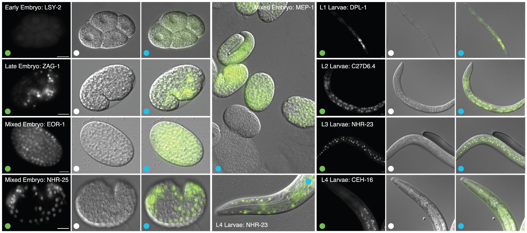
Extended Data Figure 9: Representative samples of staged, transgenic C. elegans embryos and larvae expressing GFP-tagged fusion proteins.
From: Regulatory analysis of the C. elegans genome with spatiotemporal resolution

GFP fluorescence images, differential interference contrast (DIC) images, and merged (GFP/DIC) images are labelled with green, white and blue dots, respectively. The 10-µm scale bar is shown in GFP fluorescence images. Images were selected independent of binding experiment results. Approved binding experiments include: MEP-1 (mixed embryo, L2 larvae), DPL-1 (L1 larvae), C27D6.4 (L2 larvae), NHR-23 (L3 larvae) and CEH-16 (L4 larvae) experiments.