100% found this document useful (1 vote)
369 viewsLesson 3 The Neuromotor Basis For Motor Control v2
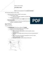
The neuromotor system forms the foundation for motor control. It includes the central nervous system (brain, spinal cord, cerebellum) and peripheral nervous system. The basic functional unit is the neuron, which transmits electrical signals through axons and dendrites. Sensory neurons carry information from receptors to the CNS, while motor neurons carry signals from the CNS to muscles. The primary motor cortex initiates voluntary movement, while other areas like the premotor and supplementary motor cortices plan movement. The basal ganglia and cerebellum help regulate and coordinate movement.
Uploaded by
Drift AlvinCopyright
© © All Rights Reserved
Available Formats
Download as PDF, TXT or read online on Scribd
100% found this document useful (1 vote)
369 viewsLesson 3 The Neuromotor Basis For Motor Control v2
The neuromotor system forms the foundation for motor control. It includes the central nervous system (brain, spinal cord, cerebellum) and peripheral nervous system. The basic functional unit is the neuron, which transmits electrical signals through axons and dendrites. Sensory neurons carry information from receptors to the CNS, while motor neurons carry signals from the CNS to muscles. The primary motor cortex initiates voluntary movement, while other areas like the premotor and supplementary motor cortices plan movement. The basal ganglia and cerebellum help regulate and coordinate movement.
Uploaded by
Drift AlvinCopyright
© © All Rights Reserved
Available Formats
Download as PDF, TXT or read online on Scribd
/ 11