Location via proxy:   [ UP ]  
[Report a bug]   [Manage cookies]                
SlideShare a Scribd company logo
PRESENTATION ON ‘TYPES OF ENDOSPERM’
GUIDED BY- PRESENTED BY-
DR. NAMITA NATH RASHMI H. RAHMAN
M.SC 2ND
SEM
DEPT OF BOTANY
ROLL NO 3
GAUHATI UNIVERSITY
Types of endosperm
INTRODUCTION
Endosperm is a nutritive tissue stored in the
seed for the embryo and it is formed by the
fusion of one male gamete with the polar
nuclei.
-Endosperm plays a main role in the diet of
human beings.
-Endosperm is found in all angiosperms except
few families like
Orchidaceae,Podostemonaceae and
Trapaceae.
-The seeds without endosperm: Non-
endospermous seeds or exalbuminous seed.
-The seed containing the endosperm:
Endospermous or albuminous seed.
EXAMPLES OF ALBUMINOUS AND
EXALBUMINOUS SEED:
Castor
Pea
TYPES OF ENDOSPERM
-Endosperm is classified into 3 types by
Schnarf(1929) on the basis of their mode of
development:
1.Nuclear endosperm
2.Cellular endosperm
3.Helobial endosperm
Fig: A-Nuclear ,B-Helobial C- Cellular
endosperm
NUCLEAR ENDOSPERM
In this type of endosperm, the division of the
primary endosperm nucleus and its further
divisions are not followed by wall formation.
Examples. Limnanthes, Oxyspora
Fig: Neuclear endosperm in Limnanthes
 In some plants, the nuclear endosperm
becomes cellular at a later stage.
 Ex. Acalypha, Phaseolus.
Fig: A-E; stages in the development of
Neuclear type of endosperm
CELLULARIZATION OF NUCLEAR ENDOSPERM
 The cellularization of nuclear endosperm
shows variations.
 In Acalypha,the endosperm becomes
cellular completely.
In Grevillea, the endosperm is cellular in the
micropylar region and nuclear in the chalazal
end. The nuclear chalazal end develops into a
vermiform appendix (Kausik,1941
Fig: Endosperm formation in Acalypha
 In coconut,the endosperm is nuclear type.
Later on it becomes cellular.The PEN
undergoes several free nuclear divisions.
 The endosperm in coconut is partly solid
and partly liquid.
Fig: Endosperm formation in Cocos nucifera
CELLULAR ENDOSPERM
 The division of the PEN and its further
divisions are followed by immediate wall
formation and the free nuclear stage is
completely absent.
 It is common in dicots.
 Haustoria are common.
Fig: Development of cellular endosperm in
Drimys winteri
Haustoria in Cellular Endosperm
The haustoria may arise at the
micropylar end or at the chalazal
end or both.
In Impatiens , micropylar haustorium
is produced.
In Nemophila both micropylar and
chalazal haustoria are found.
Fig: Endosperm in Nemophila
Fig: Micropylar haustorium in Impatiens
HELOBIAL ENDOSPERM
It is intermediate in structure.
Development is intermediate
between nuclear and cellular types.
It is more common in monocots.
Davis(1966) reported the presence of
helobial endosperm in 14 monocot
families
Fig: Development of helobial type of
endosperm in Eremurus
Types of endosperm
RUMINATE ENDOSPERM
Endosperm having irregular ridges
and furrows on the surface. E.g
Areca catechu, Myristica
Fig: Ruminate endosperm in Myristica
REFERENCE:
1. THE EMBRYOLOGY OF
ANGIOSPERMS, 5TH EDITION, S S
BHOJWANI & S P BHATNAGAR
2. DEVELOPMENTAL BOTANY AND
EXPERIMENTAL EMBRYOLOGY,
ANNIE KUMMARESAN
3. www.publishyourarticles.net
Types of endosperm

More Related Content

Types of endosperm

  • 1. PRESENTATION ON ‘TYPES OF ENDOSPERM’ GUIDED BY- PRESENTED BY- DR. NAMITA NATH RASHMI H. RAHMAN M.SC 2ND SEM DEPT OF BOTANY ROLL NO 3 GAUHATI UNIVERSITY
  • 3. INTRODUCTION Endosperm is a nutritive tissue stored in the seed for the embryo and it is formed by the fusion of one male gamete with the polar nuclei. -Endosperm plays a main role in the diet of human beings. -Endosperm is found in all angiosperms except few families like Orchidaceae,Podostemonaceae and Trapaceae. -The seeds without endosperm: Non- endospermous seeds or exalbuminous seed. -The seed containing the endosperm: Endospermous or albuminous seed.
  • 4. EXAMPLES OF ALBUMINOUS AND EXALBUMINOUS SEED: Castor Pea
  • 5. TYPES OF ENDOSPERM -Endosperm is classified into 3 types by Schnarf(1929) on the basis of their mode of development: 1.Nuclear endosperm 2.Cellular endosperm 3.Helobial endosperm Fig: A-Nuclear ,B-Helobial C- Cellular endosperm
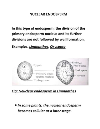
  • 6. NUCLEAR ENDOSPERM In this type of endosperm, the division of the primary endosperm nucleus and its further divisions are not followed by wall formation. Examples. Limnanthes, Oxyspora Fig: Neuclear endosperm in Limnanthes  In some plants, the nuclear endosperm becomes cellular at a later stage.
  • 7.  Ex. Acalypha, Phaseolus. Fig: A-E; stages in the development of Neuclear type of endosperm CELLULARIZATION OF NUCLEAR ENDOSPERM  The cellularization of nuclear endosperm shows variations.  In Acalypha,the endosperm becomes cellular completely. In Grevillea, the endosperm is cellular in the micropylar region and nuclear in the chalazal
  • 8. end. The nuclear chalazal end develops into a vermiform appendix (Kausik,1941 Fig: Endosperm formation in Acalypha  In coconut,the endosperm is nuclear type. Later on it becomes cellular.The PEN undergoes several free nuclear divisions.  The endosperm in coconut is partly solid and partly liquid.
  • 9. Fig: Endosperm formation in Cocos nucifera CELLULAR ENDOSPERM  The division of the PEN and its further divisions are followed by immediate wall formation and the free nuclear stage is completely absent.  It is common in dicots.  Haustoria are common.
  • 10. Fig: Development of cellular endosperm in Drimys winteri Haustoria in Cellular Endosperm The haustoria may arise at the micropylar end or at the chalazal end or both. In Impatiens , micropylar haustorium is produced. In Nemophila both micropylar and chalazal haustoria are found.
  • 11. Fig: Endosperm in Nemophila Fig: Micropylar haustorium in Impatiens
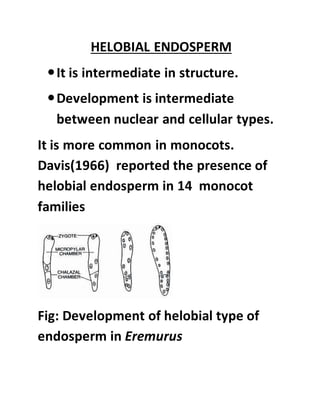
  • 12. HELOBIAL ENDOSPERM It is intermediate in structure. Development is intermediate between nuclear and cellular types. It is more common in monocots. Davis(1966) reported the presence of helobial endosperm in 14 monocot families Fig: Development of helobial type of endosperm in Eremurus
  • 14. RUMINATE ENDOSPERM Endosperm having irregular ridges and furrows on the surface. E.g Areca catechu, Myristica Fig: Ruminate endosperm in Myristica
  • 15. REFERENCE: 1. THE EMBRYOLOGY OF ANGIOSPERMS, 5TH EDITION, S S BHOJWANI & S P BHATNAGAR 2. DEVELOPMENTAL BOTANY AND EXPERIMENTAL EMBRYOLOGY, ANNIE KUMMARESAN 3. www.publishyourarticles.net