


Rhynia
- 1. Rhynia Dr. Vasanta I. Kahalkar Department of Botany M. G. Arts, Science & Late N. P. Commerce College, Armori
- 2. Rhynia • Division – Pteridophyta • Class- Psilophytopsida • Order- Psilophytales • Family- Rhyniaceae • Genus- Rhynia • Occurrence:- Rhynia is extinct plant having two species i.e R. gwynne-vaughani and R. major. It was discovered by Kidston and Lang in 1917 from Rhynia Chert Bed of middle Devonion. These plants were discovered in the form of Petrification.
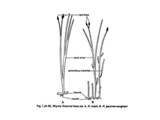
- 3. External Morphology of Rhynia • The plants of Rhynia were herbaceous. R. major was 20 cm. in height and 1.5 to 6 mm in diameter whereas R. gwynne- vaughani was only about 18 cm. in height and 1 to 3 mm in diameter. • The plant body was differentiated into a subterranean rhizome with an abruptly turned upright photosynthetic aerial shoots. Roots were absent but at places rhizome was provided with tufts of unicellular rhizoids . The aerial shoots were cylindrical and leafless with a tapering dichotomously branched system. • In R. major the aerial shoots were smooth but in case of R. gwynne-vaughani many adventitious branches were present on the aerial shoots as well as rhizome. These branches perhaps help in vegetative propagation. • The tip of the aerial branch usually bears a solitary terminal sporangium which was about 12 mm in length and about 4 mm in diameter.
- 5. Internal Structure of Rhynia:
- 6. Internal Structure of Rhynia: • Transverse section (T.S.) of Aerial shoot and Rhizome: • Anatomically, the aerial shoots and rhizome are almost similar. T. S. of aerial shoot can be differentiated into three parts: epidermis, cortex and stele. • (a) Epidermis: • It was the outer-most surrounding layer. It was one cell thick and covered by thin cuticle. In aerial shoots it was interrupted at certain places by stomata but stomata were absent in rhizome. • (b) Cortex: • Epidermis was followed by cortex. It is differentiated into outer cortex and inner cortex. The outer cortex was only 1-4 cells thick, thin walled and without intercellular spaces. The inner cortex had large intercellular spaces and its cells had chloroplast. The endodermis and pericycle layers were absent. • Stele: • The centre of the aerial shoot/rhizome was occupied by stele. The stele was a protostele (haplostele). The xylem was made up of annular tracheids and there were no sieve plates in phloem.
- 7. Reproductive Structures of Rhynia: • The sporangia were borne singly on the apices of some aerial branches, each sporangium being oval or slightly cylindrical structure with a little greater diameter than that of aerial branch on which it is developed. • A longitudinal section (L.S.) of sporangium shows that the outermost layer was 1 cell thick cuticularized epidermis. It was followed by 3 cells thick middle layers of thin walled cells. The inner-most layer was 1 cell thick tapetum. • The wall was surrounding a spacious sporangial cavity which was without columella and contained large number of spores. The spores were of same size. • It means that Rhynia was homosporous. In many specimens the sporangium contained tetrahedral tetrads of spores which suggest that they were formed by reduction division.
- 9. • THANK YOU