digestives system Physiology
- 2. LECTURES OUTLINE • The Gastrointestinal System • Control Mechanisms in Gastrointestinal Physiology • The Mouth and Esophagus • The Stomach • The Pancreas • The Liver • The Small Intestine • The Large Intestine • Enteric Motility
- 3. • The gastrointestinal system consists of the gastrointestinal tract and the accessory exocrine glands
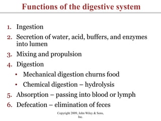
- 4. Copyright 2009, John Wiley & Sons, Inc. Functions of the digestive system 1. Ingestion 2. Secretion of water, acid, buffers, and enzymes into lumen 3. Mixing and propulsion 4. Digestion • Mechanical digestion churns food • Chemical digestion – hydrolysis 5. Absorption – passing into blood or lymph 6. Defecation – elimination of feces
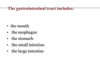
- 5. • the mouth • the esophagus • the stomach • the small intestine • the large intestine The gastrointestinal tract includes:
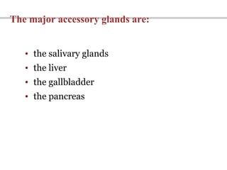
- 6. The major accessory glands are: • the salivary glands • the liver • the gallbladder • the pancreas
- 7. The Components of the Digestive System
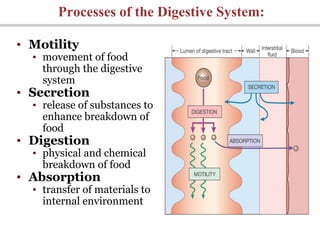
- 8. Processes of the Digestive System: • Motility • movement of food through the digestive system • Secretion • release of substances to enhance breakdown of food • Digestion • physical and chemical breakdown of food • Absorption • transfer of materials to internal environment
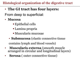
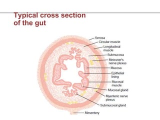
- 9. • The GI tract has four layers: From deep to superficial: • Mucosa Epithelial cells Lamina propria Muscularis mucosae • Submucosa (elastic connective tissue contains lymph and blood vessels) • Muscularis externa (smooth muscle arranged in circular and longitudinal layers) • Serosa ( outer connective tissue) Histological organization of the digestive tract
- 10. General Organization of the Gastrointestinal Tract
- 11. Typical cross section of the gut
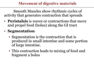
- 12. Smooth Muscles show rhythmic cycles of activity that generates contraction that spreads • Peristalsis is waves or contractions that move and propel food (bolus) along the GI tract • Segmentation • Segmentation is the contraction that is produced in small intestine and some portion of large intestine. • This contraction leads to mixing of food and fragment a bolus Movement of digestive materials
- 13. Movement of digestive materials • Peristalsis – moves • Segmentation – mixes
- 14. • Movement of materials along the digestive tract is controlled by: • Neural mechanisms • Parasympathetic and local reflexes • Hormonal mechanisms • Enhance or inhibit smooth muscle contraction • Local mechanisms • Coordinate response to changes in pH or chemical stimuli Control of the digestive system
- 15. Neural Control of Gastrointestinal Function — Enteric Nervous System
- 16. Copyright 2009, John Wiley & Sons, Inc. Neural innervation • Enteric nervous system (ENS) • Intrinsic set of nerves - “brain of gut” • Neurons extending from esophagus to anus • 2 plexuses • Myenteric plexus – GI tract motility • Submucosal plexus – controlling secretions • Autonomic nervous system • Extrinsic set of nerves • Parasympathetic stimulation increases secretion and activity by stimulating ENS • Sympathetic stimulation decreases secretions and activity by inhibiting ENS
- 17. THE ENTERIC NERVOUS SYSTEM is composed mainly of two plexuses: (1)an outer plexus lying between the longitudinal and circular muscle layers, called the myenteric plexus or Auerbach’s plexus (2) an inner plexus, called the submucosal plexus or Meissner’s plexus, that lies in the submucosa.
- 18. Copyright 2009, John Wiley & Sons, Inc. Organization of the enteric nervous system
- 19. Digestion in the Mouth. Mastication (Chewing)
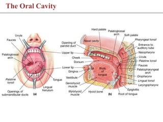
- 20. • Its functions include: • Analysis of material before swallowing • Mechanical processing by the teeth, tongue, and palatal surfaces • Lubrication • Limited digestion e.g. salivary amylase initiates digestion of complex carbohydrates The Oral Cavity The mouth opens into the oral or buccal cavity
- 21. • primary functions include: • Mechanical processing • Assistance in chewing and swallowing • Sensory analysis by touch, temperature, and taste receptors The tongue
- 22. The Oral Cavity
- 23. • There are 3 pairs of salivary glands: • Parotid • Sublingual • Submandibular SALIVARY GLANDS
- 24. • In the mouth cavity open three pair of big salivary glands and a lot of small buccal glands. • Parotid glands consist of the serum types cells and secrete serous type of saliva. • Submandibular and sublingual glands consist of the cells of serum and mucous types and secrete serous and mucus types of saliva. • Small buccal glands consist of mucous types of cells; produce mucous saliva with a big quantity of water. SALIVARY GLANDS
- 26. Quantity, composition and properties of saliva • In the case of dream produce nearly 0,05 mL/minute, in the normal condition – 0,5 mL/minute, in the case of maximal secretion – near 5 mL/minute. By day produce 0,8-1,5 L of saliva. Composition of saliva: water, organic – alfa- amilase, lipase, phosphatase, RNAase, DNAase, mucin, substances for protective – lisocim, thiocianates, antibodies – and unorganic substances – sodium, potassium, calcium, chlorites, etc.
- 27. Functions of saliva • Lubrication, moistening, and dissolving • Initiation of digestion of complex carbohydrates • Help to control bacterial population in the oral cavity
- 28. Mechanism of saliva forming • In acinars' cells produce primary saliva in which synthesis necessary amino acids, glucose, mineral substances (for example, Ca++). • In the cells of salivary glands occur passive processes, which provide moving of water and electrolits from blood to the glands’ ductus (strait). In the ductus occur reabsorption of sodium, chlorine, secretion of potassium, HCO3 –. • This is the secondary saliva. Aldosterone increase reabsorption of sodium and secretion of potassium.
- 29. Influence of vegetative nerves on secretor activity of salivary glands Stimulation of the parasympathetic nerve supply causes profuse secretion of watery saliva with a relatively low content of organic material. Stimulation of the sympathetic nerve supply causes profuse secretion of saliva with small quantity of water with a relatively big content of organic material.
- 30. Swallowing • After chewing, the food (now called a bolus) is swallowed. • Swallowing (known scientifically as deglutition) is the process in the human body that makes some things pass from the mouth, to the pharynx, and into the esophagus, while shutting the epiglottis.
- 31. • Pharynx: • Movement of food bolus in esophagus (and rest of GI tract) via peristalsis • Esophagus: • Carries solids and liquids from the pharynx to the stomach • Passes through esophageal hiatus in diaphragm • The wall of the esophagus contains mucosal, submucosal, and muscularis layers Swallowing
- 32. Stomach
- 33. • Storage - bulk storage of undigested food • Releases small amounts to small intestine • Mechanical processing of the food (forms chyme ) • Chemical processing of the food – e.g. disruption of chemical bonds via acids and enzymes • Secretion – e.g. production of intrinsic factor, HCl, pepsinogen and gastrin Stomach Functions of the stomach:
- 34. Anatomy of the stomach
- 35. Anatomically, the stomach is usually divided into two major parts: The proximal (“orad”) part: •Cardia •Fundus •Body The distal (“caudad” ) part: •Pylorus
- 36. • Cardia –portion of the stomach that connects to the esophagous • Fundus – portion superior to stomach- esophageal junction • Body – area between the fundus and the curve of the J • Pylorus – antrum and pyloric canal adjacent to the duodenum (portion of the stomach that connects to the duodenum) Anatomy of the stomach
- 39. Secretory activity of stomach • Production of stomach juice per day – near 2,5 L of juice. • Their main components – enzymes, HCl and mucin. • pH of morning saliva is neutral, after eating – sour – 0,8-1,5.
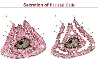
- 40. • In Fundus and Body of the stomach there are gastric pits • Each gastric pit communicate with several gastric gland • Gastric glands are dominated with two types of secretary cells: • Parietal cells • Secrete Intrinsic factor, and HCl • Intrinsic factors are needed for vitamin B12 absorption • HCl kill most acteria, denature proteins, inactivate food enzymes, breakdown plant cells and connective tissue in meat, activate pepsinogen to pepsin. • Chief cells • Secrete Pepsinogen (inactive porenzyme) that converts to pepsin (active form) in the presence of acid. • Pepsin breaks down proteins • In kids chief cells also produce lipase and rennin (help milk digestion) Gastric Secretion
- 41. • Pyloric glands • Primarily produces mucous containing several hormones • Enteroendocrine cells • G cells secrete gastrin • Gastrin stimulates paraietal cells and chief cells to secrete HCl and pepsinogen i.e. HCl and pepsinogen is produced in response to gastrin • D cells secrete somatostatin • Somatostatin inhibits gastrin secretion Gastric Secretion
- 42. Secretion of Parietal Cells
- 43. SECRETION OF HCL
- 44. Role of the hydrochloric acid in the digestion • 1. To promote the swell of protein; • 2. To promote the change of pepsinogen in pepsins; • 3. To make optimal conditions for actions of pepsins; • 4. To fulfill protective role from bacteria; • 5. To promote motor and evacuated functions of stomach; • 6. To stimulate production of duodinum hormon – secretin.
- 45. • Gastric control and production is done in 3 phase: • Cephalic phase - prepares stomach to receive ingested material • Begins when one sees, smells, tastes or thinks of food • Increases gastric juice production • Gastric phase - begins with the arrival of food in the stomach • Neural, hormonal, and local responses further increase secretion • Intestinal phase - controls the rate of gastric emptying i.e. control the rate of chime entry into duodenum • Inhibit gastric acid and pepsinogen production Regulation of gastric activity
- 46. • Preliminary digestion of proteins • Pepsin –enzyme that digests proteins • However due to time limit digestion of protein can not be completed • Permits digestion of carbohydrates by enzyme lipase until the pH falls to 4.5 • Absorption does not occur in stomach because: • Epithelial cells are not exposed to chime (covered by alkaline mucus) • Epithelial cells lack transport mechanism • Gastric lining is impermeable to water • Digestion is not completed • Some drugs, however, are absorbed Digestion and absorption in the stomach
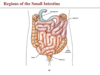
- 48. • Plays Important role in digestion and absorption • Mucosa of SI produce few enzymes, and buffer to neutralize chime • Divided into three part: • Duodenum – receives chime from stomach, bile from gall bladder, and digestive secretion from pancreas • Digestion continues in duodenum • Jejunum – digestion and absorption takes place here • Ileum • Ileocecal sphincter • Transition between small and large intestine SMALL INTESTINE (SI)
- 49. Regions of the Small Intestine
- 52. 1. Peristalsis the wave-like form of muscle contraction, moves chyme along the intestine and causes only a small amount of mixing. These contractions are weak and slow in the small intestine so that time is sufficient for complete digestion and absorption of the chyme as it moves forward. 2. Segmentation mixes chyme with digestive juices and exposes it to the intestinal mucosa for absorption. This form of motility causes only a small degree of forward movement of the chyme along the small intestine INTESTINAL MOVEMENTS
- 53. Composition and properties of intestine juice • Mucine • Enzymes: • Proteolytic • Lipolytic • Amilolytic • Nuclealytic • Immunoglobulins; ions; leukocytes • pH of intestine juice is 7,5-8,0; production per day – near 1,8-2,5 L. • Functions: ending hydrolyses of all nutritive substances; protective of mucus wall; support of chyme in fluid condition; formed of base reaction of intestine contents. Increase secretion: parasympathetic nerves, secretine, and glucagons.
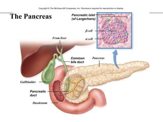
- 54. • Pancreas is both an endocrine and exocrine gland • Endocrine functions • Secretes Insulin and glucagons • Exocrine functions • Produces majority of pancreatic secretions • Pancreatic juice secreted into small intestine contain THE PANCREAS
- 55. 55 From liver Gallbladder Pancreas Duodenum b cell Pancreatic islet (of Langerhans) a cell Common bile duct Pancreatic duct Copyright © The McGraw-Hill Companies, Inc. Permission required for reproduction or display. The Pancreas
- 56. • Performs metabolic and hematological regulation and produces bile • Histological organization • Consist of two lobes • Each lobe is divided into lobules • Lobule is the functional unit of liver • Lobules unite to form common hepatic duct • Duct meets cystic duct to form common bile duct The liver
- 57. The Anatomy of the Liver
- 58. Liver lobule is the basic functional unit of the liver • Hepatocytes form irregular plates arranged in spoke-like fashion • Bile canaliculi carry bile to the bile ductules • Bile ductules lead to portal areas
- 59. Liver Histology
- 60. BILIARY SYSTEM • Bile is a complex fluid secreted by the liver and consists of organic molecules in an alkaline solution. The biliary tract consists of a series of ducts that convey bile from the liver to the duodenum. Bile has three functions: • It facilitates the assimilation of dietary lipid; approximately 50% of dietary lipid appears in feces if bile is excluded from the small intestine. Bile facilitates fat digestion by promoting its emulsification and solubilization. This is achieved through the formation of mixed micelles, which enhance lipase action and then assist in the delivery of the digestive products to be absorbed by the enterocytes. • It provides a pathway to excrete hydrophobic molecules that may not be readily excreted by the kidney. • It assists in neutralizing gastric acid because it is an alkaline solution.
- 61. Composition of bile: • bilirubin, • bile acids, • cholesterol, • leukocytes, • some epitheliocytes, • cristalls of bilirubin, • calcium.
- 62. BILE PRODUCTION AND BILE SECRETOIN • Secretion of bile occur all time and increase by influences of bile acids, cholecystokinin- pancreasemin, secretin. • Bile secretion in the duodenum depends from take food (minerals water, HCl, fatty acids increase bile formation). • It depends of nervus vagus (increase bile formation) and humoral influences – concentration of cholecystokinin-pancreasemin (increase bile formation and ejection), secretin, gastrin.
- 63. • Functions of gall bladder are that it: • Stores bile • modifies bile and • concentrates bile • Bile slat break large droplet of fat for digestion THE GALLBLADDER Animation: Accessory Organ (see tutorial)
- 64. The Gallbladder
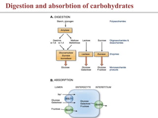
- 65. Digestion and absorbtion of carbohydrates
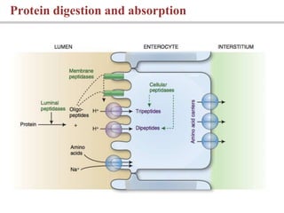
- 66. Protein digestion and absorption
- 67. • Begins in the mouth • Salivary and pancreatic enzymes: • Oligosachrides, Disaccharides and trisaccharides • Brush border enzymes • Monosaccharides • Absorption of monosaccharides occurs across the intestinal epithelia Carbohydrate digestion and absorption
- 68. Absorption and intracellular processing of fats A. Diffusion of micelles. B. Intracellular synthesis of triglyceride and formation of chylomicrons. C. Exocytosis of chylomicrons to lymphatic vessels.
- 69. •Bile salts improve chemical digestion by emulsifying lipid drops (lipids interact with bile salts and form emulsification droplets) •pancreatic lipases break emulsified fat into free fatty acids and monoglycerides •Lipid-bile salt complexes called micelles are formed •Micelles diffuse into intestinal epithelial cells • Reform triglycerides in epithelial cells • Combined w/ protein to form chylomicrons • Chylomicrons released via exocytosis into lymphatic system • Then its transported to blood Lipid digestion and absorption
- 70. • Water • Nearly all that is ingested is reabsorbed via osmosis • Ions • Absorbed via diffusion, cotransport, and active transport • Vitamins • Water soluble vitamins are absorbed by diffusion • Fat soluble vitamins are absorbed as part of micelles • Vitamin B12 requires intrinsic factor ABSORPTION WATER, IONS, VITAMINS
- 71. LARGE INTESTINE • is the region of the digestive tract from the ileocecal valve to the anus. Approximately 1.5 m in length, it has a larger diameter than the small intestine. The mucosa of the large intestine is composed of absorptive cells and mucus-secreting goblet cells and • does not form villi. The large intestine consists of the following structures: Cecum collects and stores material from ileum Appendix Colon consist of four regions: ascending colon, transverse colon, descending colon and sigmoid colon Rectum – it is expandable
- 73. FUNCTIONS OF THE LARGE INTESTINE • Major functions: 1. absorption of water 2. absorption of vitamins produced by bacteria 3. storage of fecal material prior to defecation • The colon’s absorption of most of the water and salt from the chyme results in this “drying” or concentrating process. As a result, only about 100 ml of water is lost through this route daily. The remaining contents, now referred to as feces, are “stored” in the large intestine until it can be eliminated by way of defecation.
- 74. Role of the microflora of large intestine • 1. Ending decompose of all nutritive substances, which are do not digested in small intestines. • 2. Synthesis of some vitamins – of B group, vitamin K. • 3. Take place in metabolic processes. • 4. Stimulates absorption of water and amino acids. • 5. Inactivate enzymes of small intestines. • 6. Forms normal reaction in large intestine. • 7. Decompose organic substances of chyme. • 8. Have protective role to the pathogenic microorganisms.
- 75. QUESTIONS 1. Functions of the gastrointestinal tract. Structure of the GI. 2. Types of Movement of digestive materials. 3. Oral cavity. 4. Salivary glands. Composition of saliva. 5. Mechanism of secretion of saliva. Regulation of Salivary secretion. 6. Chewing. 7. Esophagus. 8. Stomach. Structure and innervation of the stomach. 9. Gastric secretion. Types of gland cells. 10. Composition of gastric juice. 11. Mechanism of hydrochlolic acid secretion. Substances that stimulate and ihibite of HCL secretion. 12. Regulation of gastric secretion. 13. Small intestine. Structure. Functions. Types of motility in SI. 14. Digestion and absorption carbohydrates, proteins, lipids in SI. 15. Large intestine. Structure. Functions. Role of the microflora of large intestine.
