Location via proxy:   [ UP ]  
[Report a bug]   [Manage cookies]                
SlideShare a Scribd company logo

1

Conduction System of the Heart

2

ThesinoatrialnodeNormal cardiac conduction is initiated by the dominant pacemaker of the heart, the sinoatrial(SA) node.Dr. Carlos Augusto Azañero Inope    www.carlosvirtual.com

3

ThesinoatrialnodeThe SA node is located at the junction of the right atrium and superior vena cava, and its vascular supply is from the SA nodal artery, which originates from the right coronary artery in 55% of patients and the left circumflex artery in the remaining 45% of patients.Dr. Carlos Augusto Azañero Inope    www.carlosvirtual.com

4

ThesinoatrialnodeThe SA node is innervated by parasympathetic fibers from the vagus nerve and sympathetic fibers from the thoracic sympathetic trunk.Dr. Carlos Augusto Azañero Inope    www.carlosvirtual.com

5

ThesinoatrialnodeIts normal discharge rate is between 60 and 100 times per minute.Dr. Carlos Augusto Azañero Inope    www.carlosvirtual.com

6

TheatrioventricularnodeIn the normal heart, conduction proceeds through the atrial fibers to the atrioventricular(AV) node, which is located beneath the right atrialendocardium directly above the insertion of the septal leaflet of the tricuspid valve.Dr. Carlos Augusto Azañero Inope    www.carlosvirtual.com

7

TheatrioventricularnodeOntheelectrocardiogram(ECG), atrial depolarization is represented bythe P wave.Dr. Carlos Augusto Azañero Inope    www.carlosvirtual.com

8

TheatrioventricularnodeThe AV nodal artery provides the blood supply for the AV node, arising in the majority of cases (90%) from therightcoronaryartery.Dr. Carlos Augusto Azañero Inope    www.carlosvirtual.com

9

TheatrioventricularnodePhysiologically, the AV node slows conduction velocity to allow greater time for ventricular fillingduringdiastole.Dr. Carlos Augusto Azañero Inope    www.carlosvirtual.com

10

TheatrioventricularnodeIn addition, itslong refractory period protects the ventricles from excessively rapid stimulation which could cause inadequate diastolic filling time and acute cardiac failure.Dr. Carlos Augusto Azañero Inope    www.carlosvirtual.com

11

TheatrioventricularnodeThe AV node is innervated by the same parasympathetic and sympathetic fibers as the SA node.Dr. Carlos Augusto Azañero Inope    www.carlosvirtual.com

12

TheatrioventricularnodeOn the ECG, the PR interval represents the time between the onset of depolarization in the atria and the onset of depolarization in the ventricles, and is used as an estimation of AV nodal conductiontime.Dr. Carlos Augusto Azañero Inope    www.carlosvirtual.com

13

TheatrioventricularnodeThe normal PR interval is between 0.12 and 0.20 seconds. Prolongation of the PR interval may occur as a result of excessive vagalstimulation, drugs affecting the AV node, AV nodal ischemia, or underlying conduction systemdisease.Dr. Carlos Augusto Azañero Inope    www.carlosvirtual.com

14

TheHis–PurkinjesystemDepolarization proceeds from the AV node to the bundle of His, which is composed of rapidly conducting Purkinjefibers.Dr. Carlos Augusto Azañero Inope    www.carlosvirtual.com

15

TheHis–PurkinjesystemThe bundle divides in the muscular interventricular septum into two major branches: the left and right bundle branches, which innervate the left and right ventricle (LV and RV), respectively.Theleftbundlebranchdivides into the left anterior and left posterior fascicles.Dr. Carlos Augusto Azañero Inope    www.carlosvirtual.com

16

TheHis–PurkinjesystemVentricular conductionand depolarizationthrough the His–Purkinje system are represented on the surface ECG by the QRS complex. Normal QRS width is 0.06–0.10 seconds.Dr. Carlos Augusto Azañero Inope    www.carlosvirtual.com

17

TheHis–PurkinjesystemWidening of the QRS complexbeyond 0.12 secondsrepresentsventricular conduction delay, which can occur as a result of bundle branch blocks, aberrant conduction, electrolyteabnormalities, drugsaffectingthe myocardium, or rhythms that originate in theventricular myocardium.Dr. Carlos Augusto Azañero Inope    www.carlosvirtual.com

18

Ventricular repolarizationRepolarization of the ventricular myocardium is representedbytheT-wave.Dr. Carlos Augusto Azañero Inope    www.carlosvirtual.com

19

Ventricular repolarizationThe QT interval, which represents ventricular depolarization and repolarizationtime, is dependent to some extent onheartrate.Dr. Carlos Augusto Azañero Inope    www.carlosvirtual.com

20

Ventricular repolarizationA corrected QT interval (QTc) is obtained by dividing the measured QT interval by the square root of the RR interval. AnormalQTcis lessthan 0.47 seconds.Dr. Carlos Augusto Azañero Inope    www.carlosvirtual.com

21

Ventricular repolarizationProlongation of theQT interval can occur secondary to drug effects, electrolyte abnormalities, and congenitalabnormalities; a prolonged repolarizationperiod increases the “vulnerable period” of the ventricle, during which premature ventricular contraction can trigger a reentrant ventricular tachydysrhythmia.Dr. Carlos Augusto Azañero Inope    www.carlosvirtual.com

22

Dr. Carlos Augusto Azañero Inope    www.carlosvirtual.com

23

Dr. Carlos Augusto Azañero Inope    www.carlosvirtual.comwww.carlosvirtual.comCopyright © 2007- 2011- Perú

More Related Content

Conduction System of the Heart

  • 2. ThesinoatrialnodeNormal cardiac conduction is initiated by the dominant pacemaker of the heart, the sinoatrial(SA) node.Dr. Carlos Augusto Azañero Inope www.carlosvirtual.com
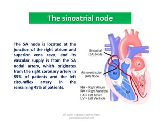
  • 3. ThesinoatrialnodeThe SA node is located at the junction of the right atrium and superior vena cava, and its vascular supply is from the SA nodal artery, which originates from the right coronary artery in 55% of patients and the left circumflex artery in the remaining 45% of patients.Dr. Carlos Augusto Azañero Inope www.carlosvirtual.com
  • 4. ThesinoatrialnodeThe SA node is innervated by parasympathetic fibers from the vagus nerve and sympathetic fibers from the thoracic sympathetic trunk.Dr. Carlos Augusto Azañero Inope www.carlosvirtual.com
  • 5. ThesinoatrialnodeIts normal discharge rate is between 60 and 100 times per minute.Dr. Carlos Augusto Azañero Inope www.carlosvirtual.com
  • 6. TheatrioventricularnodeIn the normal heart, conduction proceeds through the atrial fibers to the atrioventricular(AV) node, which is located beneath the right atrialendocardium directly above the insertion of the septal leaflet of the tricuspid valve.Dr. Carlos Augusto Azañero Inope www.carlosvirtual.com
  • 7. TheatrioventricularnodeOntheelectrocardiogram(ECG), atrial depolarization is represented bythe P wave.Dr. Carlos Augusto Azañero Inope www.carlosvirtual.com
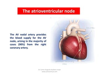
  • 8. TheatrioventricularnodeThe AV nodal artery provides the blood supply for the AV node, arising in the majority of cases (90%) from therightcoronaryartery.Dr. Carlos Augusto Azañero Inope www.carlosvirtual.com
  • 9. TheatrioventricularnodePhysiologically, the AV node slows conduction velocity to allow greater time for ventricular fillingduringdiastole.Dr. Carlos Augusto Azañero Inope www.carlosvirtual.com
  • 10. TheatrioventricularnodeIn addition, itslong refractory period protects the ventricles from excessively rapid stimulation which could cause inadequate diastolic filling time and acute cardiac failure.Dr. Carlos Augusto Azañero Inope www.carlosvirtual.com
  • 11. TheatrioventricularnodeThe AV node is innervated by the same parasympathetic and sympathetic fibers as the SA node.Dr. Carlos Augusto Azañero Inope www.carlosvirtual.com
  • 12. TheatrioventricularnodeOn the ECG, the PR interval represents the time between the onset of depolarization in the atria and the onset of depolarization in the ventricles, and is used as an estimation of AV nodal conductiontime.Dr. Carlos Augusto Azañero Inope www.carlosvirtual.com
  • 13. TheatrioventricularnodeThe normal PR interval is between 0.12 and 0.20 seconds. Prolongation of the PR interval may occur as a result of excessive vagalstimulation, drugs affecting the AV node, AV nodal ischemia, or underlying conduction systemdisease.Dr. Carlos Augusto Azañero Inope www.carlosvirtual.com
  • 14. TheHis–PurkinjesystemDepolarization proceeds from the AV node to the bundle of His, which is composed of rapidly conducting Purkinjefibers.Dr. Carlos Augusto Azañero Inope www.carlosvirtual.com
  • 15. TheHis–PurkinjesystemThe bundle divides in the muscular interventricular septum into two major branches: the left and right bundle branches, which innervate the left and right ventricle (LV and RV), respectively.Theleftbundlebranchdivides into the left anterior and left posterior fascicles.Dr. Carlos Augusto Azañero Inope www.carlosvirtual.com
  • 16. TheHis–PurkinjesystemVentricular conductionand depolarizationthrough the His–Purkinje system are represented on the surface ECG by the QRS complex. Normal QRS width is 0.06–0.10 seconds.Dr. Carlos Augusto Azañero Inope www.carlosvirtual.com
  • 17. TheHis–PurkinjesystemWidening of the QRS complexbeyond 0.12 secondsrepresentsventricular conduction delay, which can occur as a result of bundle branch blocks, aberrant conduction, electrolyteabnormalities, drugsaffectingthe myocardium, or rhythms that originate in theventricular myocardium.Dr. Carlos Augusto Azañero Inope www.carlosvirtual.com
  • 18. Ventricular repolarizationRepolarization of the ventricular myocardium is representedbytheT-wave.Dr. Carlos Augusto Azañero Inope www.carlosvirtual.com
  • 19. Ventricular repolarizationThe QT interval, which represents ventricular depolarization and repolarizationtime, is dependent to some extent onheartrate.Dr. Carlos Augusto Azañero Inope www.carlosvirtual.com
  • 20. Ventricular repolarizationA corrected QT interval (QTc) is obtained by dividing the measured QT interval by the square root of the RR interval. AnormalQTcis lessthan 0.47 seconds.Dr. Carlos Augusto Azañero Inope www.carlosvirtual.com
  • 21. Ventricular repolarizationProlongation of theQT interval can occur secondary to drug effects, electrolyte abnormalities, and congenitalabnormalities; a prolonged repolarizationperiod increases the “vulnerable period” of the ventricle, during which premature ventricular contraction can trigger a reentrant ventricular tachydysrhythmia.Dr. Carlos Augusto Azañero Inope www.carlosvirtual.com
  • 22. Dr. Carlos Augusto Azañero Inope www.carlosvirtual.com
  • 23. Dr. Carlos Augusto Azañero Inope www.carlosvirtual.comwww.carlosvirtual.comCopyright © 2007- 2011- Perú