04 joints
•Download as PPT, PDF•
5 likes•357 views

The document summarizes the main types of joints in the body - fibrous joints, cartilaginous joints, and synovial joints. It then describes the key characteristics and examples of different classes of synovial joints, including ball-and-socket, hinge, gliding, pivot, condyloid, and saddle joints. Finally, it provides details on specific synovial joints - the shoulder, elbow, wrist, hip, knee, and ankle joints, outlining their structural features, movements, and associated muscles.
Report
Share


04 joints
- 1. Prof.Sunil Chavan Prin.K.M.Kundnani Pharmacy Polytechnic JOINTS • A joint is the site at any two or more bones articulates or come together • Joints allow flexibility and movement of the skeleton and allow attachment between bones JOINTS FIBROUS CARTILAGINOUS SYNOVIAL JOINTS JOINTS JOINTS
- 2. Prof.Sunil Chavan Prin.K.M.Kundnani Pharmacy Polytechnic FIBROUS JOINTS • Bones forming these joints are linked with tough, fibrous material. • Joint does not allow any or limited movement • Joints between skull bones, sutures and joint between tooth and alveolar socket does not permit movement joint between tibia and fibula allows a limited movement. CARTILAGINOUS JOINT • Joint is formed by a pad of fibrocartilage that acts as a shock absorber • Joint allows limited degree of movement. • e.g. joints between body of vertebrates, pubis symphysis
- 3. Prof.Sunil Chavan Prin.K.M.Kundnani Pharmacy Polytechnic SYNOVIAL JOINTS Characteristics: • Capsule: Joint is surrounded & enclosed in fibrous tissue capsule which holds bones together. • Articular cartilage: Part of bones which are in capsule are covered with hyaline cartilage. This provides smooth articular surface & it is strong enough to absorb compression forces and bear weight of the body. • Synovial Membrane: It is composed of synovial (epithelial) cells. It lines the capsule, covers those part of bones not covered by cartilage and intracapsular structures that do not bear weight. It secretes synovial fluid. • Synovial Fluid: Sticky fluid provides nourishment for structures within capsule, acts as lubricant and maintains joint stability. • Intracapsular structures within capsule but outside synovial membrane e.g. fat pads & menisci in knee joint. • Extracapsular structures: ligaments, muscles or tendons that gives additional stability to joint
- 4. Prof.Sunil Chavan Prin.K.M.Kundnani Pharmacy Polytechnic Movements Possible at synovial Joints Movement Definition • Flexion Bending, usually forward but occasionally backward, e.g. knee joint • Extension Straightening or bending backward • Abduction Movement away from the midline of the body • Adduction Movement towards the midline of the body • Circumduction Combination of flexion, extension, • abduction and adduction • Rotation Movement round the long axis of a bone • Pronation Turning the palm of the hand down • Supination Turning the palm of the hand up • Inversion Turning the sole of the foot inwards • Eversion Turning the sole of the foot outwards
- 5. Prof.Sunil Chavan Prin.K.M.Kundnani Pharmacy Polytechnic Types of Synovial Joints Ball and socket: The head or ball of one bone articulates with a socket of another and the shape of the bones allows for a wide range of movements. Those possible are flexion, extension, adduction, abduction, rotation and circumduction. Examples are the shoulder and hip. Hinge joints: These allow the movements of flexion and extension only. They are the elbow, knee, ankle, the joints between the atlas and the occipital bone, and the interphalangeal joints of the fingers and toes.
- 6. Prof.Sunil Chavan Prin.K.M.Kundnani Pharmacy Polytechnic Gliding joints: The articular surfaces glide over each other, e.g. sternoclavicular joints, acromioclavicular joints and joints between the carpal bones and those between the tarsal bones. Pivot joints: Movement is round one axis (rotation), e.g. proximal and distal radioulnar joints and the joint between the atlas and the odontoid process of the axis. Condyloid joints: Movements take place round two axes, permitting flexion, extension, abduction, adduction and circumduction, e.g. the wrist, temporomandibular,metacarpophalangeal and metatarsophalangeal joints. Saddle Joints: Articulating bones fit together like man sitting on a saddle. e,g. joint between trapezium & first metacarpal bone. Range of movements is similar to condyloid but with additional flexibility.
- 7. Prof.Sunil Chavan Prin.K.M.Kundnani Pharmacy Polytechnic Shoulder joint • A ball and socket joint • Formed by the glenoid cavity of scapula and the head of humerus and is well padded with protective tissue. • Joint is stabilized by glenohumoral, coracohumeral, transverse humeral ligaments and muscles , their tendons present in shoulder. • Movements and muscles: • Flexion: coracobrachialis, deltoid and pectoralis major. • Extension: teres major, latissimus dorsi and deltoid. • Abduction: deltoid. • Adduction: combined action of flexors and extensors. • Circumduction: flexors, extensors, abductors and adductors acting in series. • Medial rotation: pectoralis major, latissimus dorsi, teres major and anterior fibres of deltoid. • Lateral rotation: posterior fibres of deltoid.
- 8. Prof.Sunil Chavan Prin.K.M.Kundnani Pharmacy Polytechnic
- 9. Prof.Sunil Chavan Prin.K.M.Kundnani Pharmacy Polytechnic Elbow Joint • Hinge Joint • Formed by trochlea and capitulum of humerus and trochlear notch of ulna and head of radius • Extracapsular Structures: anterior, posterior and lateral strengthening ligaments • Movements and muscles: • Flexion: biceps and brachialis. • Extension: triceps.
- 10. Prof.Sunil Chavan Prin.K.M.Kundnani Pharmacy Polytechnic
- 11. Prof.Sunil Chavan Prin.K.M.Kundnani Pharmacy Polytechnic Wrist Joint • Condyloid joint • Formed by distal end of radius and proximal ends of scaphoid, lunate and triquetral. A disc of fibrocartilage separates ulna from joint capsule. • Extracapsular structure-medial & lateral ligaments and anterior & posterior radiocarpal ligamnets. • Movements and muscles: • Flexion: flexor carpi radialis and the flexor carpi ulnaris. • Extension: extensors carpi radialis (longus and brevis) and the extensor carpi ulnaris. • Abduction: flexor and extensors carpi radialis. • Adduction: flexor and extensor carpi ulnaris.
- 12. Prof.Sunil Chavan Prin.K.M.Kundnani Pharmacy Polytechnic
- 13. Prof.Sunil Chavan Prin.K.M.Kundnani Pharmacy Polytechnic Hip Joint • Ball & socket joint • Formed by cup-shaped acetabulum of hip (innominate) bone & spherical head of femur • A ring of fibrocartilage attached to rim of acetabulum stabilises joint • Extracapsular structures:Iliofemoral, pubofemoral & ischiofemoral ligaments • Movements and muscles: • Flexion: psoas, iliacus, rectus femoris and sartorius. • Extension: gluteus maximus and the hamstrings. • Abduction: gluteus medius and minimus, sartorius. • Adduction: adductor group. • Lateral rotation: mainly gluteal muscles and adductor group. • Medial rotation: gluteus medius and minimus
- 14. Prof.Sunil Chavan Prin.K.M.Kundnani Pharmacy Polytechnic
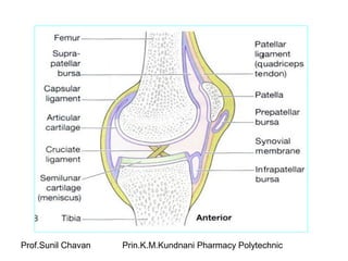
- 15. Prof.Sunil Chavan Prin.K.M.Kundnani Pharmacy Polytechnic Knee Joint • Hinge Joint-largest & most complex • Formed by condyles of femur, condyles of tibia & posterior surface of patella. • Intracapsular structures-two cruciate ligaments, many burae & pads of fat. • External ligaments-patellar ligament, an extension ofquadriceps tendon, popliteal ligament. • Movements and muscles: • Flexion: psoas, iliacus, rectus femoris and sartorius. • Extension: gluteus maximus and the hamstrings. • Abduction: gluteus medius and minimus, sartorius. • Adduction: adductor group. • Lateral rotation: mainly gluteal muscles and adductor group. • Medial rotation: gluteus medius and minimus
- 16. Prof.Sunil Chavan Prin.K.M.Kundnani Pharmacy Polytechnic
- 17. Prof.Sunil Chavan Prin.K.M.Kundnani Pharmacy Polytechnic Ankle Joint • Hinge joint • Formed by distal end of tibia & its medial malleolus, distal end of fibula and talus • External structures-deltoid and anterior,posterior, medial and lateral ligaments • Movements and muscles: • Flexion (dorsiflexion): anterior tibialis assisted by the muscles which extend the toes. • Extension (plantarflexion): gastrocnemius and soleus assisted by the muscles which flex the toes.
- 18. Prof.Sunil Chavan Prin.K.M.Kundnani Pharmacy Polytechnic