


#36391 Topic Homeland Recovery and Continuity of OperationsNum.docx
- 1. #36391 Topic: Homeland Recovery and Continuity of Operations Number of Pages: 4 (Double Spaced) Number of sources: 4 Writing Style: APA Type of document: Essay Academic Level:Undergraduate Category: Criminology Language Style: English (U.S.) Order Instructions: ATTACHED The National Security Presidential Directive-51/Homeland Security Presidential Directive-20 (NSPD-51/HSPD-20) and the National Continuity Policy Implementation Plan (NCPIP), is an effort within individual executive departments and agencies to ensure that Primary Mission Essential Functions (PMEFs) continue to be performed during a wide range of emergencies, including localized acts of nature, accidents and technological or attack-related emergencies. (https://www.fema.gov/continuity-operations) 1.Define Homeland Recovery and Continuity of Operations, including a brief overview (1- or 2-sentence summary) of each of the four phases of COOP.
- 2. 2.Select and discuss in detail one of the four phases of COOP. Assignment Expectations Assignments should be at least 3 full pages double-spaced, not counting the cover or reference page. Paper format: (a) Cover page, (b) Header, (c) Body. Submit your assignment by the last day of this module. #36415 Topic: Discussion 1 Number of Pages: 1 (Double Spaced) Number of sources: 1 Writing Style: APA Type of document: Essay Academic Level:Master Category: Psychology Language Style: English (U.S.) Order Instructions: ATTACHED I will upload the instruction
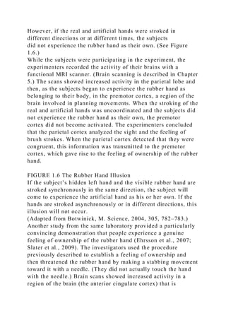
- 3. Module Chapter 5 wk6 p630 chapter 5 Methods and Strategies of Research Outline · ■ Experimental Ablation Evaluating the Behavioral Effects of Brain Damage Producing Brain Lesions Stereotaxic Surgery Histological Methods Tracing Neural Connections Studying the Structure of the Living Human Brain Section Summary · ■ Recording and Stimulating Neural Activity Recording Neural Activity Recording the Brain’s Metabolic and Synaptic Activity Stimulating Neural Activity Section Summary · ■ Neurochemical Methods Finding Neurons That Produce Particular Neurochemicals Localizing Particular Receptors Measuring Chemicals Secreted in the Brain Section Summary · ■ Genetic Methods Twin Studies Adoption Studies Genomic Studies Targeted Mutations Antisense Oligonucleotides Section Summary In July 1982, several young people began showing up at neurology clinics in northern California displaying dramatic, severe symptoms (Langston et al., 1983). The most severely affected patients were almost totally paralyzed. They were unable to speak intelligibly, they drooled constantly, and their eyes were open with a fixed stare. Others, less severely affected, walked with a slow, shuffling gait and moved slowly
- 4. and with great difficulty. The symptoms looked like those of Parkinson’s disease, but that disorder has a very gradual onset. In addition, it rarely strikes people before late middle age, and the patients were all in their twenties or early thirties. The common factor linking these patients was intravenous drug use; all of them had been taking a “new heroin,” a synthetic opiate related to meperidine (Demerol). Some detective work revealed that the illicit drug was contaminated with MPTP, a toxic chemical that damaged dopaminergic neurons and caused the neurological symptoms. Because the symptoms looked like those of Parkinson’s disease, the patients were given L-DOPA, the drug that is used to treat this disease, and they all showed significant improvement in their symptoms. Unfortunately, the improvement was temporary; the drug lost its effectiveness. Fetal transplantation, an experimental neurosurgical method of treating parkinsonism, has shown some promise. The rationale for the procedure is this: The symptoms of parkinsonism, whether from Parkinson’s disease or the toxic effects of MPTP, are caused by the lack of dopamine in the caudate nucleus and putamen. There is at present no way to induce the brain to grow new dopaminergic neurons. However, if dopamine-secreting neurons can be introduced into the caudate nucleus and putamen and if they survive and secrete dopamine, then perhaps the parkinsonian symptoms will diminish. Because the implanted neurons must be healthy and vigorous and not trigger the recipient’s immune system, the logical source for them is aborted human fetuses—or, perhaps someday, cultures of stem cells that have been induced to become dopamine-secreting neurons. At least one of the people with MPTP poisoning received such a transplant. (Let’s call him Mr. B.) Before the operation took place, Mr. B. was given an injection of radioactive L-DOPA, the precursor for dopamine. Then, one hour later, he was wheeled into a small room that housed a PET scanner. His head was positioned in the scanner, and for the next several minutes the machine gathered data from subatomic particles that were
- 5. emitted as the radioactive L-DOPA in his head broke down. A few weeks later, Mr. B. entered the hospital for his surgery. Technicians removed dopaminergic neurons from the substantia nigra of several fetal brains and prepared them for implantation into Mr. B.’s brain. Mr. B. was anesthetized, and the surgeon made cuts in his scalp to expose parts of his skull. He attached the frame of a stereotaxic apparatus to Mr. B’s skull, made some measurements, and then drilled several holes. He used the stereotaxic apparatus to guide the injections of the fetal neurons into Mr. B.’s caudate nucleus and putamen. Once the injections were complete, the surgeon removed the stereotaxic frame and sutured the incisions he had made in the scalp. The operation was quite successful; Mr. B. recovered much of his motor control. A little more than a year later, he was again given an injection of radioactive L-DOPA, and again his head was placed in the PET scanner. The results of the second scan showed what his recovery implied: The cells had survived and were secreting dopamine. Study of the physiology of behavior involves the efforts of scientists in many disciplines, including physiology, neuroanatomy, biochemistry, psychology, endocrinology, and histology. Pursuing a research project in behavioral neuroscience requires competence in many experimental techniques. Because different procedures often produce contradictory results, investigators must be familiar with the advantages and limitations of the methods they employ. Scientific investigation entails a process of asking questions of nature. The method that is used frames the question. Often we receive a puzzling answer, only to realize later that we were not asking the question we thought we were. As we will see, the best conclusions about the physiology of behavior are made not by any single experiment but by a program of research that enables us to compare the results of studies that approach the problem with different methods. An enormous—and bewildering—array of research methods is available to the investigator. If I merely presented a catalog of
- 6. them, it would not be surprising if you got lost—or simply lost interest. Instead, I will present only the most important and commonly used procedures, organized around a few problems that researchers have studied. This way, it should be easier to see the types of information provided by various research methods and to understand their advantages and disadvantages. It will also permit me to describe the strategies that researchers employ as they follow up the results of one experiment by designing and executing another one. Experimental Ablation One of the most important research methods used to investigate brain functions involves destroying part of the brain and evaluating the animal’s subsequent behavior. This method is called experimental ablation (from the Latin word ablatus, a “carrying away”). In most cases, experimental ablation does not involve the removal of brain tissue; instead, the researcher destroys some tissue and leaves it in place. Experimental ablation is the oldest method used in neuroscience, and it remains in common use today. experimental ablation The removal or destruction of a portion of the brain of a laboratory animal; presumably, the functions that can no longer be performed are the ones the region previously controlled. Evaluating the Behavioral Effects of Brain Damage A lesion is a wound or injury, and a researcher who destroys part of the brain usually refers to the damage as a brain lesion. Experiments in which part of the brain is damaged and the animal’s behavior is subsequently observed are called lesion studies. The rationale for lesion studies is that the function of an area of the brain can be inferred from the behaviors that the animal can no longer perform after the area has been damaged. For example, if, after part of the brain has been destroyed, an animal can no longer perform tasks that require vision, we can conclude that the animal is blind—and that the damaged area plays some role in vision. lesion study A synonym for experimental ablation.
- 7. Just what can we learn from lesion studies? Our goal is to discover what functions are performed by different regions of the brain and then to understand how these functions are combined to accomplish particular behaviors. The distinction between brain function and behavior is an important one. Circuits within the brain perform functions, not behaviors. No one brain region or neural circuit is solely responsible for a behavior; each region performs a function (or set of functions) that contributes to performance of the behavior. For example, the act of reading involves functions required for controlling eye movements, focusing the lens of the eye, perceiving and recognizing words and letters, comprehending the meaning of the words, and so on. Some of these functions also participate in other behaviors; for example, controlling eye movement and focusing are required for any task that involves looking, and brain mechanisms used for comprehending the meanings of words also participate in comprehending speech. The researcher’s task is to understand the functions that are required for performing a particular behavior and to determine what circuits of neurons in the brain are responsible for each of these functions. The interpretation of lesion studies is complicated by the fact that all regions of the brain are interconnected. Suppose that we have a good understanding of the functions required for performance of a particular behavior. We find that damage to brain structure X impairs a particular behavior. Can we necessarily conclude that a function essential to this behavior is performed by circuits of neurons located in structure X? Unfortunately, we cannot. The functions we are interested in may actually be performed by neural circuits located elsewhere in the brain. Damage to structure X may simply interfere with the normal operation of the neural circuits in structure Y. Producing Brain Lesions How do we produce brain lesions? Usually, we want to destroy regions that are hidden away in the depths of the brain. Brain lesions of subcortical regions (regions located beneath the
- 8. cortex) are usually produced by passing electrical current through a stainless steel wire that is coated with an insulating varnish except for the very tip. We guide the wire stereotaxically so that its end reaches the appropriate location. (Stereotaxic surgery is described in the next subsection.) Then we turn on a lesion-making device, which produces radio frequency (RF) current—alternating current of a very high frequency. The passage of the current through the brain tissue produces heat that kills cells in the region surrounding the tip of the electrode. (See Figure 5.1.) Lesions produced by these means destroy everything in the vicinity of the electrode tip, including neural cell bodies and the axons of neurons that pass through the region. A more selective method of producing brain lesions employs an excitatory amino acid such as kainic acid, which kills neurons by stimulating them to death. (As we saw in Chapter 4, kainic acid stimulates glutamate receptors.) Lesions produced in this way are referred to as excitotoxic lesions. When an excitatory amino acid is injected through a cannula (a small metal tube) into a region of the brain, the chemical destroys neural cell bodies in the vicinity but spares axons that belong to different neurons that happen to pass nearby. (See Figure 5.2.) This selectivity permits the investigator to determine whether the behavioral effects of destroying a particular brain structure are caused by the death of neurons located there or by the destruction of axons that pass nearby. For example, some researchers discovered that RF lesions of a particular region in the brain stem abolished REM sleep; therefore, they believed that this region was involved in the production of this stage of sleep. (REM sleep is the stage of sleep during which dreaming occurs.) But later studies showed that when kainic acid was used to destroy the neurons located there, the animals’ sleep was notaffected. Therefore, the RF lesions must have altered sleep by destroying the axons that pass through the area. excitotoxic lesion (ek sigh towtoksik) A brain lesion produced by intracerebral injection of an excitatory amino acid, such as
- 9. kainic acid. FIGURE 5.1 Radio Frequency Lesion The arrows point to very small lesions produced by passing radio frequency current through the tips of stainless steel electrodes placed in the medial preoptic nucleus of a rat brain. The oblong hole in the middle of the photograph is the third ventricle. (Frontal section, cell-body stain.) (From Turkenburg, J. L., Swaab, D. F., Endert, E., et al. Brain Research Bulletin, 1988, 21, 215–224. Reprinted with permission.) Even more specific methods of targeting and killing particular types of neurons are available. For example, molecular biologists have devised ways to conjugate (attach together) saporin, a toxic protein, and antibodies that will bind with particular proteins found only on certain types of neurons in the brain. The antibodies target these proteins, and the saporin kills the cells to which the proteins are attached. FIGURE 5.2 Excitotoxic Lesion (a) Section through a normal hippocampus of a rat brain. (b) A lesion produced by infusion of an excitatory amino acid in a region of the hippocampus. Arrowheads mark the ends of the region in which neurons have been destroyed. (Courtesy of Benno Roozendaal, University of California, Irvine.) Note that, when we produce subcortical lesions by passing RF current through an electrode or infusing a chemical through a cannula, we always cause additional damage to the brain. When we pass an electrode or a cannula through the brain to get to our target, we inevitably cause a small amount of damage even before turning on the lesion maker or starting the infusion. Thus, we cannot simply compare the behavior of brain-lesioned animals with that of unoperated control animals; the incidental damage to the brain regions above the lesion may actually be responsible for some of the behavioral deficits we see. What we
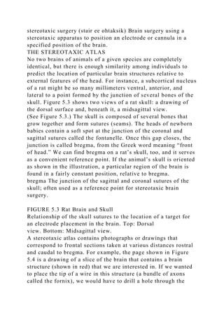
- 10. do is operate on a group of animals and produce sham lesions. To do so, we anesthetize each animal, put it in the stereotaxic apparatus (described below), cut open the scalp, drill the holes, insert the electrode or cannula, and lower it to the proper depth. In other words, we do everything we would do to produce the lesion except turn on the lesion maker or start the infusion. This group of animals serves as a control group; if the behavior of the animals with brain lesions is different from that of the sham-operated control animals, we can conclude that the lesions caused the behavioral deficits. (As you can see, a sham lesion serves the same purpose as a placebo does in a pharmacology study.) sham lesion A placebo procedure that duplicates all the steps of producing a brain lesion except the one that actually causes the brain damage. Most of the time, investigators produce permanent brain lesions, but sometimes it is advantageous to disrupt the activity of a particular region of the brain temporarily. The easiest way to do so is to inject a local anesthetic or a drug called muscimol into the appropriate part of the brain. The anesthetic blocks action potentials in axons entering or leaving that region, thus effectively producing a temporary lesion (usually called a reversible brain lesion). Muscimol, a drug that stimulates GABA receptors, inactivates a region of the brain by inhibiting the neurons located there. (You will recall that GABA is the most important inhibitory neurotransmitter in the brain.) Stereotaxic Surgery So how do we get the tip of an electrode or cannula to a precise location in the depths of an animal’s brain? The answer is stereotaxic surgery. Stereotaxis literally means “solid arrangement”; more specifically, it refers to the ability to locate objects in space. A stereotaxic apparatus contains a holder that fixes the animal’s head in a standard position and a carrier that moves an electrode or a cannula through measured distances in all three axes of space. However, to perform stereotaxic surgery, one must first study a stereotaxic atlas.
- 11. stereotaxic surgery (stair ee ohtaksik) Brain surgery using a stereotaxic apparatus to position an electrode or cannula in a specified position of the brain. THE STEREOTAXIC ATLAS No two brains of animals of a given species are completely identical, but there is enough similarity among individuals to predict the location of particular brain structures relative to external features of the head. For instance, a subcortical nucleus of a rat might be so many millimeters ventral, anterior, and lateral to a point formed by the junction of several bones of the skull. Figure 5.3 shows two views of a rat skull: a drawing of the dorsal surface and, beneath it, a midsagittal view. (See Figure 5.3.) The skull is composed of several bones that grow together and form sutures (seams). The heads of newborn babies contain a soft spot at the junction of the coronal and sagittal sutures called the fontanelle. Once this gap closes, the junction is called bregma, from the Greek word meaning “front of head.” We can find bregma on a rat’s skull, too, and it serves as a convenient reference point. If the animal’s skull is oriented as shown in the illustration, a particular region of the brain is found in a fairly constant position, relative to bregma. bregma The junction of the sagittal and coronal sutures of the skull; often used as a reference point for stereotaxic brain surgery. FIGURE 5.3 Rat Brain and Skull Relationship of the skull sutures to the location of a target for an electrode placement in the brain. Top: Dorsal view. Bottom: Midsagittal view. A stereotaxic atlas contains photographs or drawings that correspond to frontal sections taken at various distances rostral and caudal to bregma. For example, the page shown in Figure 5.4 is a drawing of a slice of the brain that contains a brain structure (shown in red) that we are interested in. If we wanted to place the tip of a wire in this structure (a bundle of axons called the fornix), we would have to drill a hole through the
- 12. skull immediately above it. (See Figure 5.4.) Each page of the stereotaxic atlas is labeled according to the distance of the section anterior or posterior to bregma. The grid on each page indicates distances of brain structures ventral to the top of the skull and lateral to the midline. To place the tip of a wire in the fornix, we would drill a hole above the target and then lower the electrode through the hole until the tip was at the correct depth, relative to the skull height at bregma. (Compare Figures 5.3 and 5.4.) Thus, by finding a neural structure (which we cannot see in our animal) on one of the pages of a stereotaxic atlas, we can determine the structure’s location relative to bregma (which we can see). Note that, because of variations in different strains and ages of animals, the atlas gives only an approximate location. We always have to try out a new set of coordinates, slice and stain the animal’s brain, see the actual location of the lesion, correct the numbers, and try again. (Slicing and staining of brains are described later.) stereotaxic atlas A collection of drawings of sections of the brain of a particular animal with measurements that provide coordinates for stereotaxic surgery. FIGURE 5.4 Stereotaxic Atlas This sample page from a stereotaxic atlas of the rat brain shows the target (the fornix) in red. Labels have been removed for the sake of clarity. (Adapted from Swanson, L. W. Brain Maps: Structure of the Rat Brain. New York: Elsevier, 1992.) THE STEREOTAXIC APPARATUS A stereotaxic apparatus operates on simple principles. The device includes a head holder, which maintains the animal’s skull in the proper orientation, a holder for the electrode, and a calibrated mechanism that moves the electrode holder in measured distances along the three axes: anterior–posterior, dorsal–ventral, and lateral–medial. Figure 5.5 illustrates a stereotaxic apparatus designed for small animals; various head holders can be used to outfit this device for such diverse species
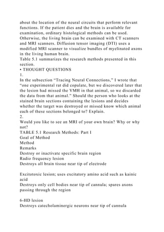
- 13. as rats, mice, hamsters, pigeons, and turtles. (See Figure 5.5.) stereotaxic apparatus A device that permits a surgeon to position an electrode or cannula into a specific part of the brain. Once we obtain the coordinates from a stereotaxic atlas, we anesthetize the animal, place it in the apparatus, and cut the scalp open. We locate bregma, dial in the appropriate numbers on the stereotaxic apparatus, drill a hole through the skull, and lower the device into the brain by the correct amount. Now the tip of the cannula or electrode is where we want it to be, and we are ready to produce the lesion. Of course, stereotaxic surgery may be used for purposes other than lesion production. Wires placed in the brain can be used to stimulate neurons as well as to destroy them, and drugs can be injected that stimulate neurons or block specific receptors. We can attach cannulas or wires permanently by following a procedure that will be described later in this chapter. In all cases, once surgery is complete, the wound is sewn together, and the animal is taken out of the stereotaxic apparatus and allowed to recover from the anesthetic. FIGURE 5.5 Stereotaxic Apparatus This apparatus is used for performing brain surgery on rats. Stereotaxic apparatuses are made for humans, by the way. Sometimes a neurosurgeon produces subcortical lesions—for example, to reduce the symptoms of Parkinson’s disease. Usually, the surgeon uses multiple landmarks and verifies the location of the wire (or other device) inserted into the brain by taking brain scans or recording the activity of the neurons in that region before producing a brain lesion. (See Figure 5.6.) Histological Methods After producing a brain lesion and observing its effects on an animal’s behavior, we must slice and stain the brain so that we can observe it under the microscope and see the location of the lesion. Brain lesions often miss the mark, so we have to verify the precise location of the brain damage after testing the animal behaviorally. To do so, we must fix, slice, stain, and examine
- 14. the brain. Together, these procedures are referred to as histological methods. (The prefix histo- refers to body tissue.) FIGURE 5.6 Stereotaxic Surgery on a Human Patient (Photograph courtesy of John W. Snell, University of Virginia Health System.) FIXATION AND SECTIONING If we hope to study the tissue in the form it had at the time of the organism’s death, we must destroy the autolytic enzymes (autolytic means “self-dissolving”), which will otherwise turn the tissue into mush. The tissue must also be preserved to prevent its decomposition by bacteria or molds. To achieve both of these objectives, we place the neural tissue in a fixative. The most commonly used fixative is formalin, an aqueous solution of formaldehyde, a gas. Formalin halts autolysis, hardens the very soft and fragile brain, and kills any microorganisms that might destroy it. fixative A chemical such as formalin; used to prepare and preserve body tissue. formalin (forma lin) The aqueous solution of formaldehyde gas; the most commonly used tissue fixative. Before the brain is fixed (that is, put into a fixative solution), it is usually perfused. Perfusion of tissue (literally, “a pouring through”) entails removal of the blood and its replacement with another fluid. The animal’s brain is perfused because better histological results are obtained when no blood is present in the tissue. The animal whose brain is to be studied is humanely killed with an overdose of a general anesthetic. Blood vessels are opened so that the blood can be drained from them and replaced with a dilute salt solution. The brain is removed from the skull and placed in a container filled with the fixative. perfusion (perfewzhun) The process by which an animal’s blood is replaced by a fluid such as a saline solution or a fixative in preparing the brain for histological examination. Once the brain has been fixed, we must slice it into thin
- 15. sections and stain various cellular structures to see anatomical details. Slicing is done with a microtome (literally, “that which slices small”). Slices prepared for examination under a light microscope are typically 10 to 80 μm in thickness; those prepared for the electron microscope are generally cut at less than l μm. (For some reason, slices of brain tissue are usually referred to as sections.) microtome (mykrow tome) An instrument that produces very thin slices of body tissues. A microtome contains three parts: a knife, a platform on which to mount the tissue, and a mechanism that advances the knife (or the platform) the correct amount after each slice so that another section can be cut. In most cases the platform includes an attachment that freezes the brain to make it hard enough to be cut into thin sections. Figure 5.7 shows a microtome. The knife holder slides forward on an oiled rail and takes a section off the top of the tissue mounted on the platform. The platform automatically rises by a predetermined amount as the knife and holder are pushed back so that the next forward movement of the knife takes off another section. (See Figure 5.7.) FIGURE 5.7 A Microtome After the tissue is cut, we attach the slices to glass microscope slides. We can then stain the tissue by putting the entire slide into various chemical solutions. Finally, we cover the stained sections with a small amount of a transparent liquid known as a mounting medium and place a very thin glass coverslip over the sections. The mounting medium keeps the coverslip in position. Simulate histological methods on MyPsychLab to see videos of these procedures. STAINING If you looked at an unstained section of brain tissue under a microscope, you would be able to see the outlines of some large cellular masses and the more prominent fiber bundles. However, no fine details would be revealed. For this reason the study of microscopic neuroanatomy requires special histological stains.
- 16. Researchers have developed many different stains to identify specific substances within and outside of cells. For verifying the location of a brain lesion, we will use one of the simplest: a cell-body stain. In the late nineteenth century Franz Nissl, a German neurologist, discovered that a dye known as methylene blue would stain the cell bodies of brain tissue. The material that takes up the dye, known as the Nissl substance, consists of RNA, DNA, and associated proteins located in the nucleus and scattered, in the form of granules, in the cytoplasm. Many dyes besides methylene blue can be used to stain cell bodies found in slices of the brain, but the most frequently used is cresyl violet. Incidentally, the dyes were not developed specifically for histological purposes; they were originally formulated for use in dyeing cloth. The discovery of cell-body stains made it possible to identify nuclear masses in the brain. Figure 5.8 shows a frontal section of a cat brain stained with cresyl violet. Note that you can observe fiber bundles by their lighter appearance; they do not take up the stain. (See Figure 5.8.) The stain is not selective for neural cell bodies; all cells are stained, neurons and glia alike. It is up to the investigator to determine which is which— by size, shape, and location. ELECTRON MICROSCOPY The light microscope is limited in its ability to resolve extremely small details. Because of the nature of light itself, magnification of more than approximately 1500 times does not add any detail. To see such small anatomical structures as synaptic vesicles and details of cell organelles, investigators must use a transmission electron microscope. A beam of electrons is passed through a thin slice of the tissue to be examined. The beam of electrons casts a shadow of the tissue on a fluorescent screen, which can be photographed or scanned into a computer. Electron photomicrographs produced in this way can provide information about structural details on the order of a few tens of nanometers. (See Figure 5.9.)
- 17. transmission electron microscope A microscope that passes a focused beam of electrons through thin slices of tissue to reveal extremely small details. FIGURE 5.8 Frontal Section of a Cat Brain The section is stained with cresyl violet, a cell-body stain. The arrowheads point to nuclei, or groups of cell bodies. (Histological material courtesy of Mary Carlson.) FIGURE 5.9 Electron Photomicrograph This electron photomicrograph shows a section through an axodendritic synapse. Two synaptic regions are indicated by arrows, and a circle indicates a region of pinocytosis in an adjacent terminal button, presumably representing recycling of vesicular membrane. T = terminal button; f = microfilaments; M = mitochondrion. (From Rockel, A. J., and Jones, E. G. Journal of Comparative Neurology, 1973, 147, 61–92. Reprinted with permission.) A scanning electron microscope provides less magnification than a standard transmission electron microscope, which transmits the electron beam through the tissue. However, it shows objects in three dimensions. The microscope scans the tissue with a moving beam of electrons. The information from the reflection of the beam is received by a detector, and a computer produces a remarkably detailed three-dimensional view. (See Figure 5.10.) scanning electron microscope A microscope that provides three- dimensional information about the shape of the surface of a small object by scanning the object with a thin beam of electrons. CONFOCAL LASER SCANNING MICROSCOPY Conventional microscopy or transmission electron microscopy requires that the tissue be sliced into thin sections. The advent of the confocal laser scanning microscope makes it possible to see details inside thick sections of tissue or even in slabs of tissue maintained in tissue cultures or in the upper layers of
- 18. tissue in the exposed living brain. The confocal microscope requires that the cells or parts of cells of interest be stained with a fluorescent dye. (This procedure, called immunocytochemistry, is described in next section of this chapter.) For example, neurons that produce a particular peptide can be labeled with a fluorescent dye. A beam of light of a particular wavelength is produced by a laser and reflected off of a dichroicmirror—a special mirror that transmits light of certain wavelengths and reflects light of other wavelengths. Lenses in the microscope focus the laser light at a particular depth in the tissue. This light triggers fluorescence in the tissue, which passes through the lenses and is transmitted through the dichroic mirror to a pinhole aperture. This aperture blocks extraneous light caused by scattering within the tissue. The light that passes through the aperture is measured by a detector. Two moving mirrors cause the laser light to scan the tissue, which provides the computer with the information it needs to form an image of a slice of tissue located at a particular depth within the sample. If multiple scans are made while the location of the aperture is moved, a stack of images of slices through the tissue—remember, this can be living tissue—can be obtained. (See Figure 5.11.) confocal laser scanning microscope A microscope that provides high-resolution images of various depths of thick tissue that contains fluorescent molecules by scanning the tissue with light from a laser beam. FIGURE 5.10 Neurons and Glia This scanning electron micrograph shows neurons and glia. (From Tissues and Organs: A Text-Atlas of Scanning Electron Microscopy, by Richard G. Kessel and Randy H. Kardon. Copyright © 1979 by W. H. Freeman and Co. Reprinted by permission of Barbara Kessel and Randy Kardon.) FIGURE 5.11 Confocal Microscope A laser scanning confocal microscope is shown in this
- 19. simplified schematic diagram. FIGURE 5.12 Branches of Dendrites These photographs of branches of dendrites of hippocampal neurons of a living mouse taken through a laser scanning confocal microscope. The images show that seizures caused by injection of drugs caused the disappearance of some dendritic spines. (From Mizrahi, A., Crowley, J. C., Shtoyerman, E., and Katz, L. C. Journal of Neuroscience, 2004, 24, 3147–3151. Copyright © 2004 by the Society for Neuroscience. Reprinted by permission of the Society for Neuroscience.) Figure 5.12 illustrates the use of confocal microscopy of the hippocampus of living anesthetized mice (Mizrahi et al., 2004). The skull was opened, and overlying cortex was dissected away so that images could be taken of the interior of the hippocampus. Molecular genetic methods had been used to insert a gene into the animals’ DNA that produced a fluorescent protein dye in certain neurons in the hippocampus. Mizrahi and his colleagues obtained images of individual dendrites of these neurons before and after they induced seizures in the animals by administering excitatory drugs. Images made before the seizures are shown in green, and images made 4 to 5 hours after the seizures are shown in red. As you can see, the animals in which the seizures were induced showed a loss of dendritic spines (arrows), but there was no loss of spines in control animals. (See Figure 5.12.) Tracing Neural Connections Let’s suppose that we were interested in discovering the neural mechanisms responsible for reproductive behavior. To start out, we wanted to study the physiology of sexual behavior of female rats. On the basis of some hints we received by reading reports of experiments by other researchers published in scientific journals, we performed stereotaxic surgery on two groups of female rats. We made a lesion in the ventromedial nucleus of the hypothalamus (VMH) of the rats in the experimental group
- 20. and performed sham surgery on the rats in the control group. After a few days’ recovery we placed the animals (individually, of course) with male rats. The females in the control group responded positively to the males’ attention; they engaged in courting behavior followed by copulation. However, the females with the VMH lesions rejected the males’ attention and refused to copulate with them. We confirmed with histology that the VMH was indeed destroyed in the brains of the experimental animals. (One experimental rat did copulate, but we discovered later that the lesion had missed the VMH in that animal, so we discarded the data from that animal.) The results of our experiment indicate that neurons in the VMH appear to play a role in functions required for copulatory behavior in females. (By the way, it turns out that these lesions do not affect copulatory behavior in males.) So where do we go from here? What is the next step? In fact, there are many questions that we could pursue. One question concerns the system of brain structures that participate in female copulatory behavior. Certainly, the VMH does not stand alone; it receives inputs from other structures and sends outputs to still others. Copulation requires integration of visual, tactile, and olfactory perceptions and organization of patterns of movements in response to those of the partner. In addition, the entire network requires activation by the appropriate sex hormones. What is the precise role of the VMH in this complicated system? Before we can hope to answer this question, we must know more about the connections of the VMH with the rest of the brain. What structures send their axons to the VMH, and to what structures does the VMH, in turn, send its axons? Once we know what the connections are, we can investigate the role of these structures and the nature of their interactions. (See Figure 5.13.) How do we investigate the connections of the VMH? The question cannot be answered by means of histological procedures that stain all neurons, such as cell-body stains. If we look closely at a brain that has been prepared by these means,
- 21. we see only a tangled mass of neurons. But in recent years, researchers have developed very precise methods that make specific neurons stand out from all of the others. TRACING EFFERENT AXONS Eventually, the VMH must affect behavior. That is, neurons in the VMH must send axons to parts of the brain that contain neurons that are responsible for muscular movements. The pathway is probably not direct; more likely, neurons in the VMH affect neurons in other structures, which influence those in yet other structures until, eventually, the appropriate motor neurons are stimulated. To discover this system, we want to be able to identify the paths followed by axons leaving the VMH. In other words, we want to trace the efferent axons of this structure. FIGURE 5.13 Tracing Neural Circuits Once we know that a particular brain region is involved in a particular function, we may ask what structures provide inputs to the region and what structures receive outputs from it. We will use an anterograde labeling method to trace these axons. (Anterograde means “moving forward.”) Anterograde labeling methods employ chemicals that are taken up by dendrites or cell bodies and are then transported through the axons toward the terminal buttons. anterograde labeling method (annter oh grade) A histological method that labels the axons and terminal buttons of neurons whose cell bodies are located in a particular region. Over the years, neuroscientists have developed several different methods for tracing the pathways followed by efferent axons. For example, to discover the destination of the efferent axons of neurons located within the VMH, we could inject a minute quantity of PHA-L (a protein found in kidney beans) into that nucleus. (We would use a stereotaxic apparatus to do so, of course.) The molecules of PHA-L are taken up by dendrites and are transported through the soma to the axon, where they travel by means of fast axoplasmic transport to the terminal buttons.
- 22. Within a few days the cells are filled in their entirety with molecules of PHA-L: dendrites, soma, axons and all their branches, and terminal buttons. Then we kill the animal, slice the brain, and mount the sections on microscope slides. A special immunocytochemicalmethod is used to make the molecules of PHA-L visible, and the slides are examined under a microscope. (See Figure 5.14.) PHA-L Phaseolus vulgaris leukoagglutinin; a protein derived from kidney beans and used as an anterograde tracer; taken up by dendrites and cell bodies and carried to the ends of the axons. Immunocytochemical methods take advantage of the immune reaction. The body’s immune system has the ability to produce antibodies in response to antigens. Antigens are proteins (or peptides), such as those found on the surface of bacteria or viruses. Antibodies, which are also proteins, are produced by white blood cells to destroy invading microorganisms. Antibodies either are secreted by white blood cells or are located on their surface, in the way neurotransmitter receptors are located on the surface of neurons. When the antigens present on the surface of an invading microorganism come into contact with the antibodies that recognize them, the antibodies trigger an attack on the invader by the white blood cells. immunocytochemical method A histological method that uses radioactive antibodies or antibodies bound with a dye molecule to indicate the presence of particular proteins of peptides. FIGURE 5.14 Using PHA-L to Trace Efferent Axons Molecular biologists have developed methods for producing antibodies to any peptide or protein. The antibody molecules are attached to various types of dye molecules. Some of these dyes react with other chemicals and stain the tissue a brown color. Others are fluorescent; they glow when they are exposed to light of a particular wavelength. To determine where the peptide or protein (the antigen) is located in the brain, the investigator places fresh slices of brain tissue in a solution that contains the
- 23. antibody/dye molecules. The antibodies attach themselves to their antigen. When the investigator examines the slices with a microscope (under light of a particular wavelength in the case of fluorescent dyes), he or she can see which parts of the brain—even which individual neurons—contain the antigen. Figure 5.15 shows how PHA-L can be used to identify the efferents of a particular region of the brain. Molecules of this chemical were injected into the VMH. Two days later, after the PHA-L had been taken up by the neurons in this region and transported to the ends of their axons, the animal was killed. Figure 5.15shows a photomicrograph of the periaqueductal gray matter (PAG). This region contains some labeled axons and terminal buttons (gold color), which proves that some of the efferent axons of the VMH terminate in the PAG. (See Figure 5.15.) To continue our study of the role of the VMH in female sexual behavior, we would find the structures that receive information from neurons in the VMH (such as the PAG) and see what happens when each of them is destroyed. Let’s suppose that damage to some of these structures also impairs female sexual behavior. We will inject these structures with PHA-L and see where their axons go. Eventually, we will discover the relevant pathways from the VMH to the motor neurons whose activity is necessary for copulatory behavior. (In fact, researchers have done so, and some of their results are presented in Chapter 10.) FIGURE 5.15 Anterograde Tracing Method PHA-L was injected into the ventromedial nucleus of the hypothalamus, where it was taken up by dendrites and carried through the cells’ axons to their terminal buttons. The section shows labeled axons and terminal buttons in the periaqueductal gray matter. (Courtesy of Kirsten Nielsen Ricciardi and Jeffrey Blaustein, University of Massachusetts.) TRACING AFFERENT AXONS Tracing efferent axons from the VMH will tell us only part of
- 24. the story about the neural circuitry involved in female sexual behavior: the part between the VMH and the motor neurons. What about the circuits before the VMH? Is the VMH somehow involved in the analysis of sensory information (such as the sight, odor, or touch of the male)? Or perhaps the activating effect of a female’s sex hormones on her behavior act through the VMH or through neurons whose axons form synapses there. To discover the parts of the brain that are involved in the “upstream” components of the neural circuitry, we need to find the inputs of the VMH—its afferent connections. To do so, we will employ a retrograde labeling method. retrograde labeling method A histological method that labels cell bodies that give rise to the terminal buttons that form synapses with cells in a particular region. Retrograde means “moving backward.” Retrograde labeling methods employ chemicals that are taken up by terminal buttons and carried back through the axons toward the cell bodies. The method for identifying the afferent inputs to a particular region of the brain is similar to the method used for identifying its efferents. First, we inject a small quantity of a chemical called fluorogold into the VMH. The chemical is taken up by terminal buttons and is transported back by means of retrograde axoplasmic transport to the cell bodies. A few days later we kill the animal, slice its brain, and examine the tissue under light of the appropriate wavelength. The molecules of fluorogold fluoresce under this light. We discover that the medial amygdala is one of the regions that provides input to the VMH. (See Figure 5.16.) fluorogold (flewroh gold) A dye that serves as a retrograde label; taken up by terminal buttons and carried back to the cell bodies. The anterograde and retrograde labeling methods that I have described identify a single link in a chain of neurons—neurons whose axons enter or leave a particular brain region. Transneuronal tracing methods identify a series of two, three, or more neurons that form serial synaptic connections
- 25. with each other. The most effective retrograde transneuronal tracing method uses a pseudorabies virus—a weakened form of a pig herpes virus that was originally developed as a vaccine. For anterograde transneuronal tracing, a variety of the herpes simplex virus, similar to the one that causes cold sores, is used. The virus is injected directly into a brain region, is taken up by neurons there, and infects them. The virus spreads throughout the infected neurons and is eventually released by the terminal buttons, passing the infection to other neurons that form synaptic connections with them. pseudorabies virus A weakened form of a pig herpes virus used for retrograde transneuronal tracing, which labels a series of neurons that are interconnected synaptically. herpes simplex virus A form of herpes virus used for anterograde transneuronal tracing, which labels a series of neurons that are interconnected synaptically. FIGURE 5.16 Retrograde Tracing Method Fluorogold was injected in the VMH, where it was taken up by terminal buttons and transported back through the axons to their cell bodies. The photograph shows these cell bodies, located in the medial amygdala. (Courtesy of Yvon Delville, University of Massachusetts Medical School.) After the animal is killed and the brain is sliced, immunocytochemical methods are used to localize a protein produced by the virus. For example, Daniels, Miselis, and Flanagan-Cato (1999) injected pseudorabies virus in the muscles responsible for female rats’ mating posture. After a few days, the rats were killed, and their brains were examined for evidence of viral infection. The study indicated that the virus found its way up the motor nerves to the motor neurons in the spinal cord, then to the reticular formation of the medulla, then to the periaqueductal gray matter, and finally to the VMH. These results confirm the results of the anterograde and retrograde labeling methods I just described. (Labeled neurons
- 26. were found in other structures as well, but they are not relevant to this discussion.) Together, anterograde and retrograde labeling methods— including transneuronal methods—enable us to discover circuits of interconnected neurons. Thus, these methods help to provide us with a “wiring diagram” of the brain. (See Figure 5.17.) Armed with other research methods (including some to be described later in this chapter), we can try to discover the functions of each component of this circuit. Studying the Structure of the Living Human Brain There are many good reasons to investigate the functions of brains of animals other than humans. For one thing, we can compare the results of studies made with different species in order to make some inferences about the evolution of various neural systems. Even if our primary interest is in the functions of the human brain, we certainly cannot ask people to submit to brain surgery for the purposes of research. But diseases and accidents do occasionally damage the human brain, and if we know where the damage occurs, we can study the people’s behavior and try to make the same sorts of inferences we make with deliberately produced brain lesions in laboratory animals. The problem is, where is the lesion? FIGURE 5.17 Results of Tracing Methods The figure shows one of the inputs to the VMH and one of the outputs, as revealed by anterograde and retrograde labeling methods. In past years a researcher might have studied the behavior of a person with brain damage and never found out exactly where the lesion was located. The only way to be sure was to obtain the patient’s brain when he or she died and examine slices of it under a microscope. But it was often impossible to do this. Sometimes the patient outlived the researcher. Sometimes the patient moved out of town. Sometimes (often, perhaps) the family refused permission for an autopsy. Because of these practical problems, study of the behavioral effects of damage to
- 27. specific parts of the human brain made rather slow progress. Recent advances in X-ray techniques and computers have led to the development of several methods for studying the anatomy of the living brain. These advances permit researchers to study the location and extent of brain damage while the patient is still living. The first method that was developed is called computerized tomography (CT) (from the Greek for tomos, “cut,” and graphein, “to write”). This procedure, usually referred to as a CT scan, works as follows: The patient’s head is placed in a large doughnut-shaped ring. The ring contains an X-ray tube and, directly opposite it (on the other side of the patient’s head), an X-ray detector. The X-ray beam passes through the patient’s head, and the detector measures the amount of radioactivity that gets through it. The beam scans the head from all angles, and a computer translates the numbers it receives from the detector into pictures of the skull and its contents. (See Figure 5.18.) computerized tomography (CT) The use of a device that employs a computer to analyze data obtained by a scanning beam of X-rays to produce a two-dimensional picture of a “slice” through the body. FIGURE 5.18 Computerized Tomography (CT) Scanner (© Larry Mulvihill/Rainbow.) Figure 5.19 shows a series of these CT scans taken through the head of a patient who sustained a stroke. The stroke damaged a part of the brain involved in bodily awareness and perception of space. The patient lost her awareness of the left side of her body and of items located on her left. You can see the damage as a white spot in the lower left corner of scan 5. (See Figure 5.19.) FIGURE 5.19 CT Brain Scans The patient has a lesion in the right occipital-parietal area (scan 5). The lesion appears white because it was accompanied by bleeding; blood absorbs more radiation than the surrounding brain tissue. Rostral is up, caudal is down; left and right are
- 28. reversed. Scan 1 shows a section through the eyes and the base of the brain. (Courtesy of J. McA. Jones, Good Samaritan Hospital, Portland, Oregon.) An even more detailed, high-resolution picture of what is inside a person’s head is provided by a process called magnetic resonance imaging (MRI). The MRI scanner resembles a CT scanner, but it does not use X-rays. Instead, it passes an extremely strong magnetic field through the patient’s head. When a person’s head is placed in this strong magnetic field, the nuclei of spinning hydrogen atoms align themselves to the magnetic field. When a pulse of a radio frequency wave is then passed through the brain, these nuclei flip at an angle to the magnetic field and then flip back to their original position at the end of the radio pulse. As they do so, they release energy that they absorbed from the radio pulse. The released energy is sensed by a coil of wire that serves as a detector. Because different tissues contain different amounts of water (and hence different concentrations of hydrogen atoms) they emit different amounts of energy. The computer associated with the MRI scanner analyzes the signal and prepares pictures of slices of the brain. (See Figure 5.20.) magnetic resonance imaging (MRI) A technique whereby the interior of the body can be accurately imaged; involves the interaction between radio waves and a strong magnetic field. FIGURE 5.20 Midsagittal MRI Scan of a Human Brain (Living Art Enterprises/Photo Researchers, Inc.) As you can see in Figure 5.20, MRI scans distinguish between regions of gray matter and white matter, so major fiber bundles (such as the corpus callosum) can be seen. However, small fiber bundles are not visible on these scans. A special modification of the MRI scanner permits the visualization of even small bundles of fibers and the tracing of fiber tracts. Above absolute zero, all molecules move in random directions because of thermal agitation: The higher the temperature, the faster the random
- 29. movement. Diffusion tensor imaging (DTI) takes advantage of the fact that the movement of water molecules in bundles of white matter will not be random but will tend to be in a direction parallel to the axons that make up the bundles. The MRI scanner uses information about the movement of the water molecules to determine the location and orientation of bundles of axons in white matter. Figure 5.21 shows a sagittal view of some of the axons that project from the thalamus to the cerebral cortex in the human brain, as revealed by diffusion tensor imaging. The computer adds colors to distinguish different bundles of axons. (See Figure 5.21.) diffusion tensor imaging (DTI) An imaging method that uses a modified MRI scanner to reveal bundles of myelinated axons in the living human brain. FIGURE 5.21 Diffusion Tensor Imaging This image shows a sagittal view of some of the axons that project from the thalamus to the cerebral cortex in the human brain, as revealed by diffusion tensor imaging. (From Wakana, S., Jian, H., Nagae-Poetscher, L. M., van Zijl, P. C. M., and Mori, S. Radiology, 2004, 230, 77–87. Reprinted with permission.) SECTION SUMMARY: Experimental Ablation The goal of research in behavioral neuroscience is to understand the brain functions required for the performance of a particular behavior and then to learn the location of the neural circuits that perform these functions. The lesion method is the oldest one employed in such research, and it remains one of the most useful. A subcortical lesion is made under the guidance of a stereotaxic apparatus. The coordinates are obtained from a stereotaxic atlas, and the tip of an electrode or cannula is placed at the target. A lesion is made by passing radio frequency current through the electrode or infusing an excitatory amino acid through the cannula, producing an excitotoxic lesion. The advantage of excitotoxic lesions is that they destroy only neural cell bodies; axons passing through the region are not damaged.
- 30. Injection of molecules of toxic saporin attached to an antibody targeted against a protein found in particular neurons in the brain produces even more specific lesions. The location of a lesion must be determined after the animal’s behavior has been observed. The animal is killed by humane means, and the brain is perfused with a saline solution, removed, and placed in a fixative such as formalin. A microtome is used to slice the brain, which is usually frozen to make it hard enough to cut into thin sections. These sections are mounted on glass slides, stained with a cell-body stain, and examined under a microscope. Light microscopes enable us to see cells and their larger organelles, but an electron microscope is needed to see small details, such as individual mitochondria and synaptic vesicles. Scanning electron microscopes provide a three-dimensional view of tissue but at a lower magnification than transmission electron microscopes. Confocal laser-scanning microscopes provide images of “slices” of tissues that can show the presence of particular molecules—even in living tissue. The next step in a research program often requires the investigator to discover the afferent and efferent connections of the region of interest with the rest of the brain. Efferent connections (those that carry information from the region in question to other parts of the brain) are revealed with anterograde tracing methods, such as the one that uses PHA-L. Afferent connections (those that bring information to the region in question from other parts of the brain) are revealed with retrograde tracing methods, such as the one that uses fluorogold. Chains of neurons that form synaptic connections are revealed by transneuronal tracing method. The pseudorabies virus can be used as a retrograde tracer, and a variety of the herpes simplex virus can be used as an anterograde tracer. Although brain lesions are not deliberately made in the human brain for the purposes of research, diseases and accidents can cause brain damage, and if we know where the damage is located, we can study people’s behavior and make inferences
- 31. about the location of the neural circuits that perform relevant functions. If the patient dies and the brain is available for examination, ordinary histological methods can be used. Otherwise, the living brain can be examined with CT scanners and MRI scanners. Diffusion tensor imaging (DTI) uses a modified MRI scanner to visualize bundles of myelinated axons in the living human brain. Table 5.1 summarizes the research methods presented in this section. ▪ THOUGHT QUESTIONS 1. In the subsection “Tracing Neural Connections,” I wrote that “one experimental rat did copulate, but we discovered later that the lesion had missed the VMH in that animal, so we discarded the data from that animal.” Should the person who looks at the stained brain sections containing the lesions and decides whether the target was destroyed or missed know which animal each of these sections belonged to? Explain. 2. Would you like to see an MRI of your own brain? Why or why not? TABLE 5.1 Research Methods: Part I Goal of Method Method Remarks Destroy or inactivate specific brain region Radio frequency lesion Destroys all brain tissue near tip of electrode Excitotoxic lesion; uses excitatory amino acid such as kainic acid Destroys only cell bodies near tip of cannula; spares axons passing through the region 6-HD lesion Destroys catecholaminergic neurons near tip of cannula
- 32. Infusion of local anesthetic or drug that produces local neural inhibition Temporarily inactivates specific brain region; animal can serve as its own control Infusion of saporin conjugated with an antibody Destroys neurons that contain the antibody; produces very precise brain lesions Place electrode or cannula in specific region within brain Stereotaxic surgery Consult stereotaxic atlas for coordinates Find location of lesion Perfuse brain; fix brain; slice brain; stain sections Identify axons leaving a particular region and the terminal buttons of these axons Anterograde tracing method, such as PHA-L Identify location of neurons whose axons terminate in a particular region Retrograde tracing method, such as fluorogold Identify chain of neurons that are interconnected synaptically Transneuronal tracing method; uses pseudorabies virus (for retrograde tracing) or herpes simplex virus (for anterograde tracing) Find location of lesion in living human brain Computerized tomography (CT scanner) Shows “slice” of brain; uses X-rays Magnetic resonance imaging (MRI scanner) Shows “slice” of brain; better detail than CT scan; uses a magnetic field and radio waves Find location of fiber bundles in living human brain
- 33. Diffusion tensor imaging (DTI) Shows bundles of myelinated axons; uses an MRI scanner Visualize details of cells in thick sections of tissue Confocal laser scanning microscopy Can be used to see “slices” of tissue in living brain; requires the presence of fluorescent molecules in the tissue Recording and Stimulating Neural Activity The first section of this chapter dealt with the anatomy of the brain and the effects of damage to particular regions. This section considers a different approach: studying the brain by recording or stimulating the activity of particular regions. Brain functions involve activity of circuits of neurons; thus, different perceptions and behavioral responses involve different patterns of activity in the brain. Researchers have devised methods to record these patterns of activity or artificially produce them. Recording Neural Activity Axons produce action potentials, and terminal buttons elicit postsynaptic potentials in the membrane of the cells with which they form synapses. These electrical events can be recorded (as we saw in Chapter 2), and changes in the electrical activity of a particular region can be used to determine whether that region plays a role in various behaviors. For example, recordings can be made during stimulus presentations, decision making, or motor activities. Recordings can be made chronically, over an extended period of time after the animal recovers from surgery, or acutely, for a relatively short period of time during which the animal is kept anesthetized. Acute recordings, made while the animal is anesthetized, are usually restricted to studies of sensory pathways. Acute recordings seldom involve behavioral observations, since the behavioral capacity of an anesthetized animal is limited, to say the least. RECORDINGS WITH MICROELECTRODES Drugs that affect serotonergic and noradrenergic neurons also affect REM sleep. Suppose that, knowing this fact, we wondered whether the activity of serotonergic and noradrenergic neurons
- 34. would vary during different stages of sleep. To find out, we would record the activity of these neurons with microelectrodes. Microelectrodes, usually made of thin wires, have a very fine tip, small enough to record the electrical activity of individual neurons. This technique is usually called single-unit recording (a unit refers to an individual neuron). microelectrode A very fine electrode, generally used to record activity of individual neurons. single-unit recording Recording of the electrical activity of a single neuron. Because we want to record the activity of single neurons over a long period of time in unanesthetized animals, we want more durable electrodes. We can purchase arrays of very fine wires, gathered together in a bundle, which can simultaneously record the activity of many different neurons. The wires are insulated so that only their tips are bare. We implant the electrodes in the brains of animals through stereotaxic surgery. We attach them to miniaturized electrical sockets and bond the sockets to the animals’ skull, using plastics that were originally developed for the dental profession. Then, after recovery from surgery, the animal can be “plugged in” to the recording system. Laboratory animals pay no heed to the electrical sockets on their skulls and behave quite normally. (See Figure 5.22.) Researchers often attach rather complex devices to the animals’ skulls when they implant microelectrodes. These devices include screw mechanisms that permit the experimenters to move the electrode—or array of electrodes—deeper into the brain so that they can record from several different parts of the brain during the course of their observations. The electrical signals detected by microelectrodes are quite small and must be amplified. Amplifiers used for this purpose work just like the amplifiers in a stereo system, converting the weak signals recorded at the brain into stronger ones. These signals can be displayed on an oscilloscope and stored in the
- 35. memory of a computer for analysis at a later time. What about the results of our recordings from serotonergic and noradrenergic neurons? As you will learn in Chapter 9, if we record the activity of these neurons during various stages of sleep, we will find that their firing rates fall almost to zero during REM sleep. This observation suggests that these neurons have an inhibitory effect on REM sleep. That is, REM sleep does not occur until these neurons stop firing. FIGURE 5.22 Implantation of Electrodes The drawing shows a permanently attached set of electrodes, with a connecting socket cemented to the rat’s skull. RECORDINGS WITH MACROELECTRODES Sometimes, we want to record the activity of a region of the brain as a whole, not the activity of individual neurons located there. To do this, we would use macroelectrodes. Macroelectrodes do not detect the activity of individual neurons; rather, the records that are obtained with these devices represent the postsynaptic potentials of many thousands—or millions—of cells in the area of the electrode. These electrodes can consist of unsharpened wires inserted into the brain, screws attached to the skull, or even metal disks attached to the human scalp with a special paste that conducts electricity. Recordings taken from the scalp, especially, represent the activity of an enormous number of neurons, whose electrical signals pass through the meninges, skull, and scalp before reaching the electrodes. macroelectrode An electrode used to record the electrical activity of large numbers of neurons in a particular region of the brain; much larger than a microelectrode. Occasionally, neurosurgeons implant macroelectrodes directly into the human brain. The reason for doing so is to detect the source of abnormal electrical activity that is giving rise to frequent seizures. Once the source has been determined, the surgeon can open the skull and remove the source of the seizures—usually scar tissue caused by brain damage that
- 36. occurred earlier in life. Usually, the electrical activity of a human brain is recorded through electrodes attached to the scalp and displayed on a polygraph. A polygraph contains a mechanism that moves a very long strip of paper past a series of pens. These pens are essentially the pointers of large voltmeters, moving up and down in response to the electrical signal sent to them by the biological amplifiers. (Often, the information is stored in a computer and displayed on a monitor rather than on strips of paper.) Figure 5.23 illustrates a record of electrical activity recorded from macroelectrodes attached to various locations on a person’s scalp. (See Figure 5.23.) Such records are called electroencephalograms (EEGs), or “writings of electricity from the head.” They can be used to diagnose epilepsy or study the stages of sleep and wakefulness, which are associated with characteristic patterns of electrical activity. electroencephalogram (EEG) An electrical brain potential recorded by placing electrodes on the scalp. Another use of the EEG is to monitor the condition of the brain during procedures that could potentially damage it. I witnessed just such a procedure several years ago. Mrs. F. had sustained one mild heart attack, and subsequent tests indicated a considerable amount of atherosclerosis, commonly referred to as “hardening of the arteries.” Many of her arteries were narrowed by cholesterol-rich atherosclerotic plaque. A clot formed in a particularly narrow portion of one of her coronary arteries, which caused her heart attack. As the months passed after her heart attack, Mrs. F. had several transient ischemic attacks, brief episodes of neurological symptoms that appear to be caused by blood clots forming and then dissolving in cerebral blood vessels. In her case, they caused numbness in her right arm and difficulty in talking. Her physician referred her to a neurologist, who ordered an angiogram. This procedure revealed that her left carotid artery was almost totally blocked. The neurologist referred Mrs. F. to a neurosurgeon, who urged her to have an
- 37. operation that would remove the plaque from part of her left carotid artery and increase the blood flow to the left side of her brain. The procedure is called a carotid endarterectomy. I was chatting with Mrs. F.’s neurosurgeon after a conference, and he happened to mention that he would be performing the operation later that morning. I asked whether I could watch, and he agreed. When I entered the operating room, scrubbed and gowned, I found Mrs. F. already anesthetized, and the surgical nurse had prepared the left side of her neck for the incision. In addition, several EEG electrodes had been attached to her scalp, and I saw that Dr. L., a neurologist who specializes in clinical neurophysiology, was seated at his EEG machine. The surgeon made an incision in Mrs. F.’s neck and exposed the carotid artery, at the point where the common carotid, coming from the heart, branched into the external and internal carotid arteries. He placed a plastic band around the common carotid artery and clamped it shut, stopping the flow of blood. “How does it look, Ken?” he asked Dr. L. “No good—I see some slowing. You’d better shunt.” The surgeon quickly removed the constricting band and asked the nurse for a shunt, a short length of plastic tubing a little thinner than the artery. He made two small incisions in the artery well above and well below the region that contained the plaque, and inserted the shunt. Now he could work on the artery without stopping the flow of blood to the brain. He made a longitudinal cut in the artery, exposing a yellowish mass that he dissected away and removed. He sewed up the incision, removed the shunt, and sutured the small cuts he had made to accommodate it. “Everything still okay?” he asked Dr. L. “Yes, her EEG is fine.” Most neurosurgeons prefer to do an endarterectomy by temporarily clamping the artery shut while they work on it. The work goes faster, and complications are less likely. Because the blood supply to the two hemispheres of the brain are interconnected (with special communicating arteries), it is often
- 38. possible to shut down one of the carotid arteries for a few minutes without causing any damage. However, sometimes the blood flow from one side of the brain to the other is insufficient to keep the other side nourished with blood and oxygen. The only way the surgeon can know is to have the patient’s EEG monitored. If the brain is not receiving a sufficient blood supply, the EEG will show the presence of characteristic “slow waves.” That is what happened when Mrs. F.’s artery was clamped shut, and that is why the surgeon had to use a shunt tube. Without it, the procedure might have caused a stroke instead of preventing one. By the way, Mrs. F. made a good recovery. MAGNETOENCEPHALOGRAPHY As you undoubtedly know, when electrical current flows through a conductor, it induces a magnetic field. This means that as action potentials pass down axons or as postsynaptic potentials pass down dendrites or sweep across the somatic membrane of a neuron, magnetic fields are also produced. These fields are exceedingly small, but engineers have developed superconducting detectors (called SQUIDs, or “superconducting quantum interference devices”) that can detect magnetic fields that are approximately one-billionth of the size of the earth’s magnetic field. FIGURE 5.23 Record from a Polygraph Magnetoencephalography is performed with neuromagnetometers, devices that contain an array of several SQUIDs, oriented so that a computer can examine their output and calculate the source of particular signals in the brain. The neuromagnetometer shown in Figure 5.24 contains 275 SQUIDs. These devices can be used clinically—for example, to find the sources of seizures so that they can be removed surgically. They can also be used in experiments to measure regional brain activity that accompanies the perception of various stimuli or the performance of various behaviors or cognitive tasks. (See Figure 5.24.)
- 39. magnetoencephalography A procedure that detects groups of synchronously activated neurons by means of the magnetic field induced by their electrical activity; uses an array of superconducting quantum interference devices, or SQUIDs. An important advantage of magnetoencephalography is its temporal resolution. Functional MRI provides excellent spatial resolution but relatively poor temporal resolution. That is, the image can accurately measure differences in activity of closely spaced regions of the brain, but the acquisition of an fMRI image takes a relatively long time compared with the rapid flow of information in the brain. The image produced by means of magnetoencephalography is much cruder than an fMRI, but it can be acquired much more rapidly and can consequently reveal fast-moving events. Recording the Brain’s Metabolic and Synaptic Activity Electrical signals are not the only signs of neural activity. If the neural activity of a particular region of the brain increases, the metabolic rate of this region increases, too, largely as a result of increased operation of ion transporters in the membrane of the cells. This increased metabolic rate can be measured. The experimenter injects radioactive 2-deoxyglucose (2-DG) into the animal’s bloodstream. Because this chemical resembles glucose (the principal food for the brain), it is taken into cells. Thus, the most active cells, which use glucose at the highest rate, will take up the highest concentrations of radioactive 2-DG. But unlike normal glucose, 2-DG cannot be metabolized, so it stays in the cell. The experimenter then kills the animal, removes the brain, slices it, and prepares it for autoradiography. 2-deoxyglucose (2-DG) (dee ox eeglookohss) A sugar that enters cells along with glucose but is not metabolized. FIGURE 5.24 Magnetoencephalography An array of SQUIDs in this neuromagnetometer detects regional changes in magnetic fields produced by electrical activity of the brain. Autoradiography can be translated roughly as “writing with
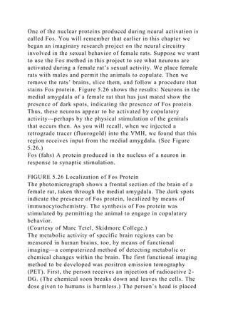
- 40. one’s own radiation.” Sections of the brain are mounted on microscope slides. The slides are then taken into a darkroom, where they are coated with a photographic emulsion (the substance found on photographic film). Several weeks later, the slides, with their coatings of emulsion, are developed, just like photographic film. The molecules of radioactive 2-DG show themselves as spots of silver grains in the developed emulsion because the radioactivity exposes the emulsion, just as X-rays or light will do. autoradiography A procedure that locates radioactive substances in a slice of tissue; the radiation exposes a photographic emulsion or a piece of film that covers the tissue. FIGURE 5.25 2-DG Autoradiogram The frontal section of a rat brain (dorsal at the top) shows especially high regions of activity in the pair of nuclei in the hypothalamus, at the base of the brain. (From Schwartz, W. J., and Gainer, H. Science, 1977, 197, 1089–1091. Reprinted with permission.) The most active regions of the brain contain the most radioactivity, showing this radioactivity in the form of dark spots in the developed emulsion. Figure 5.25 shows an autoradiograph of a slice of a rat brain; the dark spots at the bottom (indicated by the arrow) are nuclei of the hypothalamus with an especially high metabolic rate. Chapter 9 describes the function of these nuclei. (See Figure 5.25.) Simulateautoradiography on MyPsychLab to see an animation of this procedure. Another method of identifying active regions of the brain capitalizes on the fact that when neurons are activated (for example, by the terminal buttons that form synapses with them), particular genes in the nucleus called immediate early genes are turned on, and particular proteins are produced. These proteins then bind with the chromosomes in the nucleus. The presence of these nuclear proteins indicates that the neuron has just been activated.
- 41. One of the nuclear proteins produced during neural activation is called Fos. You will remember that earlier in this chapter we began an imaginary research project on the neural circuitry involved in the sexual behavior of female rats. Suppose we want to use the Fos method in this project to see what neurons are activated during a female rat’s sexual activity. We place female rats with males and permit the animals to copulate. Then we remove the rats’ brains, slice them, and follow a procedure that stains Fos protein. Figure 5.26 shows the results: Neurons in the medial amygdala of a female rat that has just mated show the presence of dark spots, indicating the presence of Fos protein. Thus, these neurons appear to be activated by copulatory activity—perhaps by the physical stimulation of the genitals that occurs then. As you will recall, when we injected a retrograde tracer (fluorogold) into the VMH, we found that this region receives input from the medial amygdala. (See Figure 5.26.) Fos (fahs) A protein produced in the nucleus of a neuron in response to synaptic stimulation. FIGURE 5.26 Localization of Fos Protein The photomicrograph shows a frontal section of the brain of a female rat, taken through the medial amygdala. The dark spots indicate the presence of Fos protein, localized by means of immunocytochemistry. The synthesis of Fos protein was stimulated by permitting the animal to engage in copulatory behavior. (Courtesy of Marc Tetel, Skidmore College.) The metabolic activity of specific brain regions can be measured in human brains, too, by means of functional imaging—a computerized method of detecting metabolic or chemical changes within the brain. The first functional imaging method to be developed was positron emission tomography (PET). First, the person receives an injection of radioactive 2- DG. (The chemical soon breaks down and leaves the cells. The dose given to humans is harmless.) The person’s head is placed
- 42. in a machine similar to a CT scanner. When the radioactive molecules of 2-DG decay, they emit subatomic particles called positrons, which meet nearby electrons. The particles annihilate each other and emit two photons, which travel in directly opposite paths. Sensors arrayed around the person’s head detect these photons, and the scanner plots the locations from which these photons are being emitted. From this information, the computer produces a picture of a slice of the brain, showing the activity level of various regions in that slice. (See Figure 5.27.) functional imaging A computerized method of detecting metabolic or chemical changes in particular regions of the brain. positron emission tomography (PET) A functional imaging method that reveals the localization of a radioactive tracer in a living brain. FIGURE 5.27 PET Scans The top row shows three horizontal scans from a person at rest. The bottom row shows three scans from the same person while he was clenching and unclenching his right fist. The scans show increased uptake of radioactive 2-deoxyglucose in regions of the brain that are devoted to the control of movement, which indicates increased metabolic rate in these areas. Different computer-generated colors indicate different rates of uptake of 2-DG, as shown in the scale at the bottom. (Courtesy of the Brookhaven National Laboratory and the State University of New York, Stony Brook.) One of the disadvantages of PET scanners is their operating cost. For reasons of safety the radioactive chemicals that are administered have very short half-lives; that is, they decay and lose their radioactivity very quickly. For example, the half-life of radioactive 2-DG is 110 minutes; the half-life of radioactive water (also used for PET scans) is only 2 minutes. Because these chemicals decay so quickly, they must be produced on site, in an atomic particle accelerator called a cyclotron. Therefore, to the cost of the PET scanner must be
- 43. added the cost of the cyclotron and the salaries of the personnel who operate it. Another disadvantage of PET scans is the relatively poor spatial resolution (the blurriness) of the images. The temporal resolution is also relatively poor. The positrons being emitted from the brain must be sampled for a fairly long time, which means that rapid, short-lived events within the brain are likely to be missed. These disadvantages are not seen in functional MRI, described in the next paragraph. However, PET scanners can do something that functional MRI scanners cannot do: measure the concentration of particular chemicals in various parts of the brain. I will describe this procedure later in this chapter. FIGURE 5.28 Functional MRI Scans These scans of human brains show localized average increases in neural activity of males (left) and females (right) while they were judging whether pairs of written words rhymed. (From Shaywitz, B. A., et al., Nature, 1995, 373, 607–609. Reprinted with permission.) The brain-imaging method with the best spatial and temporal resolution is known as functional MRI (fMRI). Engineers have devised modifications to existing MRI scanners and their software that permit the devices to acquire images that indicate regional metabolism. Brain activity is measured indirectly, by detecting levels of oxygen in the brain’s blood vessels. Increased activity of a brain region stimulates blood flow to that region, which increases the local blood oxygen level. The formal name of this type of imaging is BOLD: blood oxygen level–dependent signal. Functional MRI scans have a higher resolution than PET scans do, and they can be acquired much faster. Thus, they reveal more detailed information about the activity of particular brain regions. You will read about many functional imaging studies that employ fMRI scans in subsequent chapters of this book. (See Figure 5.28.) functional MRI (fMRI) A functional imaging method; a
- 44. modification of the MRI procedure that permits the measurement of regional metabolism in the brain, usually by detecting changes in blood oxygen level. Stimulating Neural Activity So far, this section has been concerned with research methods that measure the activity of specific regions of the brain. But sometimes we may want to artificially change the activity of these regions to see what effects these changes have on the animal’s behavior. For example, female rats will copulate with males only if certain female sex hormones are present. If we remove the rats’ ovaries, the loss of these hormones will abolish the rats’ sexual behavior. We found in our earlier studies that VMH lesions disrupt this behavior. Perhaps if we activate the VMH, we will make up for the lack of female sex hormones and the rats will copulate again. ELECTRICAL AND CHEMICAL STIMULATION How do we activate neurons? We can do so by electrical or chemical stimulation. Electrical stimulation simply involves passing an electrical current through a wire inserted into the brain, as you saw in Figure 5.22. Chemical stimulation is usually accomplished by injecting a small amount of an excitatory amino acid, such as kainic acid or glutamic acid, into the brain. As you learned in Chapter 4, the principal excitatory neurotransmitter in the brain is glutamic acid (glutamate), and both of these substances stimulate glutamate receptors, thus activating the neurons on which these receptors are located. Injections of chemicals into the brain can be done through an apparatus that is permanently attached to the skull so that the animal’s behavior can be observed several times. We place a metal cannula (a guide cannula) in an animal’s brain and cement its top to the skull. At a later date we place a smaller cannula of measured length inside the guide cannula and then inject a chemical into the brain. Because the animal is free to move about, we can observe the effects of the injection on its behavior. (See Figure 5.29.) Simulate cannula implantation on MyPsychLab to see a video of this surgical
- 45. procedure. The principal disadvantage of chemical stimulation is that it is slightly more complicated than electrical stimulation; chemical stimulation requires cannulas, tubes, special pumps or syringes, and sterile solutions of excitatory amino acids. However, it has a distinct advantage over electrical stimulation: It activates cell bodies but not axons. Because only cell bodies (and their dendrites, of course) contain glutamate receptors, we can be assured that an injection of an excitatory amino acid into a particular region of the brain excites the cells there but not the axons of other neurons that happen to pass through the region. Thus, the effects of chemical stimulation are more localized than are the effects of electrical stimulation. You might have noticed that I just said that kainic acid, which I described earlier as a neurotoxin, can be used to stimulate neurons. These two uses are not really contradictory. Kainic acid produces excitotoxic lesions by stimulating neurons to death. Whereas large doses of a concentrated solution kill neurons, small doses of a dilute solution simply stimulate them. FIGURE 5.29 An Intracranial Cannula (a) A guide cannula is permanently attached to the skull. (b) At a later time a thinner cannula can be inserted through the guide cannula into the brain. Chemicals can be infused into the brain through this device. What about the results of our hypothetical experiment? In fact (as we shall see in Chapter 10), VMH stimulation does substitute for female sex hormones. Perhaps, then, the female sex hormones exert their effects in this nucleus. We will see how to test this hypothesis in the final section of this chapter. When chemicals are injected into the brain through cannulas, molecules of the chemicals diffuse over a region that includes many different types of neurons: excitatory neurons, inhibitory neurons, interneurons that participate in local circuits, projection neurons that communicate with different regions of
- 46. the brain, and neurons that release or respond to a wide variety of neurotransmitters and neuromodulators. Stimulating a particular brain region with electricity or an excitatory chemical affects all of these neurons, and the result is unlikely to resemble normal brain activity, which involves coordinated activation and inhibition of many different neurons. Ideally, we would like to be able to stimulate or inhibit selected populations of neurons in a given brain regions. OPTOGENETIC METHODS Recent developments are providing the means to do just what I said in the previous paragraph: Optogenetic methods can be used to stimulate or inhibit particular types of neurons in particular brain regions (Boyden et al., 2005; F. Zhang et al., 2007; Baker, 2011). Photosensitive proteins have evolved in many organisms—even single-celled organisms such as algae and bacteria. Researchers have discovered that one of these proteins, Channelrhodopsin-2 (ChR2), found in green algae, controls ion channels that, when open, permit the flow of sodium, potassium, and calcium ions. When blue light strikes a ChR2-ion channel, the channel opens, and the rush of positively charged sodium and calcium ions depolarizes the membrane, causing excitation. A second photosensitive protein, Natronomonas pharaonis halorhodopsin (NpHR), is found in a bacterium. This protein controls a transporter that moves chloride into the cell when activated by yellow light. This influx of negatively charged ions hyperpolarizes the membrane, causing inhibition. The action of both of these photosensitive proteins begins and ends very rapidly when light of the appropriate wavelength is turned on and off. (See Figure 5.30.) optogenetic method The use of a genetically modified virus to insert light-sensitive ion channels into the membrane of particular neurons in the brain; can depolarize or hyperpolarize the neurons when light of the appropriate wavelength is applied. ChR2 and NpHR can be introduced into neurons by attaching the genes that code for them into the genetic material of
- 47. harmless viruses. The viruses are then injected into the brain, where they infect neurons and begin expressing the proteins, which are inserted into the cell membrane. The genes can be modified so that the proteins will be expressed only in particular types of neurons. In this way, researchers can observe the effects of turning on or off particular types of neurons in a particular region of the brain. FIGURE 5.30 Photostimulation (a) Photosensitive proteins can be inserted into neural membranes by means of genetically modified viruses. Blue light causes ChR2 ion channels to depolarize the membrane, and yellow light causes NpHR ion transporters to hyperpolarize it. (b) The graph shows the effects on the membrane potential of different wavelengths of light acting on ChR2 or NpHR proteins. (c) The graph shows action potentials elicited by pulses of blue light (blue arrows) and the inhibitory effects of the hyperpolarization caused by yellow light. (Part (a) adapted from Hausser, M., and Smith, S. L. Nature, 2007, 446, 617–619, and parts (b) and (c) adapted from Zhang, F., Wang, L. P., Brauner, M., et al. Nature, 2007, 446, 633– 639.) Of course, because ChR2 and NpHR are activated by light, researchers must be able to introduce light into the brain. If the neurons that express these photosensitive proteins are located in the cerebral cortex, a small hole can be drilled in the skull, and light-emitting diodes (LEDs) can be attached directly above the hole. To activate photosensitive proteins in the membranes of neurons deep within the brain, optical fibers can be implanted by means of stereotaxic surgery, just like electrodes or cannulas, and light can be transmitted through these fibers. For example, Tsai et al. (2009) used optogenetic methods to insert ChR2 ion channels into the membranes of dopaminergic neurons in the ventral tegmental area of rats. (Other research has shown that these neurons play an essential role in behavioral reinforcement.) The investigators found that if these neurons
- 48. were stimulated when the rats were in one of two chambers in a testing apparatus, the animals preferred to spend time in that chamber. The development of these procedures has caused much excitement among neuroscientists because they promise ways to study the functions of particular neural circuits in the brain. Some investigators are also exploring possible clinical uses of photosensitive proteins. For example, retinitis pigmentosa is a genetic disease that causes blindness in humans. People with this disease are born with normal vision, but they gradually become blind as the photoreceptor cells in their retinas degenerate. The retina contains two major categories of photoreceptors: rods, which are responsible for night vision, and cones, which are responsible for daytime vision. The rods of people with retinitis pigmentosa die, but although the cones lose their sensitivity to light, their cell bodies survive. Busskamp et al. (2010) used an optogenetic method to try to reestablish vision in mice with a genetic modification that causes them to develop retinitis pigmentosa. The investigators targeted the animals’ cones with NpHR. (Because the membranes of photoreceptors are normally hyperpolarized by light, they chose to use this protein.) Electrical recording and behavioral studies found that the treatment at least partially reestablished the animals’ vision. Furthermore, the same treatment reestablished light sensitivity in retinal tissue removed from deceased people who had suffered from retinitis pigmentosa. These findings provide hope that further research may develop a treatment for this form of blindness. TRANSCRANIAL MAGNETIC STIMULATION As we saw earlier in this chapter, neural activity induces magnetic fields that can be detected by means of magnetoencephalography. Similarly, magnetic fields can be used to stimulate neurons by inducing electrical currents in brain tissue. Transcranial magnetic stimulation (TMS) uses a coil of wires, usually arranged in the shape of the numeral 8, to stimulate neurons in the human cerebral cortex. The stimulating
- 49. coil is placed on top of the skull so that the crossing point in the middle of the 8 is located immediately above the region to be stimulated. Pulses of electricity send magnetic fields that activate neurons in the cortex. Figure 5.31 shows an electromagnetic coil used in transcranial magnetic stimulation and its placement on a person’s head. (See Figure 5.31.) transcranial magnetic stimulation (TMS) Stimulation of the cerebral cortex by means of magnetic fields produced by passing pulses of electricity through a coil of wire placed next to the skull; interferes with the functions of the brain region that is stimulated. FIGURE 5.31 Transcranial Magnetic Stimulation Pulses of electricity through the coil produce a magnetic field that stimulates a region of the cerebral cortex under the crossing point in the middle of the figure 8. (Photographs courtesy of the Kastner Lab, Princeton University, Princeton, New Jersey.) The effects of TMS are very similar to those of direct stimulation of the exposed brain. For example, as we shall see in Chapter 6, stimulation of a particular region of the visual association cortex will disrupt a person’s ability to detect movements in visual stimuli. In addition, as we will see in Chapters 15 and 16, TMS has been used to treat the symptoms of neurological and mental disorders. Depending on the strength and pattern of stimulation, TMS can either excite the region of the brain over which the coil is positioned or interfere with its functions. SECTION SUMMARY: Recording and Stimulating Neural Activity When circuits of neurons participate in their normal functions, their electrical activity and metabolic activity increase. Thus, by observing these processes as an animal perceives various stimuli or engages in various behaviors, we can make some inferences about the functions performed by various regions of the brain. Microelectrodes can be used to record the electrical
- 50. activity of individual neurons. Chronic recordings require that the electrode be attached to an electrical socket, which is fastened to the skull with a plastic adhesive. Macroelectrodes record the activity of large groups of neurons. In rare cases, macroelectrodes are placed in the depths of the human brain, but most often they are placed on the scalp, and their activity is recorded on a polygraph. Metabolic activity can be measured by giving an animal an injection of radioactive 2-DG, which accumulates in metabolically active neurons. The presence of the radioactivity is revealed through autoradiography: Slices of the brain are placed on microscope slides, covered with a photographic emulsion, left to sit for a while, and then developed like photographic negatives. When neurons are stimulated, they synthesize the nuclear protein Fos. The presence of Fos, revealed by a special staining method, provides another way to discover active regions of the brain. The metabolic activity of various regions of the living human brain can be revealed by the 2-DG method, but a PET scanner is used to detect the active regions. Functional MRI scanners depict localized brain activity by measuring regional blood oxygen levels. Functional MRI scans have much higher spatial and temporal resolution than PET scans do. Researchers can stimulate various regions of the brain by implanting a macroelectrode and applying mild electrical stimulation. Alternatively, they can implant a guide cannula in the brain; after the animal has recovered from the surgery, they insert a smaller cannula and inject a weak solution of an excitatory amino acid into the brain. The advantage of this procedure is that only neurons whose cell bodies are located nearby will be stimulated; axons passing through the region will not be affected. Viruses can be used to deliver genes for photosensitive proteins that produce depolarizations or hyperpolarizations of the membranes of specific neurons when the proteins are stimulated by light. Transcranial magnetic stimulation induces electrical activity in the human cerebral
- 51. cortex, which temporarily disrupts the functioning of neural circuits located there. Table 5.2 summarizes the research methods presented in this section. ■ THOUGHT QUESTIONS 1. Suppose that you had the opportunity to record an fMRI from a person (perhaps yourself) while the person was performing a behavior, thinking about something, or attending to a particular stimulus. Describe what you would have the person do. 2. Can you think of some ways that you could use optogenetic methods to investigate neural mechanisms involved in the control of a behavior or sensory system? TABLE 5.2 Research Methods: Part II Goal of Method Method Remarks Record electrical activity of single neurons Glass or metal microelectrodes Metal microelectrodes can be implanted permanently to record neural activity as animal moves Record electrical activity of regions of brain Metal macroelectrodes In humans, usually attached to the scalp with a special paste Record magnetic fields induced by neural activity Magnetoencephalography; uses a neuromagnetometer, which contains an array of SQUIDs Can determine the location of a group of neurons firing synchronously Record metabolic activity of regions of brain 2-DG autoradiography Measures local glucose utilization Measurement of Fos protein Identifies neurons that have recently been stimulated
- 52. 2-DG PET scan Measures regional metabolic activity of human brain Functional magnetic resonance imaging (fMRI) scan Measures regional metabolic activity of human brain; better spatial and temporal resolution than PET scan Measure neurochemicals in the living human brain PET scan Can localize any radioactive substance taken up in the human brain Stimulate neural activity Electrical stimulation Stimulates neurons near the tip of the electrode and axons passing through region Chemical stimulation with excitatory amino acid Stimulates only neurons near the tip of the cannula, not axons passing through region Transcranial magnetic stimulation Stimulate neurons in the human cerebral cortex with an electromagnet placed on the head Neurochemical Methods Sometimes we are interested not in the general metabolic activity of particular regions of the brain, but in the location of neurons that possess particular types of receptors or produce particular types of neurotransmitters or neuromodulators. We might also want to measure the amount of these chemicals secreted by neurons in particular brain regions during particular circumstances. Finding Neurons That Produce Particular Neurochemicals Suppose we learn that a particular drug affects behavior. How would we go about discovering the neural circuits that are responsible for the drug’s effects? To answer this question, let’s take a specific example. Physicians discovered several years