


Anatomyofspinalcord
- 1. INTERNAL STRUCTURE OF SPINAL CORD Iram Anwar
- 3. Gross Appearance Cylindrical in shape Foramen magnum L1/L2 (adult) L3 (newborn) Occupies upper ⅔ of vertebral canal Surrounded by 3 layers of meniges : dura mater arachnoid mater pia mater CSF in subarachnoid space
- 4. Enlargements: cervical & lumbar Conus medullaris Filum termniale Anterior median fissure Posterior median sulcus 31 pairs of spinal nerves attached to it by the anterior roots & posterior roots
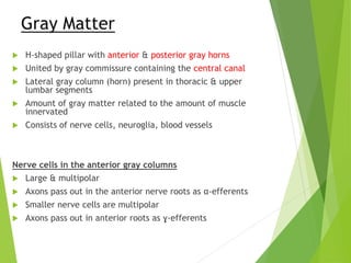
- 6. Gray Matter H-shaped pillar with anterior & posterior gray horns United by gray commissure containing the central canal Lateral gray column (horn) present in thoracic & upper lumbar segments Amount of gray matter related to the amount of muscle innervated Consists of nerve cells, neuroglia, blood vessels Nerve cells in the anterior gray columns Large & multipolar Axons pass out in the anterior nerve roots as α-efferents Smaller nerve cells are multipolar Axons pass out in anterior roots as ɣ-efferents
- 7. Nerve cells in the posterior gray columns 4 nerve cell groups Substantia gelatinosa situated at the apex throughout the length of spinal cord composed mainly of Golgi Type II neurons Nucleus proprius anterior to substantia gelatinosa present throughout the whole length of spinal cord main bulk of cells in posterior gray column
- 8. Nucleus dorsalis (Clark’s column) base of posterior column C8 – L3 / L4 associated with proprioceptive endings (neuromuscular spindles & tendon spindles) Visceral afferent nucleus lateral to nucleus dorsalis T1 – L3 receives visceral afferent info
- 9. Nerve cells in the lateral gray columns Formed by the intermediolateral group of cells T1 – L2 / L3 Cells give rise to preganglionic sympathetic fibres In S2, S3, S4; they give rise to preganglionic parasympathetic fibres The gray commissure & central canal connects the gray on each side central canal in the centre posterior gray commissure anterior gray commissure central canal present throughout superiorly continuous with the central canal of medulla oblongata inferiorly, expands as terminal ventricle terminates within the root of filum terminale
- 10. White Matter Divided into anterior white column lateral white column posterior white column Consists of nerve fibres, neuroglia, blood vessels White due to myelinated fibres TRACTS Ascending Descending Intersegmental
- 11. Ascending Tracts Fibres that ascend from spinal cord to higher centres Conduct afferent information which may or may not reach consciousness Information may be exteroceptive (pain, Tº, touch) Lateral spinothalamic tract Pain & temp pathways 1st-order neurons Pain conducted by δ A-type fibres & C-type fibres 2nd-order neurons decussate to the opposite side 3rd-order neurons ends in sensory area in postcentral gyrus Anterior spinothalamic tracts Light (crude) touch & pressure pathways
- 12. Posterior spinocerebellar tract Muscle joint sense pathways to cerebellum Unconscious proprioception Muscle joint info from muscle spindles, GTO, joint receptors of the trunk & lower limbs Info is used by the cerebellum in the coordination of movements & maintenance of posture Anterior spinocerebellar tract Majority of 2nd-order neurons cross to the opposite side Enter cerebellum through superior cerebellar peduncle Info from trunk, upper & lower limbs Also carries info from skin & subcut tissue
- 13. Descending Tracts Lower motor neurons Upper motor neurons Corticospinal tracts concerned with voluntary, discrete, skilled movements Meninges Dura mater Arachnoid mater Pia mater
- 15. Dura mater Dense, strong fibrous membrane Encloses the spinal cord & cauda equina Separated from wall of vertebral canal by the extradural space Arachnoid mater Delicate impermeable membrane Lies between pia and dura mater Separated from pia mater by subarachnoid space Pia mater Vascular membrane Closely covers spinal cord Thickened on either side between nerve roots to form the ligamentum denticulatum