Animal Cells
- 2. OBJECTIVES At the end of the discussion, the students will be expected to: i. Identify the different parts of an animal cell and know their functions. ii. Draw and label its parts after the discussion. iii. Give the importance in knowing the different parts of an animal cell.
- 3. 1665, Robert Hooke examined thin slices of cork and other plant materials contained microscopic compartments; named it cells Anton Van Leeuwenhoek made further observations of the cells of plants , animals and microorganisms Matthias Schleiden –all plants are composed cells Theodore Schwann– all animals are composed of cells Rudolf Virchow- proposed the cell theory 1840,Purkinje named the cell contents protoplasm
- 4. 1.Nucleolus 2.Nucleus 3.Ribosome 4.Vesicle 5.Rough endoplasmic reticulum 6.Golgi apparatus (or "Golgi body") 7.Cytoskeleton 8.Smooth endoplasmic reticulum 9.Mitochondrion 10.Vacuole 11.Cytosol 12.Lysosome 13.Centrosome 14.Cell membrane
- 5. largest structure in the nucleus consists of nucleolar organizers, ribosomal RNA, and proteins Function: primarily serves as the site of ribosome synthesis and assembly. .
- 6. Notable structure within the cell The genetic control centre of the cell -chromatin(network of dark-staining threads) Surrounded by nuclear envelope Function: directs cell division control protein synthesis and many of the metabolic activities of the cell
- 7. The border of the cell About 8 nm thick A semi-permeable membrane Composed of proteins and lipids Unit membrane Model-tripartite arrangement of the plasma membrane (protein-lipid-protein)
- 8. Fluid Mosaic model- protein molecules penetrated into the lipid layers and not continuous in on the surface of lipid
- 9. Function: It supports and protects the cell. Regulates the movement of material in and out of the cell Facilitated diffusion- scattering of particles, from high concentration to lower concentration Active transport- process resembling facilitated diffusion in that it involves association of molecules to be transported with a membrane area of low concentration to high concentration Pinocytosis- engulfing of particles
- 10. A large interconnecting membrane of tunnels Continuous with the nuclear envelope Two types: 1. Rough Endoplasmic Reticulum - network of interconnected flattened sacs - is studded with ribosomes Function: to make protein( secretory protein), to make more membrane channelling products both to the outside of the cell, via the membrane
- 11. 2.Smooth Endoplasmic Reticulum network of interconnected tubules lack of ribomes Function: Synthesizing and secreting of certain steroid hormones, enzymes of carbohydrate metabolism, and enzymes of lipid synthesis.
- 12. Named from Camillo Golgi, n Italian biologist and physician A series of from 3 to 20 parallel flattened sacs closely stacked together, cisternae End of the sacs bud off various vesicles Function: receives and modifies and packages the substances manufactured by ER,
- 13. Are spherical to rod- shaped structures from 0.2 to 7µm; a doubled layer membrane Cristae( complex folding of inner membrane) Mobile structures, capable of changing their shapes “powerhouse of the cell” Function: produced energy in the form of ATP
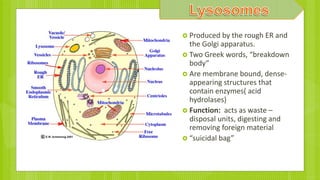
- 14. Produced by the rough ER and the Golgi apparatus. Two Greek words, “breakdown body” Are membrane bound, dense- appearing structures that contain enzymes( acid hydrolases) Function: acts as waste – disposal units, digesting and removing foreign material “suicidal bag”
- 15. enclosed compartments which are filled with water containing inorganic and organic molecules including enzymes in solution Function: Digestion; storage of chemicals, cell enlargement; water balance
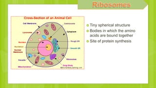
- 16. Tiny spherical structure Bodies in which the amino acids are bound together Site of protein synthesis
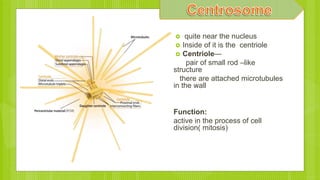
- 17. quite near the nucleus Inside of it is the centriole Centriole— pair of small rod –like structure there are attached microtubules in the wall Function: active in the process of cell division( mitosis)
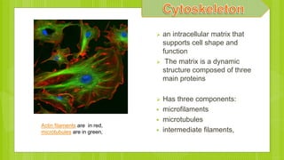
- 18. an intracellular matrix that supports cell shape and function The matrix is a dynamic structure composed of three main proteins Has three components: microfilaments microtubules intermediate filaments, Actin filaments are in red, microtubules are in green,
- 19. Microtubules- tiny cylindrical elements of animal cells about 20 to 25 nm in diameter composed of tubulins function: provide rigidity and shape in one area Tracts for organelle movement within the cell Basis of ciliary and flagellar movement Microtubules in gel fixated cell
- 20. Microfilaments-smaller than microtubules ranging in diameter from 4 to 7 nm Solid helical rods composed of actin Function:providing motive force for cell contraction amoeboid movement, and possibly intracellular transport
- 21. Protoplasm that surrounds the nucleus A semi-liquid substance that composes the foundation of the cell Within the cytoplasm are a number of different organelles
- 23. Biology: Concepts and Connections by Campbell, Mitchelle and Reece General Zoology by Storer,Usinger, Stibbens E-biology: The Next Generation by Santos, Danac and Ocampo Biology for non-sience majors by Reyes et. Al Wikipedia.org Enchanted Learning.com
Editor's Notes
- Englisman hooke, reminded the monastery,cell Contained many partitions of many cavities The cell as the basic unit of life Cells are the basic units of organisms All cells come from preexisting cells through cell division. Unicellular organisms are made of one cell only Protoplasm—used to describe the living substance of the cell, it compasses the whole array of organelles
- Most living things can be divided into two. Do you know what they are? To answer this question, look at these two pictures. Left is a picture of a plant cell and right is of an animal cell.
- Nucleolar-certain chromosomes Malfunction of nucleoli can be the cause of several human diseases Nucleoli are made of proteins and RNA and form around specific chromosomal regions functions like assembly of signal recognition particles playing a role in the cell's response to stress Nuclear organizers- certain
- Ince in a low power light of microscope, visible Nuclear envelope has two memebrane
- Good transmission electron microscope Lipid- molecules that are assymetrical-electric charge hydrophilic. Uncharge hydrophobic
- According to the fluid mosaic model of S. J. Singer and G. L. Nicolson (1972), which replaced the earlier model of Davson and Danielli, biological membranes can be considered as a two-dimensional liquid in which lipid and protein molecules diffuse more or less easily.[8] Although the lipid bilayers that form the basis of the membranes do indeed form two-dimensional liquids by themselves, the plasma membrane also contains a large quantity of proteins, which provide more structure. Examples of such structures are protein-protein complexes, pickets and fences formed by the actin-based cytoskeleton, and potentially lipid rafts. Diagram of the arrangement of amphipathic lipid molecules to form a lipid bilayer. The yellow polar head groups separate the grey hydrophobic tails from the aqueous cytosolic and extracellular environments Frreexe-etch ttechnique--
- Secretory protein are– annti body, defensive molecule Means of communicatio
- Cisternae- Receiving dock for transport vesicles Warehouse and finishing factory
- Fold contain enzymes that are used to convert food energy
- The rough er puts enzymes and membranes together, then the golgi apparatus chemically refines the enzymes A lysosome (derived from the Greek words lysis, meaning "to loosen", and soma, "body") is a membrane-bound cell organelle found in most animal cells (they are absent in red blood cells). Structurally and chemically, they are spherical vesicles containing hydrolytic enzymes capable of breaking down virtually all kinds of biomolecules, including proteins, nucleic acids, carbohydrates, lipids, and cellular debris. They are known to contain more than 50 different enzymes, which are all optimally active at an acidic environment of about pH 4.5 (about the pH of black coffee). Thus lysosomes act as the waste disposal system of the cell by digesting unwanted materials in the cytoplasm, both from outside of the cell and obsolete components inside the cell. For this function they are popularly referred to as "suicide bags" or "suicide sacs" of the cell.[1][2] Furthermore, lysosomes are responsible for cellular homeostasis for their involvements in secretion, plasma membrane repair, cell signalling and energy metabolism, which are related to health and diseases
- Vacuoles are formed by the fusion of multiple membrane vesicles and are effectively just larger forms of these.[3] The organelle has no basic shape or size; its structure varies according to the needs of the cell.
- Active in the formation of cilia and are self duplicating organelles Lacks centriole then it cannot undergo the process of cell division 9 evenly spaces
- , which are capable of rapid assembly or disassembly dependent on the cell's requirements.[1 microfilaments (made of the protein actin) and microtubules (made of the protein tubulin) .[3][5] By contrast intermediate filaments, which have more than 60 different building block proteins, have been found so far only in animal cells (apart from one non-eukaryotic bacterial intermediate filament crescentin).
- Property of contractility
- Protoplasm– a living dsubstance
