Anthoceros
•Download as PPTX, PDF•
58 likes•44,523 views

1. Anthoceros is a genus of hornworts that includes about 200 species found worldwide in shady, moist tropical and temperate areas. 2. The plant body is a gametophyte that consists of a small, dark green, lobed thallus containing chloroplasts and rhizoids. 3. Reproduction can occur vegetatively through tubers, gemmae, and persistent apices, or sexually through antheridia that produce sperm and archegonia containing eggs leading to fertilization and formation of a sporophyte.
Report
Share


Anthoceros
- 3. Habitat • Distributed world wide • Grows in shady and moist areas of tropical and temperate regions • About 200 species, and in Pakistan 3 common species are found in Himalayas 1. Anthoceros himalayensis 2. Anthoceros erectus 3. Anthoceros chambensis
- 4. Vegetative morphology Thallus • The plant body is gametophyte and consist of small, dark green, prostrate thallus • It is rosette like, and with lobes of whose margins are divided into small lobed segments • Dichotomous system is present • Surface of thallus is smooth, velvety , and contains ridges and spines • Numerous thread like rhizoids are present on ventral surface of thallus
- 5. Thallus
- 6. Internal structure of thallus • There is no internal differentiation • Parenchymatous cells are present • Each contain single lens shaped chloroplast • Deep cells contain 2-8 chloroplasts • Each chloroplast contains single pyrenoid • There are two surfaces, upper and lower epidermis • Lower epidermis contains mucilaginous cavities that opens through a pore called slime pore • In these cavities blue green alga resides.
- 9. Vegetative reproduction 1. Death of older plants: Vegetative reproduction takes place by the death of older parts. Younger parts form new thallus. 2. Tuber: Some thallus forms tubers. These tubers are rich in stored fats and proteins. These tubers germinate to on the margin of the lobes. They can survive long periods of drought. Tuber detach and from new plants.
- 10. Vegetative reproduction 1. Gemmae: Gemma are also produced on short stalks on the upper surface of the thallus. These are also act as vegetative reproductive bodies. 2. Persistent apices : in some species the thallus dies except the apices , there are called persistent apices , they remain buried in soil and develop into new plant body during favorable conditions
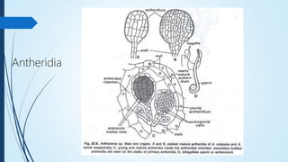
- 12. Antheridia Antheridia develops on cavities called antheridia chamber Present on the dorsal surface 1 to 25 antheridia may develop in each antheridial chambers Mature antheridium consist of an ovoid body with stalk. There is a mass of spermatogenous cells surrounded by jacket of sterile cells
- 13. Antheridia
- 14. Dehiscence: At maturity, the roof of the antheridial chamber ruptures, exposing the antheridia. The apical cell of the antheridial wall, on absorbing water, ruptures by apical aperture. The antherozoids are now liberated to the covering film of water. The antherozoids: The antherozoid is spindle like and biciliate. The cilia are attached to the anterior end of the body. Sometimes just near the attaching point of the flagella to the body, the blepharoplasty ( flagellated cell or basal body) is visible. The antherozoids swim in the water by the lashing moment of their flagella.
- 15. Archegonium • Archegonia are produced close to the growing point. Archegonia are embedded in the tissue of the thallus. • Each archegonium consists of an egg and a ventral canal cell four neck canal cells. The canal of the archegonium is closed at the top by four cover cells. • These cells project slightly above the general surface of the thallus. Development of Archegonium
- 16. Fertilization: • Water is essential for fertilization. In the mature archegonium, the venter canal cell, neck canal cells disintegrate and form a mucilaginous mass. • It absorbs water, swells up and becomes out of the archegonial neck by pushing the cover cells apart. • This mucilaginous mass becomes continuous with the mucilage mound and in this way an open passage down to egg is formed.
- 17. Fertilization The mucilaginous mass consists of chemical substances. Many antherozoids caught in the mucilage enter in the archegonial neck because of the chemotactic response, reach up to the egg, and fertilization is effected. Prior to fertilization, egg enlarges and fills the cavity of the venter. Fusion of both male and female nuclei results in the formation of diploid zygote or oospore. Fertilization ends the gametophytic phase.
- 18. Sporophyte The sporophyte of Anthoceros has certain unique features. Sporogonium is borne on the gametophyte. But mature sporogonium does not totally dependent on the gametophyte. The mature sporophyte consist a bulbous foot and a smooth, slender, erect, cylindrical, structure called capsule. Capsule varies in length from two to fifteen centimeter in different species. The Sporogonium appears like a ‘bristle’ or ‘horn’, hence, the species are called ‘hornworts It has following parts 1: foot 2: seta 3: capsule
- 19. Structure of sporophyte The Foot: It is the basal part of the sporophyte which is a rounded bulbous structure deeply embedded in the tissue of the thallus . The lowermost cells of the foot are haustorial which absorb water and mineral nutrients from the gametophyte for the developing sporophyte.
- 20. Meristematic zone The Intermediate Meristematic Zone: This is a narrow zone of meristematic cells located in-between the foot and the capsule . It regenerates the capsule from the base, thus the capsules are always in different stages of growth.
- 21. Capsule The Capsule: The capsule forms the major and conspicuous part of the sporophyte. It is a slender smooth upright cylindrical structure that slightly tapers at the apex. It consists of 1. capsule wall, 2. sporogenous tissue 3. collumela
- 22. Capsule Wall: The capsule wall is made up of 4-6 layers of parenchymatous cells. The cells of the outermost layer, which form the epidermis, are heavily cutinised, vertically elongated and interrupted by the stomata. Below the epidermal layer is the green parenchyma- tous, photosynthetic tissue containing chloroplasts. Thus, the sporophyte is capable of manufacturing their own food by photosynthesis, except for the water and minerals for which it depends upon the gametophyte.
- 23. Sporogenous Tissue The sporogenous tissue (archesporium) of Anthoceros is situated in between the jacket and the collumela. At maturity it differentiates into 1. spore mother cells 2. pseudoelaters.
- 24. Collumela Collumela: the central part of capsule is collumela. It contains 4 rows of elongated vertical cells. But layer it becomes 16 rows of cells In transverse section, the columella appears to be a solid square. The columella provides mechanical support to the capsule. It also helps the spores to disperse and is associated with the conduction of water and minerals.
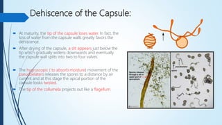
- 25. Dehiscence of the Capsule: At maturity, the tip of the capsule loses water. In fact, the loss of water from the capsule walls greatly favors the dehiscence. After drying of the capsule, a slit appears just below the tip which gradually widens downwards and eventually the capsule wall splits into two to four valves. The hygroscopic ( to absorb moisture) movement of the pseudoelaters releases the spores to a distance by air current and at this stage the apical portion of the capsule looks twisted. The tip of the collumela projects out like a flagellum.