Location via proxy:   [ UP ]  
[Report a bug]   [Manage cookies]                
SlideShare a Scribd company logo
Apoptosis
APOPTOSIS
Introduction
Detail pathway
Hina zamir
Roll # 04
OVERVIEW
• Apoptosis
• Characteristics
• Importance of apoptosis
• Apoptosis : morphology of cell
• Apoptosis vs necrosis
• Classical stages of apoptosis
• Pathway/mechanism of apoptosis
• Schematic representation
intrinsic pathway
extrinsic pathway
perforin/granzyme pathway
APOPTOSIS
 Apoptosis derived from Greek means "falling off"
 It is a process of programmed cell death that occurs in multicellular
organisms
 A physiological way for a cell to die
 Physiological cell death
 Cell suicide
 Cell deletion
 The Between 50 and 70 billion cells die each day due to apoptosis in the
average human adult
 History : German scientist Karl Vogt was first to describe the principle of
apoptosis in 1842
 Characteristics: An active and energy dependent cytological process
 It is programmed or controlled by genetic protocol
 It may be triggered by intrinsic or extrinsic stimuli
 It occurs in almost all living creatures
Importance of Apoptosis
1) Crucial for development
Examples:
The resorption of the tadpole tail
The formation of the fingers and toes of the fetus
Errors in Apoptosis can lead to Birth Defects
2) Important for maintaining homeostasis
Cell death is balanced with mitosis to regulate cell number.
3) Apoptosis is needed to destroy cells that represent threat to
the integrity of the organism
Examples:
• Cells infected with viruses
• Cells of the immune system
• Cells with DNA damage
• Cancer cells
Morphology
• Biochemical events lead to characteristic cell changes (morphology) and death.
These changes include
• Early: Cell shrinkage
(condensation of cytoplasm)
• Chromatic condensation
• Later: Nuclear fragmentation
• Breakdown of mitochondria
• Cell membrane blebbing
• Cell Fragmentation
• apoptotic body formation
• End : Phagocytosis
membrane
blebbing &
changes
mitochondrial
leakage
organelle
reduction
cell
shrinkage
nuclear
fragmentation
chromatin
condensation
APOPTOSIS: Morphology
Bleb
Blebbing & Apoptotic bodies Phagocytosis
The control retained over the cell membrane &
cells bleb and eventually break apart without
releasing contents cytoskeleton allows intact
pieces of the cell to separate for recognition &
phagocytosis by MFs
Apoptotic body
Blebbing of WBC
How Apoptosis Differs from Necrosis?
Healthy cell
1. DEATH SIGNAL
Committment to death by extracellular or intracellular triggers/signals
2. EXECUTION (Cell killing by activation of intracellular proteases
(caspases) )
Dead cell (condensed, crosslinked)
3. ENGULFMENT (by macrophages, neighboring cells)
4. DEGRADATION
Four stages of apoptosis:
Mechanisms of Apoptosis
• The mechanisms of apoptosis are highly complex and sophisticated,
involving an energy-dependent cascade of molecular events
• 2 main pathways (caspase-dependant):
1. The extrinsic (death receptor pathway) and
2. The intrinsic (mitochondrial pathway)
• 1 additional pathway (caspase independent ): T-cell mediated
cytotoxicity and perforin-granzyme-dependent pathway.
• all three apoptotic pathways converge on the same terminal,
or execution pathway.
• Caspases:
• The activation of initiator caspases ( 2 8 9 10 )
requires binding to specific oligomeric adaptor
protein.
• Effector caspases (3 6 7 ) are then activated by these
active initiator caspases.
Schematic representation of apoptotic events
Mechanisms of Apoptosis
• Each pathway requires specific triggering signals to begin pathway
• Each pathway activates its own initiator caspase (8, 9, 10) which in
turn will converge on the same terminal, or execution pathway by
activating the executioner caspase-3
• The execution pathway results in characteristic cytomorphological
features including
• cell shrinkage, chromatin condensation, formation of cytoplasmic
blebs and apoptotic bodies , finally phagocytosis of the apoptotic
bodies by adjacent phagocytes
A. DEATH SIGNAL
1. Extrinsic Pathway:
 Extrinsic pathway also known as the “death receptor pathway”. This receptor
mediated pathway included death receptors and adaptors
• Death Receptors (DRs) are Cell surface Receptors.
• They transmit Apoptotic signals initiated by specific ligands.
• They belong to the superfamily of TNFR (Tumor Necrosis Factor Receptor).
• Members of the TNF receptor family have a cytoplasmic domain called the
“death domain” The best-characterized Death Receptors are Fas and TNFR1
(Tumor Necrosis Factor Receptor-1).
• Adaptors: cytoplasmic protein molecules
• FADD (Fas-Associated Death Domain)
• TRADD (Tumor Necrosis Factor Receptor-1-Associated Death Domain)
• The Adapter-molecules contain Death Domains so that they can interact
with the DRs and transmit the Apoptotic signal to the death-machinery.
Extrinsic pathway
1. The extrinsic pathway begins
outside the cell through
activation of death receptors on
the cell surface by ligands
2. Upon ligand binding, adapters
molecules are recruited whose
corresponding death domains
bind with the death
receptors. The binding of Fas
ligand to Fas receptor results in
the binding of the adapter
protein FADD
3. binding of TNF ligand to TNF
receptor results in the binding of
the adapter protein TRADD with
recruitment of FADD and RIP.
4. FADD then recruit procaspase-
8
Extrinsic pathway
1. At this point, a death-
inducing signaling complex
(DISC) is formed, resulting in
the auto-catalytic activation
of procaspase-8 to caspase-8
(active )
2. The DISC is composed of the
death receptor, FADD, and
caspase 8
3. This activated initiator
caspase activate the effector
caspase (execution caspase 3
) committing the cell to
Apoptosis by entering into
execution pathway
A. DEATH SIGNAL
2. Intrinsic pathway
• mitochondrial pathway
• Stimuli for the intrinsic pathway include:
viral infections
damage to the cell by toxins, free radicals
radiation damage to the cellular DNA
• permeabilization of the mitochondria: These stimuli induce changes
in the inner mitochondrial membrane that result in the loss of
transmembrane potential, causing the release of 2 groups of pro-
apoptotic proteins into the cytosol
• Group 1: cytochrome c, Smac and the serine protease Omi
• Group 2: AIF, endonuclease G and CAD
• Bcl-2 family (pro and anti apoptotic proteins)
Example: P53-mediated apoptosis
• P53 detects damage in DNA ( IF SEVERE DAMAGE )
• Bax protein (proapoptotic protein)
transcription by p53 + dissociation of bax from
bax- Bcl-2 complex
• High concentration of bax in cytosol
• Oligomerization of bax
• Formation of hole in mitochondria membrane
• Release of mitochondrial content (2 groups)
• 1. Smac & serine protease Omi:
promote apoptosis by inhibiting IAP activity
IAP (inhibitors of apoptosis proteins) suppress
the activity of caspases
Example: P53-mediated apoptosis
• 2. cytochrome c
14 cytochrome c + 7 Apaf-1 = apoptosome
+ initiator procaspase 9 = active initiator caspase 9
This activated initiator caspase-9 activate the effector
caspase (execution caspase 3 ) committing the cell to
Apoptosis by entering into execution pathway
• 3. AIF (Apoptosis Inducing Factor),
endonuclease G (apoptotic Dnase)
and CAD (Caspase-Activated Dnase):
translocate to nucleus and involved in chromatin
condensation (DNA fragmentation)
A. DEATH SIGNAL
3. Perforin/granzyme Pathway
• Tcells and NK cells : the granzymes are packaged in cytotoxic
granules with perforin
• cytoplasmic granules containing granzyme A and granzyme B enter
in the target cells through the newly formed pores by perforin
• Granzyme A induces the cleavage of the SET complex
• cleavage of the SET complex allows the endonuclease and the
exonuclease to cause DNA to damage in the target cell and gradual
destruction of chromatin integrity.
• Granzyme B can activate the effector caspase-3
directly or
 through caspase-10.
• Furthermore, granzyme B can use the intrinsic pathway (through
specific cleavage of Bid (bax activator) and induction of
Cytochrome c release) to amplify of the death signal
B. Executional pathway
• final pathway of apoptosis involves activation of the execution
caspases that cleaves various substrates that ultimately cause the
morphological and biochemical changes seen in apoptotic cells.
• effector or “executioner” caspases:
Caspase-3, caspase-6, and caspase-7
• Caspase-3 is considered to be the most important of the executioner caspases and is activated by
any of the initiator caspases (caspase-8, caspase-9, or caspase-10
• Execution caspases activate
cytoplasmic endonuclease, which degrades nuclear material,
proteases that degrade the nuclear and cytoskeletal proteins.
E.g iCAD: iCAD-CAD = CAD
C. Phagocytosis:
• Phagocytic uptake of apoptotic cells is the last
component of apoptosis.
• hallmark of this phase :
• Phospholipid asymmetry
• externalization of phosphatidylserine on the surface of apoptotic
cells and their fragments
• The appearance of phosphotidylserine on the outer leaflet of
apoptotic cells facilitates noninflammatory phagocytic recognition,
allowing for their early uptake and disposal.
• This process of early and efficient uptake with no release of cellular
constituents, results in essentially no inflammatory response
D. ENGULFMENT OF APOPTOTIC CELL: OOPSONIZATION
References:
• Bax-induced apoptotic cell death. John Pawlowski and Andrew S. Kraft PNAS 2000 97 (2) 529-
531; doi:10.1073/pnas.97.2.529
• Elmore, Susan. “Apoptosis: A Review of Programmed Cell Death.” Toxicologic pathology 35.4
(2007): 495–516. PMC. Web. 22 Nov. 2016.
• Ooi, Hsu Kiang, and Lan Ma. “Modeling Heterogeneous Responsiveness of Intrinsic Apoptosis
Pathway.” BMC Systems Biology 7 (2013): 65. PMC. Web. 22 Nov. 2016.
• Apoptosis. (2016, November 19). In Wikipedia, The Free Encyclopedia. Retrieved 05:28,
November 19, 2016, from
https://en.wikipedia.org/w/index.php?title=Apoptosis&oldid=750354570
THANK YOU

More Related Content

Apoptosis

  • 3. OVERVIEW • Apoptosis • Characteristics • Importance of apoptosis • Apoptosis : morphology of cell • Apoptosis vs necrosis • Classical stages of apoptosis • Pathway/mechanism of apoptosis • Schematic representation intrinsic pathway extrinsic pathway perforin/granzyme pathway
  • 4. APOPTOSIS  Apoptosis derived from Greek means "falling off"  It is a process of programmed cell death that occurs in multicellular organisms  A physiological way for a cell to die  Physiological cell death  Cell suicide  Cell deletion  The Between 50 and 70 billion cells die each day due to apoptosis in the average human adult  History : German scientist Karl Vogt was first to describe the principle of apoptosis in 1842  Characteristics: An active and energy dependent cytological process  It is programmed or controlled by genetic protocol  It may be triggered by intrinsic or extrinsic stimuli  It occurs in almost all living creatures
  • 5. Importance of Apoptosis 1) Crucial for development Examples: The resorption of the tadpole tail The formation of the fingers and toes of the fetus Errors in Apoptosis can lead to Birth Defects 2) Important for maintaining homeostasis Cell death is balanced with mitosis to regulate cell number. 3) Apoptosis is needed to destroy cells that represent threat to the integrity of the organism Examples: • Cells infected with viruses • Cells of the immune system • Cells with DNA damage • Cancer cells
  • 6. Morphology • Biochemical events lead to characteristic cell changes (morphology) and death. These changes include • Early: Cell shrinkage (condensation of cytoplasm) • Chromatic condensation • Later: Nuclear fragmentation • Breakdown of mitochondria • Cell membrane blebbing • Cell Fragmentation • apoptotic body formation • End : Phagocytosis
  • 8. Bleb Blebbing & Apoptotic bodies Phagocytosis The control retained over the cell membrane & cells bleb and eventually break apart without releasing contents cytoskeleton allows intact pieces of the cell to separate for recognition & phagocytosis by MFs Apoptotic body Blebbing of WBC
  • 9. How Apoptosis Differs from Necrosis?
  • 10. Healthy cell 1. DEATH SIGNAL Committment to death by extracellular or intracellular triggers/signals 2. EXECUTION (Cell killing by activation of intracellular proteases (caspases) ) Dead cell (condensed, crosslinked) 3. ENGULFMENT (by macrophages, neighboring cells) 4. DEGRADATION Four stages of apoptosis:
  • 11. Mechanisms of Apoptosis • The mechanisms of apoptosis are highly complex and sophisticated, involving an energy-dependent cascade of molecular events • 2 main pathways (caspase-dependant): 1. The extrinsic (death receptor pathway) and 2. The intrinsic (mitochondrial pathway) • 1 additional pathway (caspase independent ): T-cell mediated cytotoxicity and perforin-granzyme-dependent pathway. • all three apoptotic pathways converge on the same terminal, or execution pathway. • Caspases: • The activation of initiator caspases ( 2 8 9 10 ) requires binding to specific oligomeric adaptor protein. • Effector caspases (3 6 7 ) are then activated by these active initiator caspases.
  • 12. Schematic representation of apoptotic events
  • 13. Mechanisms of Apoptosis • Each pathway requires specific triggering signals to begin pathway • Each pathway activates its own initiator caspase (8, 9, 10) which in turn will converge on the same terminal, or execution pathway by activating the executioner caspase-3
  • 14. • The execution pathway results in characteristic cytomorphological features including • cell shrinkage, chromatin condensation, formation of cytoplasmic blebs and apoptotic bodies , finally phagocytosis of the apoptotic bodies by adjacent phagocytes
  • 15. A. DEATH SIGNAL 1. Extrinsic Pathway:  Extrinsic pathway also known as the “death receptor pathway”. This receptor mediated pathway included death receptors and adaptors • Death Receptors (DRs) are Cell surface Receptors. • They transmit Apoptotic signals initiated by specific ligands. • They belong to the superfamily of TNFR (Tumor Necrosis Factor Receptor). • Members of the TNF receptor family have a cytoplasmic domain called the “death domain” The best-characterized Death Receptors are Fas and TNFR1 (Tumor Necrosis Factor Receptor-1). • Adaptors: cytoplasmic protein molecules • FADD (Fas-Associated Death Domain) • TRADD (Tumor Necrosis Factor Receptor-1-Associated Death Domain) • The Adapter-molecules contain Death Domains so that they can interact with the DRs and transmit the Apoptotic signal to the death-machinery.
  • 16. Extrinsic pathway 1. The extrinsic pathway begins outside the cell through activation of death receptors on the cell surface by ligands 2. Upon ligand binding, adapters molecules are recruited whose corresponding death domains bind with the death receptors. The binding of Fas ligand to Fas receptor results in the binding of the adapter protein FADD 3. binding of TNF ligand to TNF receptor results in the binding of the adapter protein TRADD with recruitment of FADD and RIP. 4. FADD then recruit procaspase- 8
  • 17. Extrinsic pathway 1. At this point, a death- inducing signaling complex (DISC) is formed, resulting in the auto-catalytic activation of procaspase-8 to caspase-8 (active ) 2. The DISC is composed of the death receptor, FADD, and caspase 8 3. This activated initiator caspase activate the effector caspase (execution caspase 3 ) committing the cell to Apoptosis by entering into execution pathway
  • 18. A. DEATH SIGNAL 2. Intrinsic pathway • mitochondrial pathway • Stimuli for the intrinsic pathway include: viral infections damage to the cell by toxins, free radicals radiation damage to the cellular DNA • permeabilization of the mitochondria: These stimuli induce changes in the inner mitochondrial membrane that result in the loss of transmembrane potential, causing the release of 2 groups of pro- apoptotic proteins into the cytosol • Group 1: cytochrome c, Smac and the serine protease Omi • Group 2: AIF, endonuclease G and CAD • Bcl-2 family (pro and anti apoptotic proteins)
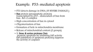
  • 19. Example: P53-mediated apoptosis • P53 detects damage in DNA ( IF SEVERE DAMAGE ) • Bax protein (proapoptotic protein) transcription by p53 + dissociation of bax from bax- Bcl-2 complex • High concentration of bax in cytosol • Oligomerization of bax • Formation of hole in mitochondria membrane • Release of mitochondrial content (2 groups) • 1. Smac & serine protease Omi: promote apoptosis by inhibiting IAP activity IAP (inhibitors of apoptosis proteins) suppress the activity of caspases
  • 20. Example: P53-mediated apoptosis • 2. cytochrome c 14 cytochrome c + 7 Apaf-1 = apoptosome + initiator procaspase 9 = active initiator caspase 9 This activated initiator caspase-9 activate the effector caspase (execution caspase 3 ) committing the cell to Apoptosis by entering into execution pathway • 3. AIF (Apoptosis Inducing Factor), endonuclease G (apoptotic Dnase) and CAD (Caspase-Activated Dnase): translocate to nucleus and involved in chromatin condensation (DNA fragmentation)
  • 21. A. DEATH SIGNAL 3. Perforin/granzyme Pathway • Tcells and NK cells : the granzymes are packaged in cytotoxic granules with perforin • cytoplasmic granules containing granzyme A and granzyme B enter in the target cells through the newly formed pores by perforin • Granzyme A induces the cleavage of the SET complex • cleavage of the SET complex allows the endonuclease and the exonuclease to cause DNA to damage in the target cell and gradual destruction of chromatin integrity. • Granzyme B can activate the effector caspase-3 directly or  through caspase-10. • Furthermore, granzyme B can use the intrinsic pathway (through specific cleavage of Bid (bax activator) and induction of Cytochrome c release) to amplify of the death signal
  • 22. B. Executional pathway • final pathway of apoptosis involves activation of the execution caspases that cleaves various substrates that ultimately cause the morphological and biochemical changes seen in apoptotic cells. • effector or “executioner” caspases: Caspase-3, caspase-6, and caspase-7 • Caspase-3 is considered to be the most important of the executioner caspases and is activated by any of the initiator caspases (caspase-8, caspase-9, or caspase-10 • Execution caspases activate cytoplasmic endonuclease, which degrades nuclear material, proteases that degrade the nuclear and cytoskeletal proteins. E.g iCAD: iCAD-CAD = CAD
  • 23. C. Phagocytosis: • Phagocytic uptake of apoptotic cells is the last component of apoptosis. • hallmark of this phase : • Phospholipid asymmetry • externalization of phosphatidylserine on the surface of apoptotic cells and their fragments • The appearance of phosphotidylserine on the outer leaflet of apoptotic cells facilitates noninflammatory phagocytic recognition, allowing for their early uptake and disposal. • This process of early and efficient uptake with no release of cellular constituents, results in essentially no inflammatory response D. ENGULFMENT OF APOPTOTIC CELL: OOPSONIZATION
  • 24. References: • Bax-induced apoptotic cell death. John Pawlowski and Andrew S. Kraft PNAS 2000 97 (2) 529- 531; doi:10.1073/pnas.97.2.529 • Elmore, Susan. “Apoptosis: A Review of Programmed Cell Death.” Toxicologic pathology 35.4 (2007): 495–516. PMC. Web. 22 Nov. 2016. • Ooi, Hsu Kiang, and Lan Ma. “Modeling Heterogeneous Responsiveness of Intrinsic Apoptosis Pathway.” BMC Systems Biology 7 (2013): 65. PMC. Web. 22 Nov. 2016. • Apoptosis. (2016, November 19). In Wikipedia, The Free Encyclopedia. Retrieved 05:28, November 19, 2016, from https://en.wikipedia.org/w/index.php?title=Apoptosis&oldid=750354570