


BhavyaSharma_pptonporifera_ramjascollege
- 2. Author BY: Bhavya Sharma Course: B.Sc. (Prog.) Life Science Roll No. : 2023/ 16/ 230 Submitted to: Pushpa Ma’am
- 3. Table of contents Porifera Canal System Water Current 01 02 03 Types of Canal System 04
- 4. 01 PORIFERA
- 5. Porifera The phylum Porifera comprises the sponges. Sponges are simple invertebrate animals that live in aquatic habitats. Although the majority of sponges are marine, some species live in freshwater lakes and streams. They are found in shallow ocean environments to depths as great as five kilometers (km). All adult sponges are sessile, meaning they live permanently attached to rocks or other submerged objects and do not move about on their own.
- 7. Features of Phylum Porifera • The cells of Poriferans are loosely organized. • They are mostly found in marine water. Only a few are found in freshwater. • They are either radially symmetrical or asymmetrical. • Their body is usually cylindrical. • The scleroblast secretes spicules while spongin fibres are secreted by spongioblasts. • They have no organs in their body. • They depict cellular grade of organization. • The body comprises numerous pores known as Ostia and osculum. • The central cavity is called spongocoel or atrium which opens to the outside through the osculum. • They reproduce asexually by budding, and fragmentation. • The nutrition is holozoic. • They have neurosensory cells but are devoid of any specific nervous system. • They have the power to regenerate the lost parts. • The development is indirect and the cleavage is holoblastic.
- 8. • Simple vase-like sponges have a single large top opening, called the osculum through which water leaves the sponge. Most compound sponges have many oscula all over the body of the sponge. The oscula are surrounded by cells and are bigger than the ostia. Epithelial cells around the osculum can contract enough to close the opening, but the process is slow (up to several minutes). • The inner surface of the sponge is lined with cells called collar cells, also known as choanocytes. The collar is made of fine tubes surrounding a long whiplike thread called a flagellum. As flagella (plural of flagellum) in the collar cells move back and forth, they create a current of water that moves into the ostia and out the osculum. Several gallons of water can circulate through a fist-sized sponge in a single day, bringing in tiny food particles such as suspended bacteria, bits of plant and animal matter, and tiny drifting planktonic organisms. As the water circulates, the fine tubes of the collar cells filter out the food particles and take them into the cells for digestion. For this reason sponges are described as filter feeders.
- 9. Cells in a sponge (A) Choanocyte, (B) Amoebocyte (C) Porocyte (D) Epithelial cells.
- 10. • Between the outer surface of epithelial cells and the inner surface of collar cells is a jellylike material. In this jelly are the structures that support the sponge. There are also free-moving cells called amoebocytes, which can move throughout the jelly layer. During feeding, some of the particles taken in by the collar cells are passed on to amoebocytes, which carry them to other cells of the sponge. Several kinds of amoebocytes serve special functions, like producing the sponge skeleton, digesting and transferring nutrients, or reproducing themselves. • The skeletal elements of the sponge are produced by the amoebocytes. The amoebocytes produce spongin, the soft fiber that forms natural bath sponges. These sponges feel soft and springy to the touch because they have soft skeletons made of flexible fibrous spongin. Other sponges have a stiff skeleton that feels prickly because it is made of hard, sliver- like spicules, which are also built by the amoebocytes. Some sponges have both spicules and spongin and feel both prickly and flexible. Many species of sponges can be identified by the shape and composition of their spicules. Siliceous sponges have spicules made of silicon. Calcareous sponges have spicules made of calcium. Spicules also have many shapes and sizes. While some sponges have no spicules, others have so many that they look and feel like lacy skeletons of glass
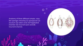
- 11. Anatomy of three different simple, vase- like sponges showing (1) spongocoel (2) osculum (3) radial canal (4) flagellated chamber (5) incurrent pore and (6) incurrent channel.
- 12. Scanning electron microscope (SEM) images illustrating the wide diversity of sponge spicule shapes Microscopic spicule lattice from Pachastrellid sponge
- 13. Classes Of Porifera Hexactinellida glass sponges Calcarea calcareous sponges Demospongiae demosponges
- 14. Calcarea • They are found in marine, shallow, and coastal water. • Their skeleton is composed of calcareous spicules made of calcium carbonate. • The body is cylindrical and exhibits radial symmetry. • The body organization is asconoid, syconoid, or leuconoid. • Eg., Clathrina, Scypha Hexactinellids • They are found in marine and the deep sea. • The skeleton is made up of six-rayed siliceous spicules. • The body is cylindrical in shape and exhibit radial symmetry. • The canal system is Sycon or Leucon. • Eg., Euplectella, Hyalonema Classification of Phylum Porifera
- 15. Desmospongiae • They are found in marine or freshwater. • The body is asymmetrical and cylindrical in shape. • The canal system is a leuconoid type. • The skeleton comprises spongin fibres, siliceous spicules, which are monoaxon and triaxon. • Eg: Spongia, Spongilla, etc.
- 16. All cells in a sponge are in contact with or near to seawater. Because each cell exchanges oxygen and carbon dioxide and discharges waste products into the seawater, a sponge has no respiratory, circulatory, or excretory system. Sponges can reproduce either asexually or sexually. Asexually reproduction (without eggs and sperm) often occurs by budding, similar to growing a new branch on a tree. Cells on the side or base of the parent begin to bulge out and form a new organism. The buds may remain attached to the parent, or they may detach and settle down nearby to form a separate organism. Sponges also reproduce sexually when specialized gametocyte cells produce sperm and eggs. Sponges undergo synchronous spawning and eject sperm and egg cells into the water. If gametes (sex cells; either sperm or egg) from the same species meet, they form a larval sponge. After a period of planktonic drifting, the larva settles to a suitable location on the bottom and grows into an adult sponge. The drifting larval stage means that sponges can colonize new locations, even though as adults they remain attached in a sessile lifestyle. Freshwater sponges can live in areas that are subject to cyclical wet and dry periods. They have a special strategy to help them deal with these harsh conditions. Freshwater sponges can produce a “resting” stage called a gemmule. A gemmule is a small, encysted bud that can tolerate being dried out for a long period of time. When the gemmule is exposed to water, it can resume development as a sponge. Organisms that can undergo a phase where they are dormant to survive harsh conditions are said to be in cryptobiosis (from the root words crypto meaning hidden and bio meaning life), because they do not appear to be living. In reality, these organisms are in a state of suspended animation.
- 17. 02 Canal System
- 18. Water Canal System The water circulatory system of sponges also called as canal system is the characteristic feature of the phylum Porifera. Canal system is also known as aquiferous system. The canal system of sponges helps in food acquisition, respiratory gas exchange and also in excretion. The numerous perforations on the body surface of the sponges for ingression and egression of water current are the main constituents of the canal system. Inside the body, the water current flows through a certain system of spaces where by the food is captured from the incoming water and the excretory material is sent out into the outgoing water.
- 19. 03 Water Current
- 20. Functions of the Water Current Water current plays the most vital role in the physiology of the sponges. The body wall of the sponges consists of two epitheloid layers the outer pinacoderm and the inner choanoderm. Pinacoderm consists of porocytes cells which bear openings called ostia. Choanoderm is composed of choanocytes or collar cells. The choanocytes have collar of microvilli around the flagellum. The water current is caused by beating of flagella of the collar cells. The following are the functions of the water current which enters the body of the sponges through the canal system: All exchanges between sponge body and external medium are maintained by means of this current. 1. Food and oxygen are brought into body through this water current 2. Also the excreta are taken out of the body with the help of this water current. 3. The reproductive bodies are carried out and into the body of the sponges by the water current.
- 21. • The spongocoel is the single largest spacious cavity in the body of the sponge. The spongocoel is lined by the flattened collar cells or choanocytes. Spongocoel opens outside through a narrow circular opening called as osculum located at the distal end and it is fringed with large monaxon spicules. • The surrounding sea water enters the canal system through the ostia. The flow of the water is maintained by the beating of the flagella of the collar cells. The rate of water flow is slow as the large spongocoel contains much water which cannot be pumped out through a single osculum.
- 22. Types Of Canal System Different sponges have different arrangement and grades of complexity of internal channels and accordingly the canal system is been divided into the following three types: Ascon type of canal system ● This canal system is the simples of all the three. It is found in asconoid type of sponges like Leucosolenia and also in some of the developmental stages of all the syconoid sponges. ● The body surface of the asconoid type of sponges is pierced by a large number of minute openings called as incurrent pores or ostia. These pores are intracellular spaces within the tube like cells called porocytes. These pores extend radially into mesenchyme and open directly into the spongocoel.
- 23. Sycon Type of Canal System Sycon type of canal system is more complex compared to the ascon type. This type of canal system is the characteristic of syconoid sponges like Scypha. Theoretically this canal system can be derived from asconoid type by horizontal folding of its walls. Also embryonic development of Scypha clearly shows the asconoid pattern being converted into syconoid pattern. Body walls of syconoid sponges include two types of canals, the radial canals and the incurrent canals paralleling and alternating with each other. Both these canals blindly end into the body wall but are interconnected by minute pores. Incurrent pores also known as dermal ostia are found on the outer surface of the body. These incurrent pores open into incurrent canals. The incurrent canals are non-flagellated as they are lined by pinacocytes and not choanocytes. The incurrent canals leas into adjacent radial canals through the minute openings called prosopyles. On the other hand radial canals are flagellated as they are lined by choanocytes. These canals open into the central spongocoel by internal ostia or apopyles. In sycon type of canal system, spongocoel is a narrow, non-flagellated cavity lined by pinacocytes. It opens to the exterior though an excurrent opening called osculum which is similar to that of the ascon type of canal system. Sycon canal system takes a more complex form in few species like Grantia, where the incurrent canals are irregular and branching forming large sub- dermal spaces. This is due to the development of cortex, involving pinacoderm and mesenchyme spreading over the entire outer surface of sponge.
- 24. Leucon type of Canal System This type of canal system results due to further folding of body wall of the sycon type of canal system. This canal system is the characteristic of the leuconoid type of sponges like Spongilla. In this type the radial symmetry is lost due to the complexity of the canal system and this result in an irregular symmetry. The flagellated chambers are small compared to that of the asconoid and syconoid type. These chambers are lined by choanocytes and are spherical in shape. All other spaces are lined by pinacocytes. The incurrent canals open into flagellated chambers through prosopyles. These flagellated chambers in turn communicate with the excurrent canals through apopyles. The excurrent canals develop as a result of shrinkage and division of spongocoel. The large and spacious spongocoel which is present in the asconoid and syconoid type of canal systems is absent here. Here the spongocoel is much reduced. This excurrent canal finally communicates with the outside through the osculum. Leucon type of canal system has the following three successive grades in its evolutionary pattern: Eurypylous type: This is the simplest and the most primitive type of leuconoid canal system. In this type the flagellated chambers directly communicate with the excurrent canal through broad apertures called the apopyles. Ex: Plakina Aphodal type: In this type of canal system the apopyles are drawn out as a narrow canal called aphodas. This connects the flagellated chambers with the excurrent canals. Ex: Geodia Diplodal type: in some of the sponges, along with aphodas another narrow tube called prosodus is present between incurrent canal and flagellated chamber. This arrangement gives rise to diplodal type of canal system. Ex: Spongilla
- 25. Leucon Type Canal System
- 27. Bibliography • https://www.studyandscore.com/studymaterial- detail/phylum-porifera-canal-system-in-sponges-types-of- canal-systems-in-sponges-functions-of-water-current • Chromeextension://efaidnbmnnnibpcajpcglclefindmkaj/https:/ /silapatharcollege.edu.in/online/attendence/classnotes/files/1 628324973.pdf • https://manoa.hawaii.edu/exploringourfluidearth/biological/in vertebrates/phylum- porifera#:~:text=The%20phylum%20Porifera%20comprises %20the,as%20five%20kilometers%20(km).
- 28. Thank You!!!