Location via proxy:   [ UP ]  
[Report a bug]   [Manage cookies]                
SlideShare a Scribd company logo

1

Cardiac Output
Maria idrees; PT

2

INTRODUCTION
• Cardiac output is the amount of blood
pumped from each ventricle.
• Usually, it refers to left ventricular output
through aorta.
• Cardiac output is the most important factor in
cardiovascular system, because rate of blood
flow through different parts of the body
depends upon cardiac output.

3

DEFINITIONS AND NORMAL VALUES
• Usually, cardiac output is expressed in three
ways:
1. Stroke volume
2. Minute volume
3. Cardiac index.

4

STROKE VOLUME
• Stroke volume is the amount of blood pumped
out by each ventricle during each beat.
• Normal value: 70 mL (60 to 80 mL) when the
heart rate is normal (72/minute).

5

MINUTE VOLUME
• Minute volume is the amount of blood
pumped out by each ventricle in one minute.
• It is the product of stroke volume and heart
rate:
Minute volume = Stroke volume × Heart rate
• Normal value: 5 L/ventricle/minute.

6

CARDIAC INDEX
• Cardiac index is the minute volume expressed
in relation to square meter of body surface
area.
• It is defined as the amount of blood pumped
out per ventricle/minute/ square meter of the
body surface area.
• Normal value: 2.8 ± 0.3 L/square meter of
body surface area/minute

7

EJECTION FRACTION
• Ejection fraction is the fraction of end diastolic
volume that is ejected out by each ventricle.
• Normal ejection fraction is 60% to 65%

8

CARDIAC RESERVE
• Cardiac reserve is the maximum amount of
blood that can be pumped out by heart above
the normal value.
• Cardiac reserve plays an important role in
increasing the cardiac output during the
conditions like exercise.
• It is essential to withstand the stress of
exercise.

9

• Cardiac reserve is usually expressed in
percentage. In a normal young healthy adult,
the cardiac reserve is 300% to 400%.
• In old age, it is about 200% to 250%.
• It increases to 500% to 600% in athletes.
• In cardiac diseases, the cardiac reserve is
minimum or nil.

10

DISTRIBUTION OF CARDIAC OUTPUT
• The whole amount of blood pumped out by
the right ventricle goes to lungs.
• But, the blood pumped by the left ventricle is
distributed to different parts of the body.
• Fraction of cardiac output distributed to a
particular region or organ depends upon the
metabolic activities of that region or organ.

11

Distribution of Blood Pumped out of
Left Ventricle
• Heart, which pumps the blood to all other
organs, receives the least amount of blood.
• Liver receives maximum amount of blood.

12

FACTORS MAINTAINING CARDIAC
OUTPUT
• Cardiac output is maintained (determined) by
four factors:
1. Venous return
2. Force of contraction
3. Heart rate
4. Peripheral resistance.

13

1. VENOUS RETURN
• Venous return is the amount of blood which is
returned to heart from different parts of the
body. When it increases, the ventricular filling
and cardiac output are increased.
• Thus, cardiac output is directly proportional to
venous return, provided the other factors
(force of contraction, heart rate and
peripheral resistance) remain constant.

14

• Venous return in turn, depends upon five
factors:
i. Respiratory pump
ii. Muscle pump
iii. Gravity
iv. Venous pressure
v. Sympathetic tone.

15

i. Respiratory Pump
• Respiratory pump is the respiratory activity
that helps the return of blood, to heart
during inspiration. It is also called
abdominothoracic pump.
• During inspiration, thoracic cavity expands
and makes the intrathoracic pressure more
negative.
• It increases the diameter of inferior vena
cava, resulting in increased venous return.

16

• At the same time, descent of diaphragm
increases the intra-abdominal pressure, which
compresses abdominal veins and pushes the
blood upward towards the heart and thereby
the venous return is increased. Respiratory
pump is much stronger in forced respiration
and in severe muscular exercise.

17

Cardiac output

18

ii. Muscle Pump
• Muscle pump is the muscular activity that
helps in return of the blood to heart. During
muscular activities, the veins are compressed
or squeezed. Due to the presence of valves in
veins, during compression the blood is moved
towards the heart. When muscular activity
increases, the venous return is more.
• When the skeletal muscles contract, the vein
located in between the muscles is
compressed.

19

• Valve of the vein proximal to the contracting
muscles is opened and the blood is propelled
towards the heart. Valve of the vein distal to
the muscles is closed by the back flow of
blood.
• During relaxation of the muscles, the valve
proximal to muscles closes and prevents the
back flow of blood. The valve distal to the
muscles opens and allows the blood to flow
upwards.

20

Cardiac output

21

iii. Gravity
• Gravitational force reduces the venous return.
When a person stands for a long period,
gravity causes pooling of blood in the legs,
which is called venous pooling.
• Because of venous pooling, the amount of
blood returning to heart decreases.

22

iv. Venous Pressure
• Venous pressure also affects the venous return.
Pressure in the venules is 12 to 18 mm Hg. In the
smaller and larger veins, the pressure gradually
decreases. In the great veins, i.e. inferior vena
cava and superior vena cava, the pressure falls to
about 5.5 mm Hg. At the junction of venae cavae
and right atrium, it is about 4.6 mm Hg.
• Pressure in the right atrium is still low and it
alters during cardiac action. It falls to zero during
atrial diastole. This pressure gradient at every
part of venous tree helps as a driving force for
venous return.

23

v. Sympathetic Tone
• Venous return is aided by sympathetic or
vasomotor tone, which causes constriction of
venules.
• Venoconstriction pushes the blood towards
heart.

24

2. FORCE OF CONTRACTION
• Cardiac output is directly proportional to the
force of contraction, provided the other three
factors remain constant. According to Frank-
Starling law, force of contraction of heart is
directly proportional to the initial length of
muscle fibers, before the onset of contraction.
Force of contraction depends upon preload
and afterload.

25

Preload
• Preload is the stretching of the cardiac muscle
fibers at the end of diastole, just before
contraction.
• It is due to increase in ventricular pressure
caused by filling of blood during diastole.
• Stretching of muscle fibers increases their
length, which increases the force of
contraction and cardiac output.

26

Afterload
• Afterload is the force against which ventricles must
contract and eject the blood. Force is determined by
the arterial pressure. At the end of isometric
contraction period, semilunar valves are opened and
blood is ejected into the aorta and pulmonary
artery. So, the pressure increases in these two
vessels.
• Now, the ventricles have to work against this
pressure for further ejection. Thus, the afterload for
left ventricle is determined by aortic pressure and
afterload for right ventricular pressure is determined
by pressure in pulmonary artery. Force of
contraction of heart and cardiac output are inversely
proportional to afterload.

27

3. HEART RATE
• Cardiac output is directly proportional to
heart rate provided, the other three factors
remain constant.
• Moderate change in heart rate does not alter
the cardiac output.
• If there is a marked increase in heart rate,
cardiac output is increased.
• If there is marked decrease in heart rate,
cardiac output is decreased.

28

4. PERIPHERAL RESISTANCE
• Peripheral resistance is the resistance offered to
blood flow at the peripheral blood vessels.
• Peripheral resistance is the resistance or load
against which the heart has to pump the blood.
• So, the cardiac output is inversely proportional to
peripheral resistance.
• Resistance is offered at arterioles so, the
arterioles are called resistant vessels. In the body,
maximum peripheral resistance is offered at the
splanchnic region.

More Related Content

Cardiac output

  • 2. INTRODUCTION • Cardiac output is the amount of blood pumped from each ventricle. • Usually, it refers to left ventricular output through aorta. • Cardiac output is the most important factor in cardiovascular system, because rate of blood flow through different parts of the body depends upon cardiac output.
  • 3. DEFINITIONS AND NORMAL VALUES • Usually, cardiac output is expressed in three ways: 1. Stroke volume 2. Minute volume 3. Cardiac index.
  • 4. STROKE VOLUME • Stroke volume is the amount of blood pumped out by each ventricle during each beat. • Normal value: 70 mL (60 to 80 mL) when the heart rate is normal (72/minute).
  • 5. MINUTE VOLUME • Minute volume is the amount of blood pumped out by each ventricle in one minute. • It is the product of stroke volume and heart rate: Minute volume = Stroke volume × Heart rate • Normal value: 5 L/ventricle/minute.
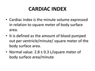
  • 6. CARDIAC INDEX • Cardiac index is the minute volume expressed in relation to square meter of body surface area. • It is defined as the amount of blood pumped out per ventricle/minute/ square meter of the body surface area. • Normal value: 2.8 ± 0.3 L/square meter of body surface area/minute
  • 7. EJECTION FRACTION • Ejection fraction is the fraction of end diastolic volume that is ejected out by each ventricle. • Normal ejection fraction is 60% to 65%
  • 8. CARDIAC RESERVE • Cardiac reserve is the maximum amount of blood that can be pumped out by heart above the normal value. • Cardiac reserve plays an important role in increasing the cardiac output during the conditions like exercise. • It is essential to withstand the stress of exercise.
  • 9. • Cardiac reserve is usually expressed in percentage. In a normal young healthy adult, the cardiac reserve is 300% to 400%. • In old age, it is about 200% to 250%. • It increases to 500% to 600% in athletes. • In cardiac diseases, the cardiac reserve is minimum or nil.
  • 10. DISTRIBUTION OF CARDIAC OUTPUT • The whole amount of blood pumped out by the right ventricle goes to lungs. • But, the blood pumped by the left ventricle is distributed to different parts of the body. • Fraction of cardiac output distributed to a particular region or organ depends upon the metabolic activities of that region or organ.
  • 11. Distribution of Blood Pumped out of Left Ventricle • Heart, which pumps the blood to all other organs, receives the least amount of blood. • Liver receives maximum amount of blood.
  • 12. FACTORS MAINTAINING CARDIAC OUTPUT • Cardiac output is maintained (determined) by four factors: 1. Venous return 2. Force of contraction 3. Heart rate 4. Peripheral resistance.
  • 13. 1. VENOUS RETURN • Venous return is the amount of blood which is returned to heart from different parts of the body. When it increases, the ventricular filling and cardiac output are increased. • Thus, cardiac output is directly proportional to venous return, provided the other factors (force of contraction, heart rate and peripheral resistance) remain constant.
  • 14. • Venous return in turn, depends upon five factors: i. Respiratory pump ii. Muscle pump iii. Gravity iv. Venous pressure v. Sympathetic tone.
  • 15. i. Respiratory Pump • Respiratory pump is the respiratory activity that helps the return of blood, to heart during inspiration. It is also called abdominothoracic pump. • During inspiration, thoracic cavity expands and makes the intrathoracic pressure more negative. • It increases the diameter of inferior vena cava, resulting in increased venous return.
  • 16. • At the same time, descent of diaphragm increases the intra-abdominal pressure, which compresses abdominal veins and pushes the blood upward towards the heart and thereby the venous return is increased. Respiratory pump is much stronger in forced respiration and in severe muscular exercise.
  • 18. ii. Muscle Pump • Muscle pump is the muscular activity that helps in return of the blood to heart. During muscular activities, the veins are compressed or squeezed. Due to the presence of valves in veins, during compression the blood is moved towards the heart. When muscular activity increases, the venous return is more. • When the skeletal muscles contract, the vein located in between the muscles is compressed.
  • 19. • Valve of the vein proximal to the contracting muscles is opened and the blood is propelled towards the heart. Valve of the vein distal to the muscles is closed by the back flow of blood. • During relaxation of the muscles, the valve proximal to muscles closes and prevents the back flow of blood. The valve distal to the muscles opens and allows the blood to flow upwards.
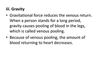
  • 21. iii. Gravity • Gravitational force reduces the venous return. When a person stands for a long period, gravity causes pooling of blood in the legs, which is called venous pooling. • Because of venous pooling, the amount of blood returning to heart decreases.
  • 22. iv. Venous Pressure • Venous pressure also affects the venous return. Pressure in the venules is 12 to 18 mm Hg. In the smaller and larger veins, the pressure gradually decreases. In the great veins, i.e. inferior vena cava and superior vena cava, the pressure falls to about 5.5 mm Hg. At the junction of venae cavae and right atrium, it is about 4.6 mm Hg. • Pressure in the right atrium is still low and it alters during cardiac action. It falls to zero during atrial diastole. This pressure gradient at every part of venous tree helps as a driving force for venous return.
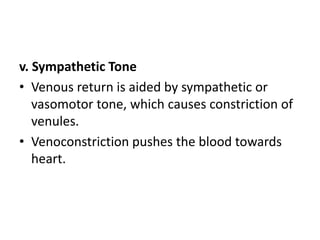
  • 23. v. Sympathetic Tone • Venous return is aided by sympathetic or vasomotor tone, which causes constriction of venules. • Venoconstriction pushes the blood towards heart.
  • 24. 2. FORCE OF CONTRACTION • Cardiac output is directly proportional to the force of contraction, provided the other three factors remain constant. According to Frank- Starling law, force of contraction of heart is directly proportional to the initial length of muscle fibers, before the onset of contraction. Force of contraction depends upon preload and afterload.
  • 25. Preload • Preload is the stretching of the cardiac muscle fibers at the end of diastole, just before contraction. • It is due to increase in ventricular pressure caused by filling of blood during diastole. • Stretching of muscle fibers increases their length, which increases the force of contraction and cardiac output.
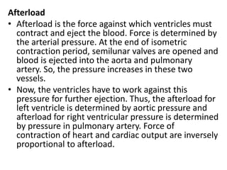
  • 26. Afterload • Afterload is the force against which ventricles must contract and eject the blood. Force is determined by the arterial pressure. At the end of isometric contraction period, semilunar valves are opened and blood is ejected into the aorta and pulmonary artery. So, the pressure increases in these two vessels. • Now, the ventricles have to work against this pressure for further ejection. Thus, the afterload for left ventricle is determined by aortic pressure and afterload for right ventricular pressure is determined by pressure in pulmonary artery. Force of contraction of heart and cardiac output are inversely proportional to afterload.
  • 27. 3. HEART RATE • Cardiac output is directly proportional to heart rate provided, the other three factors remain constant. • Moderate change in heart rate does not alter the cardiac output. • If there is a marked increase in heart rate, cardiac output is increased. • If there is marked decrease in heart rate, cardiac output is decreased.
  • 28. 4. PERIPHERAL RESISTANCE • Peripheral resistance is the resistance offered to blood flow at the peripheral blood vessels. • Peripheral resistance is the resistance or load against which the heart has to pump the blood. • So, the cardiac output is inversely proportional to peripheral resistance. • Resistance is offered at arterioles so, the arterioles are called resistant vessels. In the body, maximum peripheral resistance is offered at the splanchnic region.