CARDIOVASCULAR SYSTEM
•
1 like•44 views

This document provides an overview of the cardiovascular system and human heart. It begins with definitions and functions of the cardiovascular system. It then describes the internal and external structure of the heart, including the four chambers, valves, blood vessels, and conducting system. Key concepts covered include the cardiac cycle, blood pressure, electrocardiogram, coronary and systemic circulation. Common heart conditions such as coronary artery disease, heart attack, arrhythmias and valve disease are also summarized.
Report
Share


CARDIOVASCULAR SYSTEM
- 1. I d pharma Human anatomy and physiology Cardiovascular system Lakshman v bendre Asst. prof Dept.pharmacology
- 2. Table of Contents 1. Introduction 2. Position of heart 3. Structure of the Human Heart 4. Physiology of heart 5. Coronary circulation 6. Conducting system 7. Cardiac cycle 8. Cardiac output 9. Electrocardiogram 10. Blood pressure 11. Functions of heart 12. Types of circulation 13. Disorders of heart
- 3. CARDIO~VASCULAR SYSTEM “heart” “vessels” Definition: There is continuous flow of blood through out the human body the process is known as blood circulation. Made up of heart, blood vessels, and blood Functions Heart- pump blood Vessels- (veins, arteries, capillaries) circulate blood Blood- transports nutrients, waste, hormones, oxygen, antibodies
- 4. Introduction to the Human Heart The human heart is one of the most important organs responsible for sustaining life. It is a muscular organ with four chambers. The size of the heart is the size of about a clenched fist. The human heart functions throughout a person’s lifespan and is one of the most robust and hardest working muscles in the human body.
- 5. Position of Heart in Human Body The human heart is located between the lungs in the thoracic cavity, slightly towards the left of the sternum (breastbone). It is derived from the embryonic mesodermal germ layer.
- 6. EXTERNAL STRUCTURE OF THE HUMAN HEART The human heart is hollow muscular organ which is situated to the left of the chest and is enclosed within a fluid-filled cavity described as the pericardial cavity. The walls and lining of the pericardial cavity are made up of a membrane known as the pericardium.
- 7. • The pericardium is a fiber membrane found as an external covering around the heart. It protects the heart by producing a serous fluid, which serves to lubricate the heart and prevent friction between the surrounding organs. Apart from the lubrication, the pericardium also helps by holding the heart in its position and by maintaining a hollow space for the heart to expand itself when it is full. The pericardium has two exclusive layers— • Visceral Pericardium: It directly covers the outside of the heart. • Parietal Pericardium: : It forms a sac around the outer region of the heart that contains the fluid in the pericardial cavity.
- 8. STRUCTURE OF THE HEART WALL The heart wall is made up of 3 layers, namely: Epicardium – Epicardium is the outermost layer of the heart. It is composed of a thin-layered membrane that serves to lubricate and protect the outer section. Myocardium – This is a layer of muscle tissue and it constitutes the middle layer wall of the heart. It contributes to the thickness and is responsible for the pumping action. Endocardium – It is the innermost layer that lines the inner heart chambers and covers the heart valves. Furthermore, it prevents the blood from sticking to the inner walls, thereby preventing potentially fatal blood clots
- 9. INTERNAL STRUCTURE OF HEART The internal structure of the heart is rather intricate with several chambers and valves that control the flow of blood. Chambers of the Heart Avian and mammalian hearts consists of four chambers. Humans are mammals; hence, we have four chambers, namely: 1. Left atrium 2. Right atrium 3. Left ventricle 4. Right ventricle
- 11. Atria are thin, less muscular walls and smaller than ventricles. These are the blood-receiving chambers that are fed by the large veins. Ventricles are larger and more muscular chambers responsible for pumping and pushing blood out to the circulation. These are connected to larger arteries that deliver blood for circulation. The right ventricle and right atrium are comparatively smaller than the left chambers. The walls consist of fewer muscles compared to the left portion, and the size difference is based on their functions. The blood originating from the right side flows through the pulmonary circulation, while blood arising from the left chambers is pumped throughout the body. SEPTUM: Right and left atria are separated by inter auricular septum where as Right and left ventricles are separated by inter ventricular septum.
- 12. VALVES Valves are flaps of fibrous tissues located in the cardiac chambers between the veins. They ensure that the blood flows in a single direction (unidirectional). Flaps also prevent the blood from flowing backwards. Based on their function, valves are of two types: 1. Atrioventricular valves 2. Semilunar valves Atrioventricular valves : are between ventricles and atria. The valve between the right ventricle and right atrium is the tricuspid valve and the one which is found between the left ventricle and left atrium is known as the mitral valve (bicuspid valve) Semilunar valves : These are present in Aorta and Pulmonary artery. the valve which is located between the left ventricle and aorta called Aortic valve. It is also found between the pulmonary artery and right ventricle called Pulmonary valve.
- 13. Blood Vessels In humans with closed circulatory systems, the blood flows within vessels of varying sizes. The external structure of the heart has many blood vessels that form a network, with other major vessels emerging from within the structure. The blood vessels typically comprise the following: 1.Superior venacava and inferior venacava: They carry deoxygenated blood to the right atrium of the herat. 2.Pulmonary artery : Carries deoxygenated blood to the lung for purification 3.Pulmonary vein: They carry oxygenated blood to the left atrium. Of the heart 4.Systemic Aorta: Carries oxygenated blood to the various parts of the body from left ventricle. Capillaries are tiny, tube-like vessels which form a network between the arteries to veins.
- 14. ARTERY VEINS Involved in carrying oxygenated blood except for pulmonary arteries Involved in carrying deoxygenated blood except for pulmonary veins Red in colour. Blue in colour. Located deep within the body. Peripherally located closer to the skin. Carry blood away from the heart to various parts of the body. Carry blood towards the heart from the various parts of the body. Comparatively higher oxygen level Comparatively low oxygen level. High pressure, as the blood flows by the pumping pressure of the heart. Low pressure, as the blood flows by the capillary action of the veins.
- 16. T.S OF ARTERY AND VEIN
- 17. PATHWAY OF BLOOD CIRCULATION
- 19. MECHANISM OF BLOOD CIRCULATION Blood comes into the right atrium from the body, moves into the right ventricle and is pushed into the pulmonary arteries in the lungs. After picking up oxygen, the blood travels back to the heart through the pulmonary veins into the left atrium, to the left ventricle and out to the body's tissues through the aorta.
- 21. CORONARY CIRCULATION(Blood supply to the heart): It is the circulation of blood to the heart first bronets of aorta in the coronary artery which supplies oxygenated blood to the heart muscle it self,
- 22. CONDUCTING SYSTEM OF HEART The impulses for contracton of the heart are transmitted through the conducting system of heart. This pathway is made up of 4 elements: 1. Sino-atrial (SA) node. 2. Atrio-ventricular (AV) node. 3. Bundle of His. 4. Purkinje fibres.
- 23. 1. Sino-atrial (SA) node: It is the specialized cells in the wall of right atrium near the opening of superior venacava. The SA Node is often called as PACEMAKER because it initiates impulse of contraction of heart. This cardiac action potential spread over the right and left atria causing them to contract blood enters into ventricles. 2. Atrio-ventricular (AV) node: It is also specialized cells situated in the wall of the inter septum near the atrio- ventricular opening. Impulse of SA Node passes to this AV Node.
- 24. 3. Bundle of His: It is a specialized mass fibers that originated from AV Node which further divides into left and right branches. 4. Purkinje fibers : Right and left branches of Bundle of his breaks up into fine fibrous called Purkinje fibers. . These fibers convey the impulse of contraction from AV Node to the entire ventricle system Thus the entire sequence of conduction takes place.
- 25. Cardiac Cycle “Cardiac cycle refers to the sequence of events that take place when the heart beats.” The cardiac cycle attributes to a comprehensive heartbeat from its production to the commencement of the next beat. It comprises diastole, the systole, and the intervening pause. The occurrence of a cardiac cycle is illustrated by a heart rate, which is naturally indicated as beats per minute A healthy human heart beats 72 times per minute which states that there are 72 cardiac cycles per minute. The cardiac cycle involves a complete contraction and relaxation of both the atria and ventricles and the cycle last approximately 0.8 seconds.
- 26. Cardiac Cycle Phases Following are the different phases that occur in a cardiac cycle: 1) Atrial Diastole 2) Atrial Systole 3) Isovolumic Contraction 4) Ventricular Ejection 5) Isovolumic Relaxation 6) Ventricular Filling Stage
- 27. Duration of Cardiac Cycle In a normal person, a heartbeat is 72 beats/minute. So, the duration of one cardiac cycle can be calculated as: 1/72 beats/minute=.0139 minutes/beat At a heartbeat 72 beats/minute, duration of each cardiac cycle will be 0.8 seconds. Duration of different stages of the cardiac cycle is given below: Atrial systole: continues for about 0.1 seconds Ventricular systole: continues for about 0.3 seconds Atrial diastole: continues for about 0.7 seconds Ventricular diastole: continues for about 0.5 seconds
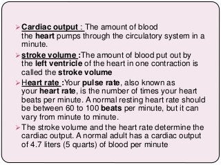
- 28. Cardiac output : The amount of blood the heart pumps through the circulatory system in a minute. stroke volume :The amount of blood put out by the left ventricle of the heart in one contraction is called the stroke volume Heart rate :Your pulse rate, also known as your heart rate, is the number of times your heart beats per minute. A normal resting heart rate should be between 60 to 100 beats per minute, but it can vary from minute to minute. The stroke volume and the heart rate determine the cardiac output. A normal adult has a cardiac output of 4.7 liters (5 quarts) of blood per minute
- 29. Cardiac output Stroke volume Heart Rate 5040 ml/min 70 ml 72/min
- 30. Electrocardiogram An electrocardiogram is a test that measures the electrical activity of the heartbeat. With each beat, an electrical impulse (or “wave”) travels through the heart. This wave causes the muscle to squeeze and pump blood from the heart. The ECG recordings are designated by letters P, Q, R, S, T. Wave ‘’P’’ represents Atrial contractions Wave ‘‘Q R S’’ represents Ventricular contraction. Wave ‘‘T’’ represents Ventricular relaxation.
- 31. Electrocardiography is the process of producing an electrocardiogram
- 32. Blood pressure Blood pressure is the pressure of circulating blood against the walls of blood vessels. the term "blood pressure" refers to the pressure in the large arteries. There are two types of blood pressure 1. Systolic blood pressure 2. Diastolic blood pressure Systolic BP- maximum pressure during systole is 120 mmHg Diastolic BP- minimum pressure during diastole is 80 mmHg Normal blood pressure 120/80 mmHg Pulse pressure- It is the difference between systolic and diastolic BP
- 33. RECORDING OF BP A sphygmomanometer is a device that measures blood pressure. It is composes of an inflatable rubber cuff, which is wrapped around the arm. A measuring device indicates the cuff's pressure. A bulb inflates the cuff and a valve releases pressure. A stethoscope is used to listen to arterial blood flow sounds. SIGNIFICANCE: a. Measuring blood pressure b. Monitoring effectiveness of medications. c. Helps to control hypertension d. It helps to detect various
- 34. ARETERY AND VENOUS SYSTEM The arterial system is the higher- pressure portion of the circulatory system, with pressure varying between the peak pressure during heart contraction ( systolic pressure ) and the minimum (diastolic) pressure between contractions when the heart expands and refills. The venous system is that part of the circulation in which the blood is transported from the periphery back to the heart. We distinguish between the superficial and the deep venous systems.
- 35. TYPES OF CIRCULATION 1. Pulmonary circulation is a portion of circulation responsible for carrying deoxygenated blood away from the heart, to the lungs and then brings oxygenated blood back to the heart. 2. Systemic circulation is another portion of circulation where the oxygenated blood is pumped from the heart to every organ and tissue in the body, and deoxygenated blood comes back again to the heart.
- 36. FUNCTIONS OF THE HEART The four main functions of the heart are: 1. Pumping oxygenated blood to the other body parts. 2. Pumping hormones and other vital substances to different parts of the body. 3. Receiving deoxygenated blood and carrying metabolic waste products from the body and pumping it to the lungs for oxygenation. 4. Maintaining blood pressure.
- 37. Heart d Conditions OR Disorders of heart 1. Coronary artery disease : Over the years, cholesterol plaques can narrow the arteries supplying blood to the heart. The narrowed arteries are at higher risk for complete blockage from a sudden blood clot (this blockage is called a heart attack). 2. Stable angina pectoris: Narrowed coronary arteries cause predictable chest pain or discomfort with exertion. The blockages prevent the heart from receiving the extra oxygen needed for strenuous activity. Symptoms typically get better with rest. 3. Unstable angina pectoris: Chest pain or discomfort that is new, worsening, or occurs at rest. This is an emergency situation as it can precede a heart attack, serious abnormal heart rhythm, or cardiac arrest. 4. Myocardial infarction (heart attack): A coronary artery is suddenly blocked. Starved of oxygen, part of the heart muscle dies. 5. Arrhythmia(dysrhythmia): An abnormal heart rhythm due to changes in the conduction of electrical impulses through the heart. Some arrhythmias are benign, but others are life-threatening. 6. Congestive heart failure: The heart is either too weak or too stiff to effectively pump blood through the body. Shortness of breath and leg swelling are common symptoms. 7. Cardiomyopathy: A disease of heart muscle in which the heart is abnormally enlarged, thickened, and/or stiffened. As a result, the heart's ability to pump blood is weakened. 8. Myocarditis: Inflammation of the heart muscle, most often due to a viral infection. 9. Pericarditis: Inflammation of the lining of the heart (pericardium). Viral
- 38. 10. Pericardial effusion: Fluid between the lining of the heart (pericardium) and the heart itself. Often, this is due to pericarditis. 11. Atrial fibrillation: Abnormal electrical impulses in the atria cause an irregular heartbeat. Atrial fibrillation is one of the most common arrhythmias. 12. Pulmonary embolism: Typically a blood clot travels through the heart to the lungs. 13. Heart valve disease: There are four heart valves, and each can develop problems. If severe, valve disease can cause congestive heart failure. 14. Heart murmur: An abnormal sound heard when listening to the heart with a stethoscope. Some heart murmurs are benign; others suggest heart disease. 15. Endocarditis: Inflammation of the inner lining or heart valves of the heart. Usually, endocarditis is due to a serious infection of the heart valves. 16. Mitral valve prolapse: The mitral valve is forced backward slightly after blood has passed through the valve. 17. Sudden cardiac death: Death caused by a sudden loss of heart function (cardiac arrest). 18. Cardiac arrest: Sudden loss of heart functionaa