


Cardiovascular System unit VIII - Copy Std.pptx
- 2. Objectives At the end of this session, the students will be able to: • Describe the location, structure and functions of the heart and its great blood vessels. • Discuss the blood flow through the heart • Describe the structure and functional features of the conducting system of the heart. • Describe the principal events of a cardiac cycle.
- 3. • Explain the structure and function of: • Arteries • Veins & • Capillaries • Describe the following types of blood circulation: • Pulmonary circulation • Systemic circulation (coronary & hepatic portal circulation).
- 4. Parts of the Circulatory System • Divided into three major parts: – Heart – Blood – Blood Vessels
- 5. Functions of cardiovascular System • Circulate blood throughout entire body for – Transport of oxygen to cells. – Transport of CO2 away from cells. – Transport of nutrients to cells. – Movement of immune system components. (cells, antibodies) – Transport of endocrine gland secretions.
- 6. Heart • Location: mediastinum and rests on the diaphragm. • Size: as much as one’s close fist. • Weight: 250 g in adult female and 300 g in male. • Structure: Cone shaped with pointed apex inferiorly to left and broad base superiorly to the right. Membranous Layers • Two Pericardium as superficial fibrous pericardium of connective tissue and deeper serous pericardium of epithelial tissue connected with the fibrous pericardium. • Serous pericardium—Parietal and visceral membrane. Pericardial cavity with pericardial fluid.
- 7. Layers of heart • —three layers Epicardium or visceral layer Myocardium—muscular layer Endocardium—endothelial layer
- 8. Serous membrane Continuous with blood vessels
- 9. Chambers of the Heart Four chambers—two pumps ─ Two atria (superior) ─ Two ventricles (inferior) Right Pump Right Atrium receives deoxygenated blood from three veins as: ─ Superior vena cava ─ Inferior vena cava ─ Interatrial septum—between right and left atrium
- 10. Right Ventricle: • Inside ridges called trabeculae carnae • Receives blood from right atrium via tricuspid valve (Right atrioventricular valve) • The cusps are connected to chordae tendineae. • Chordae tendineae are connected to papillary muscles. • The partition between right and left ventricle is interventricular septum. • Deoxygenated blood is pumped out through the pulmonary valve (Pulmonary valve) into a large artery called pulmonary trunk which divides into right and left pulmonary arteries.
- 11. Tricuspid Valve
- 12. Left Pump Left Atrium • It receives oxygenated blood from lungs through four pulmonary veins. • Passes blood to the left ventricle via bicuspid (mitral) valve or left atrioventricular valve. Left Ventricle • The thickest chamber of the heart. • Forms the apex of the heart • Blood passes from the left ventricle through the aortic valve (aortic semilunar valve) into the ascending aorta. • Some blood from the ascending aorta flows into the coronary arteries.
- 13. Great Blood vessels The great blood vessels provide a pathway for the entire cardiac circulation to proceed. •Superior and inferior vena cava. The heart receives relatively oxygen-poor blood from the veins of the body through the large superior and inferior vena cava and pumps it through the pulmonary trunk. •Pulmonary arteries. The pulmonary trunk splits into the right and left pulmonary arteries, which carry blood to the lungs, where oxygen is picked up and carbon dioxide is unloaded. •Pulmonary veins. Oxygen-rich blood drains from the lungs and is returned to the left side of the heart through the four pulmonary veins. •Aorta. Blood returned to the left side of the heart is pumped out of the heart into the aorta from which the systemic arteries branch to supply essentially all body tissues
- 14. Atrioventricular valves • The atrioventricular valves are thin, leaflike structures located between the atria and the ventricles. The right atrioventricular opening is guarded by the tricuspid valve, so called because it consists of three irregularly shaped cusps, or flaps. The leaflets consist essentially of folds of endocardium (the membrane lining the heart) reinforced with a flat sheet of dense connective tissue. At the base of the leaflets, the middle supporting flat plate becomes continuous with that of the dense connective tissue of the ridge surrounding the openings.
- 16. Semilunar valves • They have three cusps. • They are located at the base of both the pulmonary trunk (pulmonary artery) and the aorta, the two arteries taking blood out of the ventricles. • These valves permit blood to be forced into the arteries, but prevent backflow of blood from the arteries into the ventricles. • These valves do not have chordae tendineae, and are more similar to valves in veins than atrioventricular valves.
- 17. Sound of the Heart
- 18. Chambers of the heart; valves
- 19. Valvular Disorders • Stenosis (= narrowing): Failure of a valve to close completely is called insufficiency or incompetence, e.g, Mitral stenosis or aortic stenosis in which there is backflow of blood. • Rheumatic fever, an acute inflammatory disease caused by streptococcal infection, is one of the causes of valvular disorders.
- 20. Conduction System • Specialized cardiac muscle fibers called autorhythmic fibers. • Self-excitable • Generate electrical activity (action potential). • No dependent on nerves for stimulation. • Heart has its own intrinsic system.
- 21. Conduction System • Cardiac muscle cells. Cardiac muscle cells can and do contract spontaneously and independently, even if all nervous connections are severed. • Rhythms. Although cardiac muscles can beat independently, the muscle cells in the different areas of the heart have different rhythms. • Intrinsic conduction system. The intrinsic conduction system, or the nodal system, that is built into the heart tissue sets the basic rhythm. • Composition. The intrinsic conduction system is composed of a special tissue found nowhere else in the body; it is much like a cross between a muscle and nervous tissue. • Function. This system causes heart muscle depolarization in only one direction- from the atria to the ventricles; it enforces a contraction rate of
- 22. Conduction System • approximately 75 beats per minute on the heart, thus the heart beats as a coordinated unit. • Sinoatrial (SA) node. The SA node has the highest rate of depolarization in the whole system, so it can start the beat and set the pace for the whole heart; thus the term “pacemaker“. • Atrial contraction. From the SA node, the impulse spread through the atria to the AV node, and then the atria contract. • Ventricular contraction. It then passes through the AV bundle, the bundle branches, and the Purkinje fibers, resulting in a “wringing” contraction of the ventricles that begins at the heart apex and moves toward the atria. • Ejection. This contraction effectively ejects blood superiorly into the large arteries leaving the heart.
- 23. The Pathway of the Conduction System • SA node. The depolarization wave is initiated by the sinoatrial node. • Atrial myocardium. The wave then successively passes through the atrial myocardium. • Atrioventricular node. The depolarization wave then spreads to the AV node, and then the atria contract. • AV bundle. It then passes rapidly through the AV bundle. • Bundle branches and Purkinje fibers. The wave then continues on through the right and left bundle branches, and then to the Purkinje fibers in the ventricular walls, resulting in a contraction that ejects blood, leaving the heart.
- 26. Cardiac Cycle and Heart Sounds In a healthy heart, the atria contract simultaneously, then, as they start to relax, contraction of the ventricles begin. • Systole. Systole means heart contraction. • Diastole. Diastole means heart relaxation. • Cardiac cycle. The term cardiac cycle refers to the events of one complete heart beat, during which both atria and ventricles contract and then relax. • Length. The average heart beats approximately 75 times per minute, so the length of the cardiac cycle is normally about 0.8 second.
- 27. Cardiac Cycle and Heart Sounds • Mid-to-late diastole. The cycle starts with the heart in complete relaxation; the pressure in the heart is low, and blood is flowing passively into and through the atria into the ventricles from the pulmonary and systemic circulations; the semilunar valves are closed, and the AV valves are open; then the atria contract and force the blood remaining in their chambers into the ventricles. • Ventricular systole. Shortly after, the ventricular contraction begins, and the pressure within the ventricles increases rapidly, closing the AV valves; when the intraventricular pressure is higher than the pressure in the large arteries leaving the heart, the semilunar valves are forced open, and blood rushes through them out of the ventricles; the atria are relaxed, and their chambers are again filling with blood.
- 28. Cardiac Cycle and Heart Sounds • Early diastole. At the end of systole, the ventricles relax, the semilunar valves snap shut, and for a moment the ventricles are completely closed chambers; the intraventricular pressure drops and the AV valves are forced open; the ventricles again begin refilling rapidly with blood, completing the cycle. • First heart sound. The first heart sound, “lub”, is caused by the closing of the AV valves. • Second heart sound. The second heart sound, “dub”, occurs when the semilunar valves close at the end of systole.
- 29. Cardiac Output • Cardiac output is the amount of blood pumped out by each side of the heart in one minute. It is the product of the heart rate and the stroke volume. • Stroke volume. Stroke volume is the volume of blood pumped out by a ventricle with each heartbeat. • Regulation of stroke volume. According to Starling’s law of the heart, the critical factor controlling stroke volume is how much the cardiac muscle cells are stretched just before they contract; the more they are stretched, the stronger the contraction will be; and anything that increases the volume or speed of venous return also increases stroke volume and force of contraction. • Factors modifying basic heart rate. The most important external influence on heart rate is the activity of the autonomic nervous system, as well as physical factors (age, gender, exercise, and body temperature).
- 30. Blood Vessels • Blood circulates inside the blood vessels, which form a closed transport system, the so-called vascular system. • Arteries. As the heart beats, blood is propelled into large arteries leaving the heart. • Arterioles. It then moves into successively smaller and smaller arteries and then into arterioles, which feed the capillary beds in the tissues. • Veins. Capillary beds are drained by venules, which in turn empty into veins that finally empty into the great veins entering the heart.
- 31. Tunics
- 32. Tunics • Tunica intima. The tunica intima, which lines the lumen, or interior, of the vessels, is a thin layer of endothelium resting on a basement membrane and decreases friction as blood flows through the vessel lumen. • Tunica media. The tunica media is the bulky middle coat which mostly consists of smooth muscle and elastic fibers that constrict or dilate, making the blood pressure increase or decrease. • Tunica externa. The tunica externa is the outermost tunic composed largely of fibrous connective tissue, and its function is basically to support and protect the vessels.
- 33. Major Arteries of the Systemic Circulation Arterial Branches of the Ascending Aorta • The aorta springs upward from the left ventricle of heart as the ascending aorta. • Coronary arteries. The only branches of the ascending aorta are the right and left coronary arteries, which serve the heart. • Arterial Branches of the Aortic Arch • The aorta arches to the left as the aortic arch. • Brachiocephalic trunk. The brachiocephalic trunk, the first branch off the aortic arch, splits into the right common carotid artery and right subclavian artery.
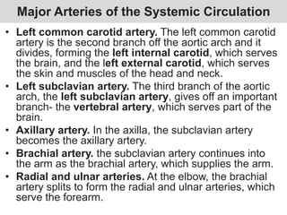
- 34. Major Arteries of the Systemic Circulation • Left common carotid artery. The left common carotid artery is the second branch off the aortic arch and it divides, forming the left internal carotid, which serves the brain, and the left external carotid, which serves the skin and muscles of the head and neck. • Left subclavian artery. The third branch of the aortic arch, the left subclavian artery, gives off an important branch- the vertebral artery, which serves part of the brain. • Axillary artery. In the axilla, the subclavian artery becomes the axillary artery. • Brachial artery. the subclavian artery continues into the arm as the brachial artery, which supplies the arm. • Radial and ulnar arteries. At the elbow, the brachial artery splits to form the radial and ulnar arteries, which serve the forearm.
- 36. Arterial Branches of the Thoracic Aorta • The aorta plunges downward through the thorax, following the spine as the thoracic aorta. • Intercostal arteries. Ten pairs of intercostal arteries supply the muscles of the thorax wall. Arterial Branches of the Abdominal Aorta • Finally, the aorta passes through the diaphragm into the abdominopelvic cavity, where it becomes the abdominal aorta. • Celiac trunk. The celiac trunk is the first branch of the abdominal aorta and has three branches: the left gastric artery supplies the stomach; the splenic artery supplies the spleen, and the common hepatic artery supplies the liver.
- 37. Arterial Branches of the Abdominal Aorta • Superior mesenteric artery. The unpaired superior mesenteric artery supplies most of the small intestine and the first half of the large intestine or colon. • Renal arteries. The renal arteries serve the kidneys. • Gonadal arteries. The gonadal arteries supply the gonads, and they are called ovarian arteries in females while in males they are testicular arteries. • Lumbar arteries. The lumbar arteries are several pairs of arteries serving the heavy muscles of the abdomen and trunk walls. • Inferior mesenteric artery. The inferior mesenteric artery is a small, unpaired artery supplying the second half of the large intestine. • Common iliac arteries. The common iliac arteries are the final branches of the abdominal aorta.
- 38. Veins Draining into the Superior Vena Cava • Veins draining into the superior vena cava are named in a distal-to-proximal direction; that is, in the same direction the blood flows into the superior vena cava. • Radial and ulnar veins. The radial and ulnar veins are deep veins draining the forearm; they unite to form the deep brachial vein, which drains the arm and empties into the axillary vein in the axillary region. • Cephalic vein. The cephalic vein provides for the superficial drainage of the lateral aspect of the arm and empties into the axillary vein. • Basilic vein. The basilic vein is a superficial vein that drains the medial aspect of the arm and empties into the brachial vein proximally.
- 39. Veins Draining into the Superior Vena Cava • Median cubital vein. The basilic and cephalic veins are joined at the anterior aspect of the elbow by the median cubital vein, often chosen as the site for blood removal for the purpose of blood testing. • Subclavian vein. The subclavian vein receives venous blood from the arm through the axillary vein and from the skin and muscles of the head through the external jugular vein. • Vertebral vein. The vertebral vein drains the posterior part of the head. • Internal jugular vein. The internal jugular vein drains the Dural sinuses of the brain. • Brachiocephalic veins. The right and left brachiocephalic veins are large veins that receive venous drainage from the subclavian, vertebral, and internal jugular veins on their respective sides. • Azygos vein. The azygos vein is a single vein that drains the thorax and enters the superior vena cava just before it joins the heart.
- 41. Veins Draining into the Inferior Vena Cava • The inferior vena cava, which is much longer than the superior vena cava, returns blood to the heart from all body regions below the diaphragm. • Tibial veins. The anterior and posterior tibial veins and the fibular vein drain the leg; the posterior tibial veins becomes the popliteal vein at the knee and then the femoral vein in the thigh; the femoral vein becomes the external iliac vein as it enters the pelvis. • Great saphenous veins. The great saphenous veins are the longest veins in the body; they begin at the dorsal venous arch in the foot and travel up the medial aspect of the leg to empty into the femoral vein in the thigh.
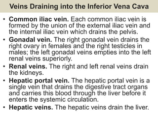
- 42. Veins Draining into the Inferior Vena Cava • Common iliac vein. Each common iliac vein is formed by the union of the external iliac vein and the internal iliac vein which drains the pelvis. • Gonadal vein. The right gonadal vein drains the right ovary in females and the right testicles in males; the left gonadal veins empties into the left renal veins superiorly. • Renal veins. The right and left renal veins drain the kidneys. • Hepatic portal vein. The hepatic portal vein is a single vein that drains the digestive tract organs and carries this blood through the liver before it enters the systemic circulation. • Hepatic veins. The hepatic veins drain the liver.
- 43. CORONARY CIRCULATION • Heart is supplied by TWO CORONARY arteries: 1- Left coronary artery---(LCA) 2- Right coronary artery---(RCA) • These coronary arteries arise at the root of the aorta. 43
- 44. Coronary arteries & their branches LCA---- it passes under the left atrium and divides into two branches: 1. Left Anterior Descending (LAD) • It gives off smaller branches to the interventricular septum and anterior walls of both ventricles. 2. Circumflex Artery • It continues around the left side of the heart and supplies blood to the left atrium and posterior wall of the left ventricle. 44
- 45. Coronary arteries RCA ---- It gives off two branches: 1. Marginal Artery • It supplies blood to the lateral aspect of the right atrium and ventricle. 2. Posterior descending artery • It supplies blood to the posterior walls of both ventricles. 45
- 46. Diagram of coronary circulation 46
- 48. Venous return of Heart Most of the venous blood return to heart occurs through the coronary sinus and anterior cardiac veins, which drain into the right atrium 48
- 49. Blood flow to Heart during Systole & Diastole • During systole when heart muscle contracts it compresses the coronary arteries therefore blood flow is less to the left ventricle during systole and more during diastole. • To the subendocardial portion of Left ventricle it occurs only during diastole 49
- 50. 50
- 51. 51 Special features of veins • Valves – Prevent backflow – Most abundant in legs (where blood has to travel against gravity) • Muscular contraction – Aids the return of blood to heart in conjunction with valves
- 52. 52 Exercise helps circulation (because muscles contract and squeeze blood back to the heart)
- 53. 53 Vascular System (Blood vessels of the body) • Two circulations – Systemic – Pulmonary • Arteries and veins usually run together • Often nerves run with them
- 54. 54 Pulmonary Circulation • Pulmonary trunk branches – Right and left pulmonary arteries – Division into lobar arteries • 3 on right • 2 on left – Smaller and smaller arterioles, into capillaries surrounding alveoli • Gas exchange
- 55. 55 Pulmonary Circulation • After gas exchange blood enters venules • Larger and larger into Superior and Inferior Pulmonary veins • Four Pulmonary Veins empty into left atrium
- 56. 56
- 57. 57 Systemic Circulation • Oxygenated blood to body • Leaves LV through Ascending Aorta – Only branches are the 2 coronary arteries to the heart • Aortic Arch has three arteries branching from it: 1. Brachiocephalic trunk, has 2 branches: • Right common carotid a. • Right subclavian a. 2. Left common carotid a. 3. Left subclavian a. Ligamentum arteriosum connecting to pulmonary a.
- 58. 58 • Hepatic portal system – Picks up digested nutrients from stomach & intestines and delivers them to liver for processing and storage • Storage of nutrients • Detoxification of toxins, drugs, etc. Tributaries of hepatic portal vein: -superior mesenteric vein -splenic vein -inferior mesenteric vein
- 59. 59
- 60. References: • Gerard J, Tortora/Brayan Derrickson edition 2016.
Editor's Notes
- Fiberous pericardium is the upper toughest membrane
- Endothelium: simple squamous epithelium
- Trabeculae carnae (little beam; carnae= fleshy)
- bishop's mitre (a type of hat) Aorta= to suspend, because the aorta once was believed to lift up the heart.