Cell DIVISION/cycle
•Download as PPTX, PDF•
11 likes•910 views

Uphold the importance of cell cycle and reproduction especially in the perpetuation of generation of species.
Report
Share


Cell DIVISION/cycle
- 1. Cell Cycle
- 2. Learning Objectives Infer the significance of cell division. Differentiate a DNA molecule, a chromosome, and a chromatid. Characterize the phases of the cell cycle and their control points. Describe the major events associated with stages of mitosis. Explain the process of cytokinesis. Describe the role of apoptosis in the life cycle of a cell. Relate cancer as a result of the malfunction of the cell during the cell cycle.
- 3. Values Uphold the importance of cell cycle and reproduction especially in the perpetuation of generation of species. Advocate preservation of life
- 4. Why must cells divide?
- 6. Cell division plays important roles in the lives of organism
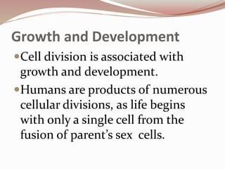
- 7. Growth and Development Cell division is associated with growth and development. Humans are products of numerous cellular divisions, as life begins with only a single cell from the fusion of parent’s sex cells.
- 8. How come bacteria and other minute organisms reproduce faster and live in almost everywhere on Earth? Reproduction is common process among life forms to make new organism from one or two parent organism. Sexual Reproduction- involves two specialized cells, called gametes, coming from the parents that will result to a unique offspring.
- 9. Asexual Reproduction Is the production of offspring from single parent without the involvement of gametes. The offspring is genetically identical with each other and to the single parent. Most Bacteria and other unicellular organisms, reproduction involves a simple cell division. Binary Fission- the cell pinches apart, splits into two , then new cell wall forms between two daughter cells.
- 11. Where do cells get the information they need to function? The cell’s genetic material is organized in tightly-coiled structures called chromosomes. A chromosome is simply a long, continuous thread of DNA wounded together by DNA- associated proteins, referred as histones.
- 12. DNA is loosely organized like scattered spaghetti on a plate.
- 13. Chromatin efficiently packages the DNA into small volume and fits inside the nucleus. Chromatin undergoes further condensation, forming the chromosome
- 14. The complex set of macromolecules that contain loose DNA, proteins, and RNA is called chromatin. Chromatin is responsible for packaging the DNA efficiently into smaller volume so that it fits the nucleus of a cell to protect the DNA structure and sequence, to prevent DNA damage, to control gene expression and DNA replication, and to reinforce the DNA molecule to allow mitosis and meiosis
- 15. Chromatid refers to each strand of the duplicated chromosomes. Together they are called sister chromatids, which are held together by centromere – a region of condensed pinched chromosomes. Located at the centromere is a group of proteins called kinetochore, which is attached to the spindle fibers during cell division.
- 16. At the ends of the DNA molecules are structures referred as telomeres that contain repeated nucleotides, which contain genetic information that do not translate into pairs. Their role is to prevent the ends of chromosomes from accidentally attaching to one another and prevent the loss genes.
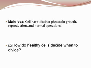
- 17. Main Idea: Cell have distinct phases for growth, reproduction, and normal operations. EQ: How do healthy cells decide when to divide?
- 18. THE CELL CYCLE Cells follow definite stages of growth, duplication, and division known collectively as the cell cycle.
- 19. STAGES OF CELL CYCLE
- 21. INTERPHASE Interphase is the growth period in the cell cycle and is divided into three parts
- 22. Gap 1 (G1) Gap 1 is the first part of the cell cycle wherein the cell carries out its normal metabolic functions. During Gap 1 phase , an intestinal cell performs its primary function to absorb nutrients, while a red blood cells shuttles oxygen to the rest of the body. During this phase, cells also increase their size, as their organelles increase in number. Cells have a required size limit.
- 23. Synthesis (S) Synthesis is the second part of cell cycle, which refers to the time that the cell makes a copy of the genetic material in the form of nuclear DNA. During S phase, the cell spends considerable amount of time and energy to make copies of its chromosomes.
- 24. Gap 2 (G2) During this stage, cells continue to carry out their normal functions and also undergo further growth. This stage contain a critical checkpoint before transitioning to the next stage. The cell make sure that everything is in order, including growing to its correct size and duplicating DNA without damage.
- 25. Mitosis (M) Mitosis involves the division of the nucleus and the genetic material. Parts of Mitosis o Prophase o Metaphase o Anaphase o Telophase During M stage, the hereditary materials of parent cell is given to the daughter cells.
- 26. This leads to the formation of two daughter cells containing the identical genetic materials. The goal of Mitosis is to distribute an identical set of genetic instructions- one copy of each chromosome to the two daughter cells. In M stage the cell’s nuclear membrane disintegrates, while the DNA condensed, forming two nuclei.
- 27. Cytokinesis Cytokinesis is basically divides the cytoplasm of the cell. Cytokinesis early during telophase and continues after the nuclei have formed in the daughter cells.
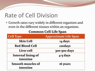
- 28. Rate of Cell Division Growth rates vary widely in different organism and even in the different tissues within an organism. Common Cell Life Span Cell Type Approximate Life Span Skin Cell 14 days Red Blood Cell 12odays Liver cell 300-500 days Internal lining of intestine 4-5 days Smooth muscles of intestine 16 years
- 29. Some parts of the body where cell divides are rarely believed by scientists to have entered a stage known as gap zero or G0. In this stage, cell are unlikely to divide but still continue to perform normal function. Such cells, like neuron cells and heart muscle cell that are highly differentiated or specialized and that body cannot easily replace, are said to be permanently in G0.
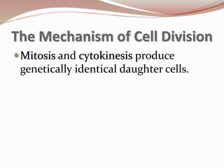
- 30. The Mechanism of Cell Division Mitosis and cytokinesis produce genetically identical daughter cells.
- 31. Mitosis Is the division of nucleus into two genetically identical nuclei containing the same full set of DNA. Mitosis occurs in body cells, except the sex cells. Mitosis prepares the cells for cytokinesis. Phases of Mitosis Prophase Metaphase Anaphase telophase
- 32. Prophase: Start of Mitosis Prophase (pro means “before”). During prophase, several distinct events occur. In prophase, the nucleus disappears. Chromatin condenses into chromosomes and the mitotic spindles is formed. Spindle fibers are organized microtubules.
- 34. Metaphase: Chromosomes at the Center Metaphase (meta means “near”, the spindle fibers attached to the kinetochore of sister chromatids facilitates the movement of chromosomes toward the middle of the cell. During metaphase, chromosomes align at the metaphase plate.
- 36. Anaphase: Chromatids to opposites Poles Anaphase ( ana means “up” or “back”), sister chromatids are tightly paired due to the centromere and protein cohesion, which is responsible for holding DNA molecules together in its entire length. In anaphase, chromatids separated toward opposite poles.
- 38. Telophase: reformation of Nuclei Telophase (telo means “end”), the two complete sets of identical chromosomes are now positioned at each pole of the cell, and the reverse of the major events in prophase happen. In telophase, new nuclear envelope forms. Chromosomes unfold back into chromatin, the nucleoli reappear, and the cell continues to elongates.
- 41. Love of Lab Locating the Stages of Mitosis on page 96A.
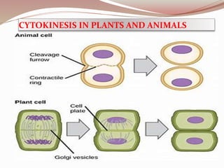
- 42. Cytokinesis This stage involves splitting of the cytoplasm into two cells and completes the entire cytoplasm divides. Cytokinesis typically starts to occur in late anaphase or telophase. It also differs in plants and animal cell because of some differences in cellular structure.
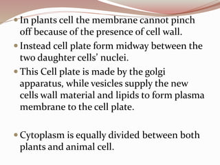
- 43. In animals cells, cytokinesis starts with the formation of cleavage furrow or trench that is pulled inward by tiny strands of protein actin called microfilaments like a drawstring Slowly membrane begins to pinch roughly in half and close off, forming a separate cell around each nucleus. Daughter cells receive equal portions of parents cell’s plasma content.
- 44. In plants cell the membrane cannot pinch off because of the presence of cell wall. Instead cell plate form midway between the two daughter cells’ nuclei. This Cell plate is made by the golgi apparatus, while vesicles supply the new cells wall material and lipids to form plasma membrane to the cell plate. Cytoplasm is equally divided between both plants and animal cell.
- 45. CYTOKINESIS IN PLANTS AND ANIMALS
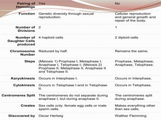
- 46. Meiosis Mitosis Type of Reproduction Sexual Asexual Occurs in Humans, animals, plants, fungi. All organisms. Genetically Different Identical Crossing Over Yes, mixing of chromosomes can occur. No, crossing over cannot occur. Definition A type of cellular reproduction in which the number of chromosomes are reduced by half through the separation of homologous chromosomes, producing two haploid cells. A process of asexual reproduction in which the cell divides in two producing a replica, with an equal number of chromosomes in each resulting diploid cell.
- 47. Pairing of Homologs Yes No Function Genetic diversity through sexual reproduction. Cellular reproduction and general growth and repair of the body. Number of Divisions 2 1 Number of Daughter Cells produced 4 haploid cells 2 diploid cells Chromosome Number Reduced by half. Remains the same. Steps (Meiosis 1) Prophase I, Metaphase I, Anaphase I, Telophase I; (Meiosis 2) Prophase II, Metaphase II, Anaphase II and Telophase II. Prophase, Metaphase, Anaphase, Telophase. Karyokinesis Occurs in Interphase I. Occurs in Interphase. Cytokinesis Occurs in Telophase I and in Telophase II. Occurs in Telophase. Centromeres Split The centromeres do not separate during anaphase I, but during anaphase II. The centromeres split during anaphase. Creates Sex cells only: female egg cells or male sperm cells. Makes everything other than sex cells. Discovered by Oscar Hertwig Walther Flemming
- 48. Cell cycle must be regulated for healthy cell growth. Both internal and external factors work together to regulate cell division Group of proteins active in controlling cell cycle & in initiating DNA synthesis Any various enzymes that catalyze the transfer of phosphate group from high energy phosphate.
- 49. External Factors External factors from outside the cell that are in the form of message from nearby cells or from remote parts of the organism’s body. Physical and Chemical External factor help regulate the cell cycle. Physical signs, such as cell-to-cell contact, exist between cells. Mammalian cells experiments grown in in vitro (Latin,within the glass) laboratory set up grow until a single layer covers the entire surface of the culture dish.
- 50. When cell touches another cell, it stops dividing- a phenomenon called contact inhibition. Another study shown cell that shown cell only grow if surface is available and stop dividing when detached from the culture dish – a phenomenon called anchorage dependent. One explanation for this is there are receptors found in neighboring cell binding each other and causing the cell’s cytoplasm to form structures that can block the signal, stopping the continuous division.
- 51. Chemical signals Released by the cell such as growth factors provide instructions for other cells to grow. Growth factor are proteins that stimulates cell division, which have the ability to activate specific genes to trigger cell growth.
- 52. Internal factor Come from inside the cell that include several types of molecules in its cytoplasm. The most well-studied internal factors in eukaryotic cell cycle are kinases and cyclins. Kinase is an enzyme that transfers a phosphate group from one molecule to the target molecules. Cyclins are group of proteins active in controlling cell cycle & in initiating DNA synthesis.
- 53. Apoptosis Orderly programmed cell death or suicide. Apoptosis is needed by the body to ensure proper cell functioning. Apoptosis occurs in body’s webbed fingers during embryonic period to allow detachment of fingers and thumbs before baby is born.
- 54. What happens to the body when there is uncontrolled cell growth?
- 55. CANCER: Growing Out Of Control Cancer refers to a group of diseases characterized by uncontrolled and abnormal cell division. It occurs when there is a disruption in the cell cycle. Instead of stopping and starting at appropriate points, cancerous cell divide continuously until a disorganized solid mass of cells called tumor is formed.
- 56. Tumors can be categorized as benign or malignant. Benign Tumors are cancer cells that remain clustered together which may be harmless or not and can probably be cured when removed out of the body. Tumors that break away or metastasize are called malignant tumors. Their ability to break away from their present location allows them to be transported to the bloodstream of the lymphatic system to other parts of the body parts to form more tumors.
- 58. Standard cancer treatment option Local surgery radiation Systematic chemotherapy immunotherapy Involves removal of the cancerous body part Involves exposure to X-rays to kill cancer cell and shrink the tumor cells Uses certain drug to kill actively dividing cells Uses substances either from the body or laboratory to improve the body’s defense to fight cancer
- 59. The human Life Cycle and Sexual Reproduction Long- Term survival on Earth requires some form of genetic variation to increase survival rate of organisms in unstable environment. Human life cycle starts with the union of two sex cells- a sperm cell or spermatozoon (plural; spermatozoa) from father and an egg or ovum (plural: ova) from mother through sexual reproduction.
- 60. Both sperm cell and the egg cell are produced in a type of cell division called meiosis. The sex cells produced in meiosis are haploid cells because they contain only half the number of chromosomes. The haploid cell is represented with the symbol n. As a result of the union spermatozoon (n) and a haploid ovum (n), a diploid (2n) zygote is formed.
- 62. The process of cell division by mitosis is a continuing process for the rest of an organism as a way to form new tissues and organs, and to repair damaged and old tissues. As a result of mitosis, each somatic cell , also called body cell, of the human body contains diploid number of chromosomes, except sex cell- 23 chromosomes
- 63. In human life cycle, the process of meiosis is important to reduce the number of chromosomes from diploid (2n= 46) to haploid (n=23) All cell in our body have specialized functions. But all these cells can be divided into two major group: somatic cells (Greek word soma, meaning body) and germ cells. Somatic cells or body cells compose you body tissues and organs.
- 65. Each diploid cell contain 23 pairs of homologous chromosomes. Homologous chromosomes refer to a set of chromosomes having the same length and appearance that was inherited from the parent. Scientist assigned a number to each pair of homologous chromosome according to their size from largest to smallest. Viewing the arranged chromosomes can be done in the process called karyotyping with the use of a diagram known as a karyogram.
- 67. Karyotype of human showing the pairs of homologous chromosomes arranged into autosomes and sex chromosomes. Chromosomes pairs 1 to 22 are designated as autosomes, which are chromosomes associated with genes not directly related to the sex of an organism. The twenty-third pair is a special one called sex chromosomes, which are directly linked to the development of sexual characteristics of an organism. Humans have two different sex chromosomes, known as the X and Y chromosomes.
- 68. Humans and mammals use the XY system to determine the gender. An organism having two X chromosome is female, while an organism having X and Y is male. The X and Y chromosome, although paired with 23 set, are not homologous. The X chromosome is larger containing more genes and other unrelated ones to sexual characteristics, The Y chromosome is the smallest chromosome
- 69. Meiosis -Is important for sexual reproduction, is the form of cell division that involves the formation of sex cells. Meiosis divides the number of the gametes, the process is also called reduction division. Meiosis involves two divisions of the nucleus known as meiosis I and meiosis II
- 70. MEIOSIS 1
- 71. 1. Prophase I Is characterized by he breaking down of nuclear envelope. Spindle fiber begin to assemble. Duplicated chromosomes condense, while the homologous chromosomes pair and line-up by gene precisely in its length. Aside from pairing along their length, they cross over at a point called chiasma.
- 72. 2. Metaphase I The pairs of homologous chromosomes are randomly moved by the spindle to the equator of the cell. This result in the lining up of 23 chromosomes alongside each other in the middle of the cell.
- 73. 3. Anaphase I Homologous chromosomes separate from each other. Like in mitosis, chromosomes of each pair are pulled to the opposite sides of the cell by the action of spindle fibers. Sister chromatids remain together and don not separate at their centromeres throughout meiosis.
- 74. 4. Telophase 1 The individual chromosomes that have pulled in opposite directions now gather at each pole. Both poles contain one chromosomes from each pair of the homologous chromosomes.
- 75. Meiosis II The goal of meiosis II is to divide sister chromatids, resulting in sex cell with only half the chromosome.
- 76. 5. Prophase II New spindle forms around the chromosome. The nuclear envelope breaks down with chromosomes pulled at opposite sides of the cell by the spindle fibers.
- 77. 6. Metaphase II Chromosomes line up along the equator through the spindle fibers. At this stage, each chromosome has sister chromatids still attached to the centromere.
- 78. 7. Anaphase II Centromeres divide and sister chromatids are individually pulled apart, then move to opposite poles of the cell.
- 79. 8. Telophase II Nuclear envelopes forms around each set of chromosomes at opposite ends of the cell. The spindle fibers breaks down and the cell undergoes cytokinesis. The result of meiosis is four haploid cells with a recombination of the chromosomes both from the parents.
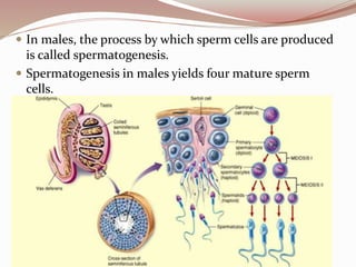
- 80. Gametogenesis Meiosis is the process of gamete formation. The formation of sperm cells is called spermatogenesis while the formation of egg cells is called oogenesis.
- 81. In males, the process by which sperm cells are produced is called spermatogenesis. Spermatogenesis in males yields four mature sperm cells.
- 82. In females, the process of gamete formation is called oogenesis Only one of the resulting cells gets nearly all of the cytoplasm. This cell receives almost all the organelles, cytoplasm, and nutrients because it is this cell that will ultimately give rise to an egg cell. The other cell is very small called a polar body. The larger cell undergoes meiosis II, and the division of the egg cell during meiosis is again unequal.
- 83. The large cell develops into a gamete called an ovum or egg. The polar body may divide again, but its cell will not survive and the polar body will die in a process called apoptosis. Oogenesis yields a single mature egg and nonfunctional polar bodies
- 84. Importance of Meiosis Three mechanisms that contribute to the genetic variation 1. Independent assortment 2. Crossing –over 3. Random fertilization
- 85. Independent Assortment The random distribution of homologous chromosomes during meiosis is called independent assortment.
- 86. Crossing-Over and Random Fertilization Another factor that contributes to genetic variation is crossing –over. This occurs during prophase I of meiosis, where chromosomes line up in the process called synapsis, while sections of their DNA are exchanged. DNA exchange during crossover adds more recombination probabilities to the independent assortment of chromosomes that occur later in meiosis.
- 88. Chromosomal errors are associated with several factors such as error in mitosis, meiosis, environment, and age of the mother. Mistakes during the process of meiosis can create an abnormal number of chromosomes. If meiosis does not occur properly, the sperm or egg cell may end up with more or less than required number of chromosomes.
- 89. The process where chromosomes or chromatids fail to separate during meiosis is called nondisjunction. Nondisjunction can occur either in meiosis I (happens when homologous pairs fail to separate) or meiosis II (involves normal separation of chromosomes in meiosis I, but sister chromatids fail to separate during anaphase I.
- 91. Turner Syndrome (45,XO) Turner syndrome is a chromosomal condition seen in female individuals. Affected females have small and underdeveloped ovaries, uterus, and oviducts. In early childhood, individuals with Turner Syndrome loses the ability of normal ovarian function. They do not experience puberty at a normal age. Affected individual have the following characteristics: short, broad chest, webbed neck, and puffiness or swelling of hands and feet. They also have normal intelligence and can live fairly normal lives.
- 93. Down Syndrome (Trisomy 21) The most common disorder of trisomy is Down Syndrome, wherein the 21th chromosome has three instead of two chromosomes. It occurs in one out of 800 newborns. Most cases of Down Syndrome is not due to inheritance but on random mistakes during formation of reproductive cells of one of the parents.
- 95. Affected offspring has the following physical attributes: short stature, round head, flat face, slanting eyes, stubby fingers, wide gap between the first and second toes, short neck, short arms,and short legs. They have muscle tone and loose joints that improve with age. They are born with a number of birth defects in the heart, intestine or breathing. Delayed development and behavioral problem are also observed.
- 97. Klinefelter Syndrome (47, XXY) Klinefelter Syndrome is a chromosomal abnormality affecting the physical and cognitive development of males. Those affected have underdeveloped prostate gland and testes, there is shortage of testosterone production. This lead to delayed or incomplete puberty, lack of facial and body hair, breast enlargement, and unusual small penis. Affected males have big hands and feet, and unusually long arms and legs. They may have delayed speech and language development, and learning disabilities. Some variants of Klinefelter have more than extra chromosome in each cell such as in 48, XXXY or 49, XXXXY that are associated with severe forms of abnormality.
- 99. Trisomy X (47, XXX) The abnormal presence of an X chromosome is also known as Trisomy X syndrome, which affects one in 1000 females. Affected individuals usually have mild symptoms or none at all. If symptoms are more pronounced, these may include development delays and language- based learning disabilities. In addition to errors in meiosis, the age of the mother can also add up to the equation. The older the woman is during pregnancy, the older is the age of her eggs because she was born with all the eggs she will ever have. A woman of 45 years during pregnancy has a 45-year-old egg as well. Errors in meiosis have a higher risk due to the aging process.
- 104. QUIZ