Location via proxy:   [ UP ]  
[Report a bug]   [Manage cookies]                
SlideShare a Scribd company logo
1.1

IMPORTANCE OF A TRANSPORT SYSTEM
IN SOME MULTICELLULAR ORGANISMS

           By: Rumaizah Muhamad
Dimension of cube (cm)             1x1x1   2x2x2   4x4x4


Total surface area of cube (cm2)


Volume of cube (cm3)


TSA/V ratio

Calculate the TSA/V ratio.
Chapter 1: Transport (1.1 - 1.3)
• In larger multicellular organisms need a
  transport system so that dissolved materials
  could be moved rapidly to and from all parts
  of the body.
1.2

CONCEPT OF THE CIRCULATORY
SYSTEM
Circulatory system in humans and
animals
• Circulatory system (CS) includes:
  – Medium  fluid that flows in CS (eg: animal :
    blood; insects: haemolymph)
  – Vesels  arteries, veins and capillaries
  – Pump  muscular heart
Chapter 1: Transport (1.1 - 1.3)
Erythrocytes
Erythrocytes
•   Small, biconcave disc
•   Have no nucleus
•   Great quantities of haemoglobin (which contains iron)


                                                             (Cells become bright red)


•   Site of production: bone marrow
•   Life span: 120 days
•   Site of destruction: liver and spleen (by phagocytes)
•   Ratio of erythrocytes to leucocytes  1000 : 1 (in normal person)
Leucocytes (White blood cells)
                 Erythrocytes


Leucocytes
Leucocytes (White blood cells)
• Less numerous than eryhtrocytes.
• Have nuclei
• Do not have haemoglobin
• Larger than erythrocytes and do not have
  fixed shapes.
• Site of production : bone marrow
• Site of growth and development: thymus
  gland or lymph nodes
Cont.
Basic types of leucocytes:
           Granulocytes (have granular cytoplasm and
           lobed nuclei)
           • Neutrophils
           • Eusinophils
           • Basophils


           Agranulocytes (clear cytoplasm and no
           lobed)
           • Monocytes (largest)
           • Lymphocytes (smallest)
Chapter 1: Transport (1.1 - 1.3)
Quizzes
1. The neutrophils are _______
  A.   Phagocytes
  B.   Leucocytes
  C.   Lymphocytes
  D.   Phosphate

2. Which leucocytes that help to control allergic
   reaction?
  A.   Basophils
  B.   Eosinophils
  C.   Monocytes
  D.   Neutrophils
3. Basophils secrete _________ to prevent blood clotting
  A.   Warfarin
  B.   Fibrinogen
  C.   Heparin
  D.   Walfarine

4. Which leucocytes that can move to the body tissues to
   become phagocytic macrophage?
  A.   Basophils
  B.   Neutrophils
  C.   Eosinophils
  D.   Monocytes
5. Some lymphocytes produce _________ to aid
   in destruction of __________
  A.   Antibodies, homogens
  B.   Antibodies, pathogens
  C.   Hormones, pathogens
  D.   Enzymes, pathogens
Platelets


• Small, irregularly
  shaped
• F(x) : blood clotting
• Life span: 5 -9 days
Plasma

• Pale, yellow liquid
• Made up of 90% water & 10% dissolved solutes
  (gases, minerals, hormones, plasma proteins and
  excretory wastes)
• BOOD SERUM : plasma without fibrinogen (clotting
  factors).
Plasma
Function of blood in transport
• Transport oxygen from the lungs to other
  parts of the body (form 4)
• Transport absorbed food materials from the
  digestive tract to body tissues (form 4)
• Transport waste products (form 4)
  – Eg: carbon dioxide from body tissues to the lungs
  – Urea to the kidneys
• Transport heat, hormones and water
Transport of heat,
                        hormones & water
• Body T can be regulated by blood by
  distributing heat from heat-producing sites
  (eg:muscles) to the skin.
• Hormones (eg:insulin & glucagon) produced
  by endocrine glands (pancreas) transported by
  blood to target organs (liver).
• Water is important to provide medium for
  biochemical reaction.
Function of haemolymph
• Circulating blood-like fluid found in invertebrates
  with open-circulatory systems
• Tubular heart pumps the haemolymph into
  haemocoel.
• Haemolymph – bathes the tissues and internal
  organ directly.
• Nutrients and hormones diffuse from
  haemolypmh into the cells
• Waste products diffuse out from the cells into
  haemolymph.
Chapter 1: Transport (1.1 - 1.3)
Structure of human blood vessels
Blood vessels : tubes that transport blood from one part to another.

         Arteries                     Capillaries                      Veins
Transport blood away from    Connect arterioles to         Transport blood to the
the heart                    venules                       heart
Transport oxygenated         Act as the sites for          Transport deoxygenated
blood (except pulmonary      exchange of substances        blood (except pulmonary
artery)                      with the cells                vein)
Thick muscular wall          Thinnest wall, one cell       Thinner wall
                             thickness
No valves except semilunar No valves                       Valves present to prevent
valves at the base of the                                  back flow of blood
aorta and pulmonary
artery
Blood flows in pulses under No pulses. Pressure lower      No pulses. Blood flows
high pressure               than arteries but higher       under lower pressure than
                            than veins                     arteries.
Chapter 1: Transport (1.1 - 1.3)
Artery, vein and capillary
How blood is propelled through the
      human circulatory system
• Organ responsible to pump the blood : heart
Structure of the human heart
How blood is propelled through the
     human circulatory system
• VIDEO
Lungs

                                                  Pulmonary veins
      Vena cava


   Right ventricle                                Left atrium


Tricuspid                                                    Bicuspid
    valve
                     deO2 blood       O2 blood               valve


    Right atrium                                 Left ventricle


                                                     Aorta
        Pulmonary
            artery
                             Whole body
FAQ SPM
1. The diagram below shows the external view of the
   human heart.


What is the blood vessel labeled W?
A. Carotid artery
B. Coronary artery
C. Subclavian artery
D. Systemic artery
3.
The pumping of the heart
 Sinoatrial node


                                           Bundle of His
Atrio-ventricular                          containing
            node                           Purkinje tissue


                                           Interventricular
                                           septum
Conducting system of the heart
• Video
Contraction of skeletal muscles
                  around veins
Circulatory system in insects
2. Valves ensure the haemolymph flow in one direction


1. When the heart relax, haemolymph re-enters ostia


                               Material exchange occurs
                               here. Haemolymph in
                               haemocoel carry nutrients
                               and waste products
Circulatory system in fish
               • Single circulatory
                 system.
               • Deoxygenated blood
     Sinuses     leaves the heart at high
                 pressure and passes
                 through the gills where
                 the gaseous exchange
                 occurs.
               • Oxygenated blood flows
                 through the organs and
                 blood pressure drops.
Circulatory system in amphibians
               (eg:frogs)
• Double circulatory system:
  – Pulmonary circulation
  – Systemic circulation
• Have three-chambered heart (2 atria & 1
  ventricle)
• Mixing of oxygenated and deoxygenated
  blood in ventricle. The mixed blood enters the
  systemic circulation.
Chapter 1: Transport (1.1 - 1.3)
Circulatory system
in humans
FAQ SPM
1. The diagram below shows a longitudinal section
   of the human heart.


  Which of the labeled parts, A, B, C or D, receives
  blood from the lungs?
2. The diagram shows the circulatory system of
   a fish.


   What are the organs P, Q and R?
                P        Q        R
          A    Gills    Lungs   Stomach
          B   Stomach   Lungs    Gills
          C    Gills    Heart   Stomach
          D   Stomach   Heart    Gills
Circulatory system in humans
• Double circulatory system (blood passes through the
  heart twice for each circuit)
   – Pulmonary circulation
   – Systemic circulation
• Two divisions of heart:
   – Right side  pump deoxygenated blood to the lungs
   – Left side  pump oxygenated blood to the body (except
     lungs)
• Advantage: blood returns to the heart to be pumped
  again will increase the blood pressure and flow rate,
  thereby speeding up delivery O2 to the tissues and
  organs.
Assignment 1.3
1. Draw and label a longitudinal section of
   human heart.
2. List the functions of the different types of
   leucocytes.
1.3

THE MECHANISM OF BLOOD
CLOTTING
The necessities of blood clotting
• To prevent:
  – serious blood loss
  – the entry of microorganisms and foreign particles
    into the blood
• To maintain:
  – normal blood pressure
  – circulation of blood in a a closed circulatory
    system
The mechanism of blood clotting
Chapter 1: Transport (1.1 - 1.3)
VIDEO
FAQ SPM
1. The conversion of the soluble fibrinogen
   present in blood plasma into the insoluble
   fibrin is by
A. calcium ion
B. thrombin
C. thromboplastins
D. vitamin K
FAQ SPM
2. Fibrinogen, fibrin, thrombin and prothrombin
  are necessary for blood clotting. Which of the
  following is the correct sequence for the
  involvement of these four substances during
  blood clotting?
A. Prothrombin, thrombin, fibrinogen, fibrin
B. Thrombin, prothrombin, fibrin, fibrinogen
C. Fibrinogen, fibrin, prothrombin, thrombin
D. Fibrin, fibrinogen, thrombin, prothrombin
Impaired blood clotting mechanisms in
               human
                   The consequences
Chapter 1: Transport (1.1 - 1.3)
Assignment 1.4
1. List the advantages of blood clotting at the
   site of damaged blood vessels.
2. Draw a schematic diagram to illustrate the
   mechanism of blood clotting.
3. What would happen if a blood clot forms
  a) In the brain
  b) In the coronary artery

More Related Content

Chapter 1: Transport (1.1 - 1.3)

  • 1. 1.1 IMPORTANCE OF A TRANSPORT SYSTEM IN SOME MULTICELLULAR ORGANISMS By: Rumaizah Muhamad
  • 2. Dimension of cube (cm) 1x1x1 2x2x2 4x4x4 Total surface area of cube (cm2) Volume of cube (cm3) TSA/V ratio Calculate the TSA/V ratio.
  • 6. • In larger multicellular organisms need a transport system so that dissolved materials could be moved rapidly to and from all parts of the body.
  • 7. 1.2 CONCEPT OF THE CIRCULATORY SYSTEM
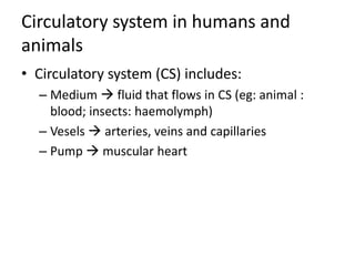
  • 8. Circulatory system in humans and animals • Circulatory system (CS) includes: – Medium  fluid that flows in CS (eg: animal : blood; insects: haemolymph) – Vesels  arteries, veins and capillaries – Pump  muscular heart
  • 12. Erythrocytes • Small, biconcave disc • Have no nucleus • Great quantities of haemoglobin (which contains iron) (Cells become bright red) • Site of production: bone marrow • Life span: 120 days • Site of destruction: liver and spleen (by phagocytes) • Ratio of erythrocytes to leucocytes  1000 : 1 (in normal person)
  • 13. Leucocytes (White blood cells) Erythrocytes Leucocytes
  • 14. Leucocytes (White blood cells) • Less numerous than eryhtrocytes. • Have nuclei • Do not have haemoglobin • Larger than erythrocytes and do not have fixed shapes. • Site of production : bone marrow • Site of growth and development: thymus gland or lymph nodes
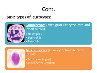
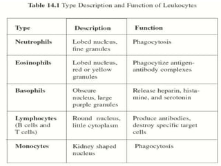
  • 15. Cont. Basic types of leucocytes: Granulocytes (have granular cytoplasm and lobed nuclei) • Neutrophils • Eusinophils • Basophils Agranulocytes (clear cytoplasm and no lobed) • Monocytes (largest) • Lymphocytes (smallest)
  • 17. Quizzes 1. The neutrophils are _______ A. Phagocytes B. Leucocytes C. Lymphocytes D. Phosphate 2. Which leucocytes that help to control allergic reaction? A. Basophils B. Eosinophils C. Monocytes D. Neutrophils
  • 18. 3. Basophils secrete _________ to prevent blood clotting A. Warfarin B. Fibrinogen C. Heparin D. Walfarine 4. Which leucocytes that can move to the body tissues to become phagocytic macrophage? A. Basophils B. Neutrophils C. Eosinophils D. Monocytes
  • 19. 5. Some lymphocytes produce _________ to aid in destruction of __________ A. Antibodies, homogens B. Antibodies, pathogens C. Hormones, pathogens D. Enzymes, pathogens
  • 20. Platelets • Small, irregularly shaped • F(x) : blood clotting • Life span: 5 -9 days
  • 21. Plasma • Pale, yellow liquid • Made up of 90% water & 10% dissolved solutes (gases, minerals, hormones, plasma proteins and excretory wastes) • BOOD SERUM : plasma without fibrinogen (clotting factors).
  • 23. Function of blood in transport • Transport oxygen from the lungs to other parts of the body (form 4) • Transport absorbed food materials from the digestive tract to body tissues (form 4) • Transport waste products (form 4) – Eg: carbon dioxide from body tissues to the lungs – Urea to the kidneys • Transport heat, hormones and water
  • 24. Transport of heat, hormones & water • Body T can be regulated by blood by distributing heat from heat-producing sites (eg:muscles) to the skin. • Hormones (eg:insulin & glucagon) produced by endocrine glands (pancreas) transported by blood to target organs (liver). • Water is important to provide medium for biochemical reaction.
  • 25. Function of haemolymph • Circulating blood-like fluid found in invertebrates with open-circulatory systems • Tubular heart pumps the haemolymph into haemocoel. • Haemolymph – bathes the tissues and internal organ directly. • Nutrients and hormones diffuse from haemolypmh into the cells • Waste products diffuse out from the cells into haemolymph.
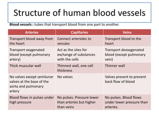
  • 27. Structure of human blood vessels Blood vessels : tubes that transport blood from one part to another. Arteries Capillaries Veins Transport blood away from Connect arterioles to Transport blood to the the heart venules heart Transport oxygenated Act as the sites for Transport deoxygenated blood (except pulmonary exchange of substances blood (except pulmonary artery) with the cells vein) Thick muscular wall Thinnest wall, one cell Thinner wall thickness No valves except semilunar No valves Valves present to prevent valves at the base of the back flow of blood aorta and pulmonary artery Blood flows in pulses under No pulses. Pressure lower No pulses. Blood flows high pressure than arteries but higher under lower pressure than than veins arteries.
  • 30. Artery, vein and capillary
  • 31. How blood is propelled through the human circulatory system • Organ responsible to pump the blood : heart
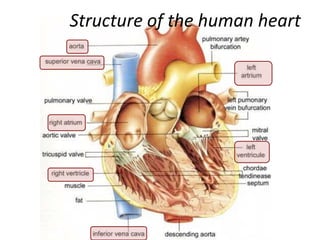
  • 32. Structure of the human heart
  • 33. How blood is propelled through the human circulatory system • VIDEO
  • 34. Lungs Pulmonary veins Vena cava Right ventricle Left atrium Tricuspid Bicuspid valve deO2 blood O2 blood valve Right atrium Left ventricle Aorta Pulmonary artery Whole body
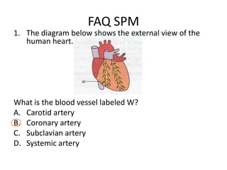
  • 35. FAQ SPM 1. The diagram below shows the external view of the human heart. What is the blood vessel labeled W? A. Carotid artery B. Coronary artery C. Subclavian artery D. Systemic artery
  • 36. 3.
  • 37. The pumping of the heart Sinoatrial node Bundle of His Atrio-ventricular containing node Purkinje tissue Interventricular septum
  • 38. Conducting system of the heart • Video
  • 39. Contraction of skeletal muscles around veins
  • 41. 2. Valves ensure the haemolymph flow in one direction 1. When the heart relax, haemolymph re-enters ostia Material exchange occurs here. Haemolymph in haemocoel carry nutrients and waste products
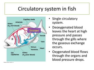
  • 42. Circulatory system in fish • Single circulatory system. • Deoxygenated blood Sinuses leaves the heart at high pressure and passes through the gills where the gaseous exchange occurs. • Oxygenated blood flows through the organs and blood pressure drops.
  • 43. Circulatory system in amphibians (eg:frogs) • Double circulatory system: – Pulmonary circulation – Systemic circulation • Have three-chambered heart (2 atria & 1 ventricle) • Mixing of oxygenated and deoxygenated blood in ventricle. The mixed blood enters the systemic circulation.
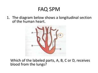
  • 46. FAQ SPM 1. The diagram below shows a longitudinal section of the human heart. Which of the labeled parts, A, B, C or D, receives blood from the lungs?
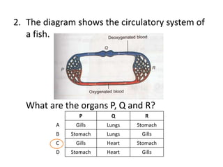
  • 47. 2. The diagram shows the circulatory system of a fish. What are the organs P, Q and R? P Q R A Gills Lungs Stomach B Stomach Lungs Gills C Gills Heart Stomach D Stomach Heart Gills
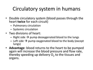
  • 48. Circulatory system in humans • Double circulatory system (blood passes through the heart twice for each circuit) – Pulmonary circulation – Systemic circulation • Two divisions of heart: – Right side  pump deoxygenated blood to the lungs – Left side  pump oxygenated blood to the body (except lungs) • Advantage: blood returns to the heart to be pumped again will increase the blood pressure and flow rate, thereby speeding up delivery O2 to the tissues and organs.
  • 49. Assignment 1.3 1. Draw and label a longitudinal section of human heart. 2. List the functions of the different types of leucocytes.
  • 50. 1.3 THE MECHANISM OF BLOOD CLOTTING
  • 51. The necessities of blood clotting • To prevent: – serious blood loss – the entry of microorganisms and foreign particles into the blood • To maintain: – normal blood pressure – circulation of blood in a a closed circulatory system
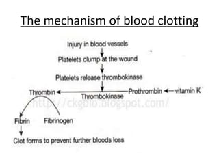
  • 52. The mechanism of blood clotting
  • 54. VIDEO
  • 55. FAQ SPM 1. The conversion of the soluble fibrinogen present in blood plasma into the insoluble fibrin is by A. calcium ion B. thrombin C. thromboplastins D. vitamin K
  • 56. FAQ SPM 2. Fibrinogen, fibrin, thrombin and prothrombin are necessary for blood clotting. Which of the following is the correct sequence for the involvement of these four substances during blood clotting? A. Prothrombin, thrombin, fibrinogen, fibrin B. Thrombin, prothrombin, fibrin, fibrinogen C. Fibrinogen, fibrin, prothrombin, thrombin D. Fibrin, fibrinogen, thrombin, prothrombin
  • 57. Impaired blood clotting mechanisms in human The consequences
  • 60. Assignment 1.4 1. List the advantages of blood clotting at the site of damaged blood vessels. 2. Draw a schematic diagram to illustrate the mechanism of blood clotting. 3. What would happen if a blood clot forms a) In the brain b) In the coronary artery

Editor's Notes

  1. Haemocoel – fluid-filled spaces
  2. Heomocel : body cavityHaemolymph : insect’s blood
  3. 1. Single circulatory system : blood flows through the heart only once for each complete circuit.2. Sinuses: collect deoxygenated blood before entering the atrium.