Location via proxy:   [ UP ]  
[Report a bug]   [Manage cookies]                
SlideShare a Scribd company logo
HETA PATEL (R2)
Outline
• What is CTEV?
• Epidemiology
• Causes
• Anatomy and pathoanatomy
• Clinical features
• X-rays
• Treatment
What is CTEV??
• Idiopathic clubfoot
• Causing CAVE
- midfoot Cavus/ increase in height
-forefoot Adductus
-hindfoot Varus
-hindfoot Equinus/ plantarflex
Hind foot equinus
Heel in varus
Midfoot cavus
Epidemiology
• Relatively common- 1 to 2 per thousand births
• Boys affected twice
• Bilateral in 1/3 of cases
Causes-unknown
• germ defect
• arrested development
• neuromuscular disorder in neurological
disorders and neural tube defect
• postural deformity
Common Types
1. Congenital
- uncommon bony problems present upon childbirth not
related to any neuromuscular factor or symptoms.
2. Teratologic
-a/w neurological conditions (eg: spina bifida)
3. Positional
- in contorted position in utero
4 Syndromic
-a/w standard hereditary issue, which includes
arthrogryposis.
Anatomy
• Hindfoot
-calcaneum, talar
• Midfoot
-cuboid, navicular,
cuneiform
• Forefoot
- metatarsals, phalanges
Pathological Anatomy
Neck of Talus
-pointing downward
and deviates
medially
Body of Talus
- Rotated outward
Posterior part of calcaneum
-held close to fibula by CF ligt
-tilted into equinus and varus
-rotated medially beneath
ankle
Navicular and forefoot
-shifted medially
-rotated into
supination
(composite varus
deformity)
Pathological Anatomy
• Skin and soft tissue of calf and medial side of
foot are short and underdeveloped
• If not corrected early, secondary growth
changes occur in the bones-PERMANENT
Clinical Features
• Heel is small and high
• Deep creases appear posteriorly and medially
• Abnormal thin calf
• Varying degree of resistance / fixed deformity
when try to dorsiflex and evert the foot
Normal baby foot
• Associated disorders
- congenital hip dislocation
- spina bifida
-arthrogryposis : absent of creases
• Look if other joints are affected
How to differentiate true and postural
clubfoot?
• True clubfoot – fixed deformity
• Postural talipes – easily correctable by gentle
passive movement
IMAGING
X-ray to assess progress of treatment
Anterioposterior view
Kite’s angle (talocalcaneal angle): normal 20-40 degree
clubfoot angle almost parallel
30 degree
plantarflex
Lateral Film (Turco view)
Normal angle : 40 degree
If less 20 degree: rocker bottom deformity
- calcaneum seem to be dorsiflexed but it had
broken at midtarsal level
Foot dorsiflex
TREATMENT
Vision & Mission
• Since CTEV can purely be therapeutic work we
would not only treat also HABILITATE club foot
to be normal.
• Increase awareness and confidence of society
with therapeutic staff.
Aim
To produce and maintain a PLANTIGRADE,
supple foot that will function well
Non Operative Operative
• Serial Manipulative and Casting
(Ponsetti’s method)
• -Posteromedial tissue release and
tendon lengthening
• -medial opening or lateral column-
shortening osteotomy, or cuboidal
decancellation
• -triple arthrodesis
• -tallectomy
Serial Manipulative and Casting
(Ponsetti’s method)
• Goal-rotate leg laterally around the fixed tallus
• Order of correction (CAVE)
-midfoot cavus
-forefoot adductus
-hindfoot varus
-hindfoot equinus
Increase the
supination
deformity of
forefoot
CTEV UG
CTEV UG
CTEV UG
DON’T SLEEP. TQ
CTEV UG

More Related Content

CTEV UG

  • 2. Outline • What is CTEV? • Epidemiology • Causes • Anatomy and pathoanatomy • Clinical features • X-rays • Treatment
  • 3. What is CTEV?? • Idiopathic clubfoot • Causing CAVE - midfoot Cavus/ increase in height -forefoot Adductus -hindfoot Varus -hindfoot Equinus/ plantarflex
  • 4. Hind foot equinus Heel in varus Midfoot cavus
  • 5. Epidemiology • Relatively common- 1 to 2 per thousand births • Boys affected twice • Bilateral in 1/3 of cases
  • 6. Causes-unknown • germ defect • arrested development • neuromuscular disorder in neurological disorders and neural tube defect • postural deformity
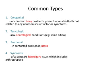
  • 7. Common Types 1. Congenital - uncommon bony problems present upon childbirth not related to any neuromuscular factor or symptoms. 2. Teratologic -a/w neurological conditions (eg: spina bifida) 3. Positional - in contorted position in utero 4 Syndromic -a/w standard hereditary issue, which includes arthrogryposis.
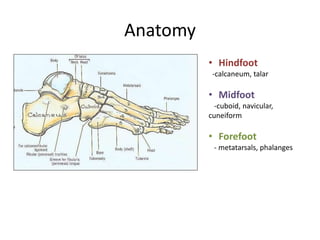
  • 8. Anatomy • Hindfoot -calcaneum, talar • Midfoot -cuboid, navicular, cuneiform • Forefoot - metatarsals, phalanges
  • 9. Pathological Anatomy Neck of Talus -pointing downward and deviates medially Body of Talus - Rotated outward Posterior part of calcaneum -held close to fibula by CF ligt -tilted into equinus and varus -rotated medially beneath ankle Navicular and forefoot -shifted medially -rotated into supination (composite varus deformity)
  • 10. Pathological Anatomy • Skin and soft tissue of calf and medial side of foot are short and underdeveloped • If not corrected early, secondary growth changes occur in the bones-PERMANENT
  • 11. Clinical Features • Heel is small and high • Deep creases appear posteriorly and medially • Abnormal thin calf
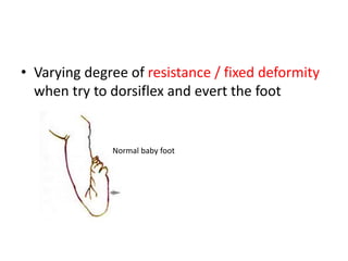
  • 12. • Varying degree of resistance / fixed deformity when try to dorsiflex and evert the foot Normal baby foot
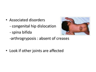
  • 13. • Associated disorders - congenital hip dislocation - spina bifida -arthrogryposis : absent of creases • Look if other joints are affected
  • 14. How to differentiate true and postural clubfoot? • True clubfoot – fixed deformity • Postural talipes – easily correctable by gentle passive movement
  • 15. IMAGING X-ray to assess progress of treatment
  • 16. Anterioposterior view Kite’s angle (talocalcaneal angle): normal 20-40 degree clubfoot angle almost parallel 30 degree plantarflex
  • 17. Lateral Film (Turco view) Normal angle : 40 degree If less 20 degree: rocker bottom deformity - calcaneum seem to be dorsiflexed but it had broken at midtarsal level Foot dorsiflex
  • 19. Vision & Mission • Since CTEV can purely be therapeutic work we would not only treat also HABILITATE club foot to be normal. • Increase awareness and confidence of society with therapeutic staff.
  • 20. Aim To produce and maintain a PLANTIGRADE, supple foot that will function well
  • 21. Non Operative Operative • Serial Manipulative and Casting (Ponsetti’s method) • -Posteromedial tissue release and tendon lengthening • -medial opening or lateral column- shortening osteotomy, or cuboidal decancellation • -triple arthrodesis • -tallectomy
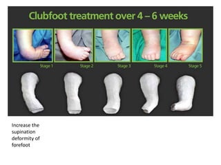
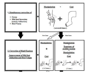
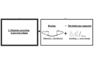
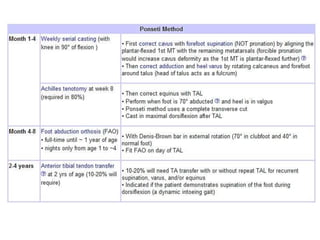
  • 22. Serial Manipulative and Casting (Ponsetti’s method) • Goal-rotate leg laterally around the fixed tallus • Order of correction (CAVE) -midfoot cavus -forefoot adductus -hindfoot varus -hindfoot equinus

Editor's Notes

  1. rot