


Facial Nerve
- 1. FACIAL NERVE Guided by Dr Anil Govindrao Ghom Dr Ajit Mishra Dr. Shweta Singh Dr. Savita Ghom Presented By Dr. Bratati Dey (PG 1ST YEAR) OMR
- 2. CONTENTSCONTENTS • Introduction • Functional components • Origin • Course and relations • Branches and distribution • Ganglia • Clinical anatomy – Upper motor neuron paralysis – Lower motor neuron paralysis (Bells’s palsy) – Clinical testing of the nerve – Facial nerve paralysis in newborn – Crocodile tears Syndrome – Ramsay-Hunt Syndrome
- 3. INTRODUCTIONINTRODUCTION • It is VII cranial nerve. • It is a mixed nerve having both motor and sensory component. It is a branch of second brancheal arch. • Has two root sensory and motor, sensory root known as Nervous Intermedius
- 4. FUNCTIONAL COMPONENTFUNCTIONAL COMPONENT 1. Special visceral or branchial efferent, 2. General visceral efferent or parasympathetic 3. General visceral afferent component 4. Special visceral afferent fibers 5. General somatic afferent fibers
- 7. AREA OF SUPPLYAREA OF SUPPLY 1. Muscle of the face, scalp, auricle, buccinator, stylohyoid, platysma, stapidius, posterior belly of digastric, motor area of facial nerve. 2. Secretomotor supply to submandibular and sublingual salivary gland 3. Sensory component bring taste sensation from anterior 2/3 of tongue except circumvallate papillae.
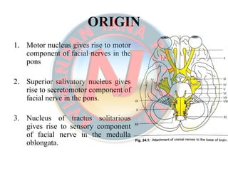
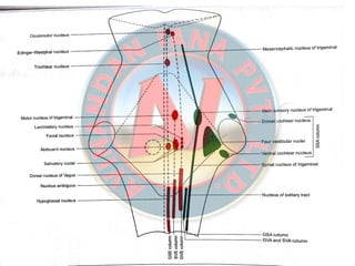
- 8. ORIGINORIGIN 1. Motor nucleus gives rise to motor component of facial nerves in the pons 2. Superior salivatory nucleus gives rise to secretomotor component of facial nerve in the pons. 3. Nucleus of tractus solitarious gives rise to sensory component of facial nerve in the medulla oblongata.
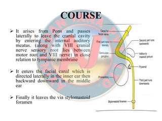
- 10. COURSECOURSE It arises from Pons and passes laterally to leave the cranial cavity by entering the internal auditory meatus. (along with VIII cranial nerve sensory root lies between motor root and VIII nerve) in close relation to tympanic membrane It enters the facial canal which is directed laterally in the inner ear then backward downward in the middle ear Finally it leaves the via stylomastoid foramen
- 11. Outside the skull the facial nerve passes along the styloid process to enter the posterio-medial surface of parotid gland. Passes transversely superficial to retro-mandibular vein & external carotid artery. Termination- It enters inside the parotid gland by dividing into 5 terminal branches.
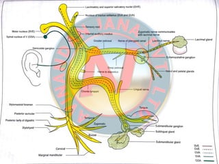
- 13. BRANCHES AND DISTRIBUTIONBRANCHES AND DISTRIBUTION A. Intra-cranial- Greater-petrosal nerve, Nerve to stapedius, Chorda tympani nerve B. At its exit from stylomastoid foramen- Posterior auricular Digastric Stylohyoid C. Terminal branches within the parotid gland- Temporal Zygomatic Buccal Marginal mandibular Cervical D. Communicating branches With adjacent cranial and spinal nerves.
- 14. GreaterGreater petrosalpetrosal nervenerve • Greater petrosal nerve originates at the geniculate ganglion • Run through pterigoid canal and carry preganglionic parasympathetic fiber to the sphenopalatine ganglion • Post ganglionic fiber start from sphenopalatine ganglion and supply the lacrimal gland.
- 15. Sensory nerve supply toSensory nerve supply to lacrimallacrimal nervenerve
- 16. Nerve toNerve to stapediusstapedius It arises opposite the pyramid of middle ear, & supplies the stapedius muscle The muscle damps excessive vibrations of the stapes caused by high-pitched sounds. Applied anatomy- In paralysis of the muscle, even normal sounds appear too loud & is known as HyperacusisHyperacusis.
- 17. ChordaChorda tympani nervetympani nerve Arises in the vertical part of the facial canal about 6cm above the stylomastoid foramen It runs upwards and forwards in a bony canal & It enters the middle ear and runs forwards in close relation to the tympanic membrane. Then it leave middle ear cavity by passing through pterigopalatine fissure to enters in the infra temporal fossa Where it joint the lingual nerve deep to muscle and distribution with it.
- 18. It carries- Preganglionic secretomotor fibers to the submandibular ganglion for supply of sub mandibular & sublingual salivary glands. Taste fibers from the anterior 2/3 of the tongue except circumvallate papillae.
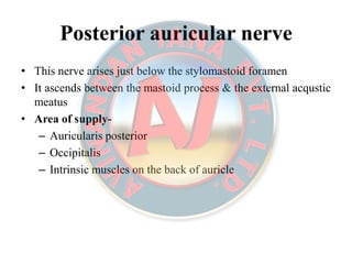
- 19. Posterior auricular nervePosterior auricular nerve • This nerve arises just below the stylomastoid foramen • It ascends between the mastoid process & the external acqustic meatus • Area of supply- – Auricularis posterior – Occipitalis – Intrinsic muscles on the back of auricle
- 20. DigastricDigastric branchbranch • It Arises close to posterior auricular nerve. • It is short and supplies the posterior belly of digastric.
- 21. StylohyoidStylohyoid branchbranch • It arises with digastric branch, is long and supplies the stylohyoid muscle
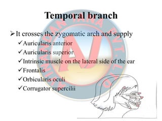
- 22. Temporal branchTemporal branch It crosses the zygomatic arch and supply Auricularis anterior Auricularis superior Intrinsic muscle on the lateral side of the ear Frontalis Orbicularis oculi Corrugator supercilii
- 23. ZygomaticZygomatic branchbranch • Zygomatic branch run across zygomatic bone and supply the orbicularis oculi.
- 24. BuccalBuccal branchbranch Buccal branches are two in number The upper buccal branch run above the parotid duct and the lower buccal branch run below the duct They supply muscles in that vicinity especially the buccinator.
- 25. MarginalMarginal mandibularmandibular branchbranch • Marginal mandibular branch run below the angle of mandible deep to the platysma. • It crosses the body of mandible and supplies muscle of the lower lip and chin.
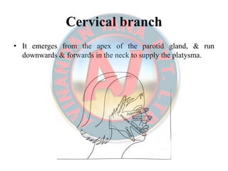
- 26. Cervical branchCervical branch • It emerges from the apex of the parotid gland, & run downwards & forwards in the neck to supply the platysma.
- 27. Communicating branchesCommunicating branches • For effective co-ordination between the movement of the muscles of the first second and third branchial arches, the motor nerves of the three arches communicate with each other • The facial nerve also communicate with the sensory nerves distributed over its motor territory.
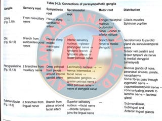
- 28. GangliaGanglia The ganglia associated with the facial nerve are as follows. 1. The Geniculate ganglion-it is a sensory ganglion. The taste fibres present in the nerve are perepheral processes of pseudounipolar neurons present in the geniculte ganglion. 2. The Submandibular ganglion- it is a parasympathetic ganglion, for relay of secretomotor fobre to the submandibular and sublingual gland. 3. The Pterigopalatine ganglion- is also a parasympathetic ganglion. Secretomotor fibers meant for the lacrimal gland relay in this ganglion.
- 30. CLINICAL ANATOMYCLINICAL ANATOMY Facial palsy lower motor neuron type Bells’s palsy and loss of taste sensation Bells’s and loss of taste sensation hyperausis Upper motor neuron type Paralysis of lower half of face Paralysis of contralateral lower half of face
- 32. Facial palsyFacial palsy UPPERUPPER MOTORMOTOR NEURONNEURON PARALYSISPARALYSIS of VII nerve results in paralysis of contralateral lower quadrant of face only. • It is seldom isolated palsy. • Affect mainly the muscles of lower part of face & is never complete. • The emotional movement are preserved. • No muscle contraction. • No reaction of regeneration. • Electromyography and nerve conduction is normal.
- 33. LOWERLOWER MOTORMOTOR NEURONNEURON PALSY/BELL’SPALSY/BELL’S PALSYPALSY - It is also called 7th nerve paraplegia or idiopathic facial paralysis. Etiology- Cold Trauma (after extraction, injection of local anaesthesia) Surgical procedure (such as removal of parotid gland tumor) Ischemia Facial canal & middle ear neoplasm Tumors Other causes (multiple sclerosis)
- 34. Sign & symptomSign & symptom Ear ache Hearing loss Pain or paraesthesia Any abnormality on otoscopy—including otitis media Associated cranial neuropathies or other neurological signs
- 35. Hypertension Lymphadenopathy pallor or bruising Vesicles in external meatus or on soft palate Single branch involvement Gradual progression of paralysis beyond 3 weeks Recurrence Mastoid swelling
- 36. Clinical features- Age & sex: women> men, middle age group. Onset: it begins abruptly as paralysis of facial musculature Prodromal symptoms: unilateral pain on ear temple & mastoid area or at the angle of jaw. Symptoms: speaking and eating difficulty, occasionally loss of taste sensation on ant. 2/3 of tongue. Eyes: Eyes can’t be closed and wrinkles disappear, watering of eye.
- 37. Mask like face. Angle of mouth drops down. Face become asymmetrical. Saliva drool from corner of mouth. Syndrome associated with melkerson-rosenthal syndrome. Lip &facial swelling VII nerve palsy Intermittent VII nerve palsy Fissure tongue
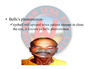
- 38. • Bells’s phenomenon- eyeball roll upward when patient attempt to close the eye, is known as bells phenomenon
- 39. Diagnosis- clinical diagnosis: Slurred speech, mask like face, drooling of saliva can diagnose this condition. lesion above origin of chorda tympani nerve show symptoms of bells palsy plus loss of taste sensation from anterior 2/3 of tongue except circumvallate papilae.
- 40. • Facial nerve can be injured at any level during its course • Lower motor neuron paralysis of VII nerve cause paralysis of ipsilateral half face i.e both upper quadrant & lower quadrant of same side as the injury.
- 41. •• ManagementManagement-- Vasodialator: like histamine Surgical decompression & anastomosis of nerve Nicotinic acid Others- systemic steroid or ACTH inj. vitamin B12, alone or in combination with steroids, recovered faster than those treated with steroids alone 100% hyperbaric oxygen recovered faster than those treated with steroids
- 42. Indication For Different Type Of SurgeryIndication For Different Type Of Surgery Early immediate nerve repair, in case of injury to the nerve Late nerve crossing by suturing perepheral branches of facial nerve to one of the following nerves Hypoglossal nerve, spinal accessory nerve Phrenic nerve Surgery to achieve movement in long standing facial palsy
- 43. •• StaticStatic procedureprocedure-- suspension of lips cheeks & angle of mouth to zygomatic bone or temporal fascia using fascia lata, palmaris longus tendon or other alloplastic material. Medial canthoplasty to reduce epiphora. Lateral tarsorrhaphy to prevent exposure keratitis due to widened palpebral fissure
- 44. •• DynamicDynamic procedureprocedure-- Muscle transfer with carefully preserved muscle nerve & vessel (temporalis muscle transfer, masseter muscle muscle transfer). Cross face nerve transplantation using sural nerve using microscope (sural nerve is sutured to the 2-3 relatively in significant branches to the VII nerve) Free neurovascular gracilis muscle graft using microvascular technique.
- 45. Clinical testing of the nerveClinical testing of the nerve • The facial nerve is examined by testing the following the facial muscle A. Frontalis- ask the patient to look upwards without moving his head, and look for the normal horizontal wrinkles of the forehead B. Dilators of mouth- showing the teeth C. Orbicularis oculi- tight closure of the eyes D. Buccinator- puffing the mouth & then blowing forcibly as is in whistling
- 46. • Platysma- – forcible pulling the angle of the mouth downwards & backwards forming prominent vertical fold of skin on the side of the neck. – The platysma contracts along with the risorius.
- 48. • In the infra-nuclear lesions of the facial nerve, known as bells palsy, the whole of the face of the same side gets paralysed – Asymmetrical face, – affected side become motionless, – eye can’t be closed – Any attempt to the smile drawn the mouth to the normal side – During mastication food accumulates between the cheek and teeth
- 49. • In the supra-nuclear lesions of the facial nerve usually a part of hemiplegia, only lower part of the opposite side of face is paralysed. • The upper part with the frontalis & orbicularis oculi escape due to its bilateral representation in the cerebral cortex.
- 50. Facial nerve paralysis in newbornFacial nerve paralysis in newborn • The mastoid process is absent in newborn & stylomastoid foramen is superficial. • Manipulation of baby’s head during delivery may damage the VII nerve. • This leads paralysis of facial muscles (buccinator) required for sucking the milk.
- 51. Crocodile tears syndromeCrocodile tears syndrome • Lacrimation during eating occurs due to aberrant regeneration after trauma. • In case of damage to facial nerve proximal to geniculate ganglia, regenerating fibre for sub mandibular salivary gland grow in endo neural sheaths of preganglionic secretomotor fibres supplying the lacrimal gland. • management- injection of botulinum toxin.
- 52. RamsayRamsay--Hunt syndromeHunt syndrome • Involvement of geniculate ganglia by herpes zoster result in this syndrome • It shows following syndrome Hyperacusis Loss of lacrimation Loss of sensation of taste in anterior two-third of tongue except vallate papillae (supplier by afferent IX cranial nerve) Bells’s palsy and lack of salivation Vesicles on the auricle.
- 53. INVESTIGATIONSINVESTIGATIONS Otoscopy is mandatory in all patients presenting with facial paralysis Where adequate auditory acuity cannot be confirmed an audiogram should be arranged. Hematological investigation- Complete blood count Radiological investigation- MRI (to identify brain stem pathology) High contrast CT Contrast enhanced magnetic resonance imaging Neurophysiological studies- measurement of fibrillation potential (part of electromyography) recording of the blink reflex
- 54. CONCLUSIONCONCLUSION • To date there is no clear evidence that any form of treatment improves outcome of idiopathic facial palsy in children. • Ninety five per cent of children will recover full function, • most within the first three weeks of the illness. • Protection of the cornea, with artificial tears and overnight patching, is normally all that is required • Neurophysiological assessment is helpful in patients with weakness persisting beyond three weeks.
- 55. REFERENCESREFERENCES BD Chaurasia’s human anatomy 354-358 Anand’s human anatomy for dental students Anil govindrao ghom, text book of oral medicine 760,1027 K rajgopal shenoy Manipal manual of surgery 307 Inamura H, Aoyagi M, Tojima H, et al. Facial nerve palsy in children: clinical aspects of diagnosis and treatment. Acta Otolaryngol Suppl 1994;511:150–2. Investigation and treatment of facial paralysis Accident and Emergency Department, Birmingham Children’s Hospital, Steelhouse Lane, Birmingham B4 6NH, UK