Final heart
- 2. 2 The Heart Mr.Sohan A Patel, Assistant Professor
- 3. Cardiovascular SystemCardiovascular System • CVS closed circulatory system. • The Principle function of the blood flow in the cvs is - provide oxygen and nutrients to the tissues of the body and - remove carbon dioxide and waste products.
- 4. Main part of CVS is Heart.
- 5. 5 HEART • The heart is a complex muscular pump that maintains blood pressure and flow through the lungs and the rest of the body. • The heart pumps about 100,000 times and moves 7200 liters (1900 gallons) of blood every day.
- 6. The Heart Left Ventricle Left Atrium Right Atrium Right Ventricle valve Vein from Lungs Artery to Head and BodyArtery to Lungs Vein from Head and Body valve
- 8. HODS - November 2006 8 HEART ANATOMY • Hollow, muscular organ • 300 grams (size of a fist) • 4 chambers • found in chest between lungs • surrounded by membrane called Pericardium • Pericardial space is fluid-filled to nourish and protect the heart.
- 9. 1. Location of heart 2. Structure of the heart 3. Layers 4. Coverings Chambers 5. Structure and function of myocardial 6. valves Different Parts of Heart
- 10. Location of the Heart • The heart is located between the lungs behind the sternum and above the diaphragm. • It is surrounded by the pericardium. • Its size is about that of a fist, and its weight is about 250-300 g. • Its center is located about 1.5 cm to the left of the midsagittal plane.
- 11. Location of the heart
- 12. 12 Heart’s position in thorax
- 13. Coverings of the heartCoverings of the heart • Pericardium -loose fitting, double layered sac • Visceral pericardium -serous membrane that is on the surface of the heart muscle • Parietal pericardium- inner layer of sac; secretes pericardial fluid • Pericardial fluid- (Serous fluid)-fluid that is between the parietal and visceral pericardium which prevents friction as the heart beats.
- 15. Pericardial Sac
- 16. Layers of heart tissueLayers of heart tissue • Epicardium • Myocardium • Endocardium
- 17. Serous membrane Continuous with blood vessels
- 18. Epicardium • Protective, outer layer of the heart wall same as the visceral pericardium • The coronary blood vessels that nourish the heart wall are located here
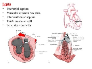
- 19. Myocardium = Middle layer made of cardiac muscle = Forms the bulk of the heart wall = Contains the septum- a thick muscular wall that completely separates the blood in the right side of the heart from the blood in the left side.
- 20. • The walls of the heart are composed of cardiac muscle, called myocardium. • It consists of four compartments: – the right and left atria and ventricles
- 21. Endocardium • Inner lining • Smooth surface that permits blood to move easily through the heart without agglutination. • Continuous with lining of blood vessels
- 22. • Epicardium – Outer, serous layer of heart • Myocardium – Strong, muscular layer of heart • Endocardium – lines the heart chambers
- 23. Serous membrane Continuous with blood vessels
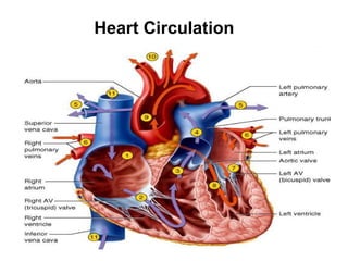
- 24. 24 HEART chambers The heart has four chambers. Two atria act as collecting reservoirs. Two ventricles act as pumps. The heart has four valves for: Pumping action of the heart. Maintaining unidirectional blood flow.
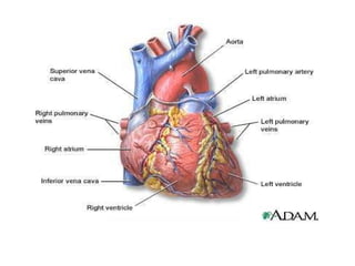
- 25. Chambers Right Atrium • Thinner wall than ventricles • Receives deoxygenated blood from vena cava • Passes blood through tricuspid valve into right Ventricle Chambers • Right Ventricle • Thicker wall than atria • Comprises most of anterior surface of heart • Circulates deoxygenated blood to lungs through the pulmonic valve into pulmonary trunk
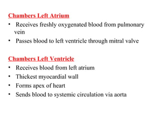
- 26. Chambers Left Atrium • Receives freshly oxygenated blood from pulmonary vein • Passes blood to left ventricle through mitral valve Chambers Left Ventricle • Receives blood from left atrium • Thickest myocardial wall • Forms apex of heart • Sends blood to systemic circulation via aorta
- 28. Chambers of the heart; valves
- 29. Septa • Interatrial septum • Muscular division b/w atria • Interventricular septum • Thick muscular wall • Seperates ventricles
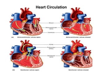
- 30. Heart Valves • Function- prevent blood from flowing backwards • Responds to changes in pressure Two types of valves in heart • Atrioventricular valves (AV) • Semi-lunar valves
- 32. Semilunar valves • Located at exit of ventricles, originiate from endothelial lining of veins Heart contains two semilunar valves • Pulmonic • Aortic
- 33. Atrioventricular Valves Left AV valve (Mitral, bicuspid) • Contains 2 cusps • Subject to abuse Right AV valve (Tricuspid) • Contains 3 cusps • Not subjected to great abuses
- 34. Atrioventricular Valves • Valve cusps are connected to papillary muscles
- 36. The Heart Valves • The tricuspid valve regulates blood flow between the right atrium and right ventricle. • The pulmonary valve controls blood flow from the right ventricle into the pulmonary arteries • The mitral valve lets oxygen-rich blood from your lungs pass from the left atrium into the left ventricle. • The aortic valve lets oxygen-rich blood pass from the left ventricle into the aorta, then to the body
- 37. S. MORRIS 2006
- 38. The circulatory system carries blood and dissolved substances to and from different places in the body. The Heart has the job of pumping these things around the body. The Heart pumps blood and substances around the body in tubes called blood vessels. The Heart and blood vessels together make up the Circulatory System. The circulatory system
- 39. lungs head & arms liver digestive system kidneys legs pulmonary artery aorta pulmonary vein main vein LeftRight How does this system work? Circulatory System
- 40. Lungs Body cells Our circulatory system is a double circulatory system. This means it has two parts parts. the right side of the system deals with deoxygenated blood. the left side of the system deals with oxygenated blood.
- 41. The Heart These are arteries. They carry blood away from the heart. This is a vein. It brings blood from the body, except the lungs. Coronary arteries, the hearts own blood supply The heart has four chambers 2 atria 2 ventricles now lets look inside the heart
- 42. The Heart Left Ventricle Left Atrium Right Atrium Right Ventricle valve Vein from Lungs Artery to Head and BodyArtery to Lungs Vein from Head and Body valve
- 43. How does the Heart work? blood from the body blood from the lungs The heart beat begins when the heart muscles relax and blood flows into the atria. STEP ONE
- 44. The atria then contract and the valves open to allow blood into the ventricles. How does the Heart work? STEP TWO
- 45. How does the Heart work? The valves close to stop blood flowing backwards. The ventricles contract forcing the blood to leave the heart. At the same time, the atria are relaxing and once again filling with blood. The cycle then repeats itself. STEP THREE
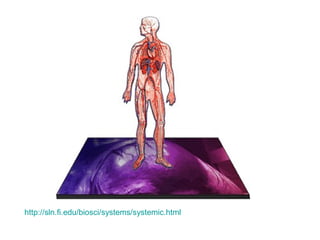
- 46. 3 Kinds of Circulation: • Pulmonary circulation • Coronary circulation • Systemic circulation
- 47. Pulmonary Circulation Movement of blood from the heart, to the lungs, and back to the heart again
- 49. Coronary Circulation Movement of blood through the tissues of the heart
- 51. Systemic Circulation Supplies nourishment to all of the tissue located throughout the body , except for the heart and lungs
- 53. Hollow tubes that circulate your blood There are 3 types of blood vessels a. ARTERY b. VEIN c. CAPILLARY Blood Vessels
- 54. Arteries • Carry blood AWAY from the heart • Heart pumps blood • Main artery called the aorta • Aorta divides and branches • Many smaller arteries • Each region of your body has system of arteries supplying it with fresh, oxygen-rich blood.
- 55. The ARTERY thick muscle and elastic fibres Arteries carry blood away from the heart. the elastic fibres allow the artery to stretch under pressure the thick muscle can contract to push the blood along.
- 58. Veins • Carry blood to the heart • Receive blood from the capillaries • Transport waste-rich/ oxygen-poor blood back to the heart • Allow blood to move in one direction
- 59. The VEIN Veins carry blood towards from the heart. thin muscle and elastic fibres veins have valves which act to stop the blood from going in the wrong direction. body muscles surround the veins so that when they contract to move the body, they also squeeze the veins and push the blood along the vessel.
- 60. Capillaries • Very thin • Only one cell thick • Connect arteries & veins
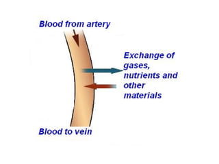
- 61. Capillaries • Food and oxygen released to the body cells • Carbon dioxide and other waste products returned to the bloodstream
- 62. The CAPILLARY Capillaries link Arteries with Veins the wall of a capillary is only one cell thick they exchange materials between the blood and other body cells. The exchange of materials between the blood and the body can only occur through capillaries.
- 63. artery vein capillaries body cell The CAPILLARY A collection of capillaries is known as a capillary bedcapillary bed.
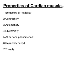
- 74. Properties of Cardiac muscle:- 1.Excitability or irritability 2.Contractility 3.Automaticity 4.Rhythmicity 5.All or none phenomenon 6.Refractory period 7.Tonicity
- 75. Coordination of chamber contraction, relaxation
- 76. I. Excitability (Irritability): = the ability of cardiac muscle to respond to adequate stimuli by generating an action potential followed by a mechanical contraction.
- 80. Electrophysiology of the cardiac muscle cell
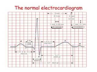
- 81. The Conduction System • Electrical signal begins in the sinoatrial (SA) node: "natural pacemaker." – causes the atria to contract. • The signal then passes through the atrioventricular (AV) node. – sends the signal to the ventricles via the “bundle of His” – causes the ventricles to contract.
- 84. Abnormalities of Heart:- 1.Hypertension 2.CHF (Conjunctive heart failure) 3.Hyperlipidemic 4.Arraythmias 5.Angina pectories
Editor's Notes
- Four types of valves regulate blood flow through your heart: The tricuspid valve regulates blood flow between the right atrium and right ventricle. The pulmonary valve controls blood flow from the right ventricle into the pulmonary arteries, which carry blood to your lungs to pick up oxygen. The mitral valve lets oxygen-rich blood from your lungs pass from the left atrium into the left ventricle. The aortic valve opens the way for oxygen-rich blood to pass from the left ventricle into the aorta, your body's largest artery, where it is delivered to the rest of your body.
- Electrical impulses from your heart muscle (the myocardium) cause your heart to beat (contract). This electrical signal begins in the sinoatrial (SA) node, located at the top of the right atrium. The SA node is sometimes called the heart's "natural pacemaker." When an electrical impulse is released from this natural pacemaker, it causes the atria to contract. The signal then passes through the atrioventricular (AV) node. The AV node checks the signal and sends it through the muscle fibers of the ventricles, causing them to contract. The SA node sends electrical impulses at a certain rate, but your heart rate may still change depending on physical demands, stress, or hormonal factors.


