Location via proxy:   [ UP ]  
[Report a bug]   [Manage cookies]                
SlideShare a Scribd company logo

1

FRACTURES  OF THE CLAVICLE  Le Kim Trong MD., Le Nghi Thanh Nhan MD.

2

General Information Clavicle fractures are common injuries Account for 5-10% of all fractures Up to 44% of injuries to the shoulder girdle Easy to recognize Majority unite uneventfully

3

Current Debate in Treatment What are the indications for early or acute surgical treatment?

4

Ossification 1 st  bone in body to ossify at 5 weeks gestation Intramembranous ossification Epiphyseal growth plates develop at both ends, only medial end is evident on radiographs and accounts for 80% of its length Sternal ossification occurs at 12-19 years of age and fuses at 22-25

5

Anatomy The clavicle is an S-shaped bone that acts as a structure between the sternum and the glenohumeral joint. The lateral third is flat and is the insertion site for the trapezius… The middle third is tubular and provides protection. The medial third is quadrangular and is the insertion for sternocleidomustoid muscle

6

Anatomy Left Clavicle:  Superior & Inferior Views Middle Third Weak to axial load Medial Third Protects brachial plexus, subclavian vessels, & superior lung Convex ventral on medial half Concave ventral on lateral half

7

Anatomy Weakest region of bone

8

Anatomy Subcutaneous along entire length  Supraclavicular nerves are only structures to cross anterior to clavicle Scapula and clavicle bound securely by the AC & CC ligaments Main nutrient artery enters just medial to CC ligaments

9

Clavicle Function Power and stability of arm Motion of the shoulder girdle Muscle attachment Protects neurovascular structures Facilitates the placement of the shoulder in a more lateral position, so the hand can be more effectively positioned to deal with a 3-D environment

10

Epidemiology Group 1  (middle one third of the clavicle - the shaft) 76% ,13 years. Group 2 (lateral one third - the acromial end) 21% ,47 years. Group 3 (medial one third - the sternal end) 3%, 59 years.

11

Mechanism of Injury Trauma  Fall against lateral shoulder (90%)  Fall on Outstretched Hand (5%) Direct blow to clavicle (5%) No trauma  (in children)  Tumor  Rickets Osteogenesis imperfecta  Physical Abuse

12

Diagnosis   History: trauma, tumor… Pain and swelling, decreased movement of the affected limb.   Bruising, tenderness, and crepitation,  pressure on the overlying skin, palpable. The arm will usually be held across the chest with the opposite limb used to support the weight of the injured limb   Observe for complications  - Neurovascular injury of affected arm , outlet thoracic - Pneumothorax, open fracture - Subcu t aneous Emphysema………… http://www.fpnotebook.com/ortho/shoulder/ClvclFrctr.htm

13

Displacement mechanism

14

Radiographs  Different angles (anteroposterior and 45° cephalic tilt): AP – evaluate superior-inferior displacement 45 o  cephalic tilt view Evaluate AP displacement Tube directed from below, upward Weighted views useful in lateral 1/3 fractures to assess CC ligaments  Chest x-ray  CT

15

VI.Classifications & Treatments   Group I - Middle third  (80-85%) Nondisplaced less than 100% displacement  nonoperative        Displaced greater than 100% displacement  nonunion rate of 4.5%  operative 

16

Group II - Lateral third (10-15%) Type I fracture occurs lateral to coracoclavicular ligaments (trapezoid, conoid) or interligamentous  usually minimally displaced  stable because conoid and trapezoid ligaments remain intact  nonoperation                       Type I Group II

17

Group II - Lateral third (10-15%) Type IIA fracture occurs medial to intact conoid and trapezoid ligament  medial clavicle unstable  up to 56% nonunion rate with nonoperative management  operative                      Type IIA G II

18

Group II - Lateral third (10-15%) Type IIB fracture occurs either between ruptured conoid and intact trapezoid ligament or lateral to bothligaments torn  medial clavicle unstable  up to 30-45% nonunion rate with nonoperative management  operative         Type IIB G II

19

Group II - Lateral third (10-15%) Type III Intraarticular fracture extending into AC joint  Conoid and trapezoid intact therefore stable injury  Patients may develop posttraumatic AC arthritis  nonoperative Type IV A physeal fracture that occurs in the skeletally immature  Displacement of lateral clavicle occurs superiorly through a tear in the thick periosteum  Clavicle pulls out of periosteal sleeve  Conoid and trapezoid ligaments remain attached to periosteum and overal the fracture pattern is stable  nonoperative Type V Comminuted fx  Conoid and trapezoid ligaments remain attached to comminuted fragment  Medial clavicle unstable  operative

20

Group III - Medial third  (5-8%) Anterior  most often non-operative  nonoperative  rare injury (2-3%)  often physeal fracture-dislocation (age < 25)  Posterior  must assess airway and great vessel compromise  operative  serendipity radiographs and CT scan to evaluate  surgical management with thoracic surgeon on standby

21

Classifications

22

Goals of Treatment Achieve bony union with minimal morbidity Minimal loss of function Minimal residual deformity

23

Nonoperative Treatment Indications:  The vast majority of fractures can be treated closed with good results. Nondisplaced Group I (middle third)  Stable Group II fractures (Type I, III, IV)   Anterior Group III (medial third)  Technique   Sling or figure-of-eight (prospective studies have not shown difference between sling and figure-of-eight).   After 2-4 weeks begin gentle range of motion exercises.  No attempt at reduction should be made.

24

Closed Reduction Techniques

25

Immobilisation

26

Researchs Stanley and Norris reviewed a consecutive series of 140 patients with fractures of the clavicle. All had been treated with either a figure-of-eight bandage or a sling. There was no difference in either the rate or speed of recovery between the groups. Stanley D, Norris SH. Recovery following fractures of the clavicle treated conservatively. Injury 1988;19:162-164

27

Researchs   Hill et al:  Evaluated 242 adult clavicle fractures treated closed 66 (27%) completely displaced middle third Nonunion 8/52=15%. Unsatisfactory result 16/52=31% Mild-moderate pain 13/52=25%. Brachial plexus irritation 15/52=29% Cosmetic complaints 28/52 with 11/52 considered corrective surgery Initial shortening at fracture of > 2cm had a significant association with nonunion and chance of unsatisfactory result Recommended ORIF of severely displaced fractures of the middle third of the clavicle in adult patients Hill JM, McGuire MH, Crosby LA. Closed treatment of displaced middle-third fractures of the clavicle gives poor results. J Bone Joint Surg Br 1997;79:537–539.

28

Researchs Wick et al: clavicle fractures with greater than 20 mm of shortening were highly predisposed to develop a nonunion. Of middle third clavicle nonunions in their series, 91% (30/33) were shortened by at least 2 cm. Wick M, Muller EJ, Kollig E, et al. Midshaft fractures of the clavicle with a shortening of more than 2 cm predispose to nonunion. Arch Orthop Trauma Surg 2001;121(4):207-211

29

Operative Treatment Indications of operative treatment       Absolute  Unstable Group II fxs (Type IIA, Type IIB, Type V).   Open fxs.  Widely displaced >= 2 cm:  increased risk for nonunion. Displaced fx with skin tenting, hypertrophic callus. Subclavian artery or vein injury.  Floating shoulder (clavicle and scapula neck fx).  Symptomatic nonunion.  Posteriorly displaced Group III fxs.  Displaced Group I (middle third).  Thoracic outlet.

30

Indications of operative treatment   (con.)     Fracture that threaten the overlying skin Bilateral clavicle fxs. With multiple ipsilateral rib fractures

31

Relative and controversial indications   -  Brachial plexus injury. - Closed head injury.  - Seizure disorder.  - Polytrauma patient.   - Contraindications of operative treatment Non-displaced fractures (no comminution   , <3mm displacement)  Infection  Elderly, low-demand, high surgical risk patients

32

Researchs   Thompson reviewed more than 100 middle-third clavicular nonunions reported in the literature and found that 90% of the original fractures had displacement greater than 100%, overriding more than 1 cm, or had severe comminution . Thompson JS. Operative Treatment of Certain Clavicle Fractures. An Orthopaedic Controversy. Orthop Trans 1988;12:141

33

Stabilization techniques include   Plate fixation  Intramedullary fixation  External fixation  Coracoclavicular ligament repair or reconstruction in Group II  Postoperative rehabilitation   Sling for 7-10 days followed by active motion  Strengthening at ~ 6 weeks when pain free motion and radiographic evidence of union  Full activity including sports at ~ 3 months

34

MIDCLAVICULAR FRACTURES

35

Close treatment Nordqvist : among 225 fractures, 197 were treated in a figure-of-eight splint for an average of 3 weeks without attempted reduction, whereas 24 were allowed immediate free shoulder motion. 185 shoulders were asymptomatic and only one patient was considered to have a poor result secondary to symptoms from thoracic outlet syndrome    comminuted fractures did not fare worse than noncomminuted fractures.  ( Nordqvist A, Petersson CJ, Redlund-Johnell I. Mid-clavicle fractures in adults: end result study after conservative treatment. J Orthop Trauma 1998;12:572-576 ). According to Neer, only 3 (0.13%) of 2235 patients with midclavicular fractures treated by closed methods failed to heal, whereas nonunion developed in 2 (4.4%) of 45 treated with immediate open reduction and fixation .  (Neer CS. Nonunion of the clavicle. JAMA 1960;172:1006-1011). Rowe found a nonunion rate of 0.8% in his series of 566 fractures.  (Rowe CR. An atlas of anatomy and treatment of midclavicular fractures. Clin Orthop 1968;58:29-42).

36

Plate fixation: Advantages: Rigid fixation Cortical compression Rotational control  Disadvantages: I ncreased soft-tissue stripping Prominence  of  hardware under skin Plate:  LC-DCP. 3.5 reconstruction plate.

37

Researchs Bostman found no difference between using 3.5-mm DCPs and 3.5-mm AO/ASIF reconstruction plates; both provided acceptable fixation and rigidity.  Any plate smaller than a 3.5-mm limited contact dynamic compression plate should be used with caution. One-third of tubular plates have a high rate of fatigue failure when used for clavicle fractures and should be avoided .   ( Bostman O, Manninen M, Pihlajamaki H. Complications of Plate Fixation in Fresh Displaced Midclavicular Fractures. J Trauma 1997;43:778 ) Superior position of the plate provides more secure fixation and anterior position is second best .( Finkemeier  C.G, Fracture and dislocation of the shoulder girdle and humerus, Chapman's Orthopaedic Surgery, 3rd Edition,  Lippincott Williams & Wilkins , 2001, 432-481.)

38

LC-DCP  Incision close : -  The periosteum and fascial layer are closed with a heavy absorbable suture in interrupted fashion.  - The subcutaneous tissue is also closed with interrupted suture.  - The skin is closed with a pullout, monofilament, subcuticular suture.

39

a) Reconstruction plate fixation at the top. b) DCP fixation at the anterior surface .

40

Researchs   Bostman et al, J Trauma, 1997 Complications of plate fixation in fresh displaced midclavicular fractures 103 fractures 9.5% of 1081 midclavicle fractures seen over 6 years One or more complications 24/103=23% Infection rate 7.8% Reoperations 14/103=14% Severely comminuted fracture and intoxication on admission markers of increased complication risk ( Bostman O, Manninen M, Pihlajamaki H. Complications of Plate Fixation in Fresh Displaced Midclavicular Fractures. J Trauma 1997;43:778 )

41

Researchs Oroka et al, Bull Hosp Jt Dis, 1999 41 patients Could not demonstrate any relationship between clavicular shortening and shoulder function Shen et al, Injury, 1999 251 fresh completely displaced middle third fractures over 2 year period received ORIF 232 had follow-up Mean time to union: 10 weeks Nonunion 7/232=3% Healing with angulation 14/232=6% Infection 5/232=2% Soreness or skin numbness 49/232=21% Hardware removal 171/232=74% 94% satisfied with procedure

42

Intramedullary fixation Advantages:   Easy of procedure Limited exposure with minimal soft-tissue disruption Satisfactory rates of healing. Implants:  a pin must be strongly and stiffly tempered to withstand the unsupported weight of the upper extremity without bending or breaking Knowles pins  Hagie pins Rockwood pins Minimally invasive titanium nails Kirschner wire  (wire breakage and migration to a variety of anatomic locations)

43

- Open intramedullary fixation  is popular: typically a threaded pin is inserted through the fracture site ,the pin is removed 3 months after surgery - Close intramedullary fixation  :Smooth titanium nail has been inserted through the medial clavicle, without opening the fracture site  Intramedullary fixation

44

McKeever intramedullary fixation of clavicle

45

Rockwood  intramedullary pin  of clavicle

46

Researchs Paffen and Jansen reviewed 73 of which required open reduction and Kirschner-wire fixation, the rate of union was 97%.  ( Paffen PJ, Jansen EW. Surgical treatment of clavicular fractures with Kirschner wires: a comparative study. Arch Chir Neerl 1978;30:43-53 ). Neviaser et al reported their results of intramedullary fixation using Knowles pins, demonstrating a healing rate of 100%.  ( Neviaser RJ, Neviaser JS, Neviaser TJ. A simple technique for internal fixation of the clavicle. A long term evaluation. Clin Orthop 1975;109:103-107 ). Zenni et al: 24 cases of clavicle fracture treated by open reduction and intramedullary wire or pin fixation, 21 of which were midshaft fractures. All fractures went on to heal in anatomic or near anatomic position.  ( Zenni EJ Jr, Krieg JK, Rosen MJ. Open reduction and internal fixation of clavicular fractures. J Bone Joint Surg Am 1981;63:147-151 ).

47

Research Jubel et al: 58 midshaft clavicle fractures . Indication: shortening greater than 2 cm, multiple trauma, additional lower extremity trauma that did not allow full weight bearing, concomitant neurovascular injury, or a floating shoulder.  Implant : elastic titanium intramedullary nail.  Hardware was removed at an average of 8 weeks postoperatively.  No infections or refracture after hardware removal occurred.  One nonunion which was treated with bone grafting and plating. Jubel A, Andermahr J, Schiffer G, et al. Elastic stable intramedullary nailing of midclavicular fractures with a titanium nail. Clin Orthop Relat Res 2003;408:279-285

48

Research Shen et al:  Comparison of 40 patients who underwent open reduction and intramedullary fixation using a 2.5-mm threaded pin vs 40 patients who were treated with a figure-of-eight bandage for an average of 6 weeks Close reduction: low rate of complications Open reduction:  higher rate of complications (35%) 3 refractures after pin removal 8 superficial infections 2 cases of delayed union with pin breakage, and two nonunions   Conclusion:  Intramedullary pin fixation be reserved for those fractures that were severely displaced Shen WJ, Liu TJ, Shen YS. Plate fixation of fresh displaced midshaft clavicle fractures. Injury 1999;30(7):497-500

49

DISTAL CLAVICULAR FRACTURES The literature is more controversial for this fracture type than for the midclavicle

50

Undisplaced Lateral-End Fractures  (Neer Type I ) Nonoperative management is the treatment of choice  Late excision of the distal segment (through either an arthroscopic or an open approach) may be used in this group of patients if the fragment is small http://www.ejbjs.org/cgi/content/full/91/2/447

51

Displaced Lateral-End Fractures  (Neer Type II ) Osteosynthesis procedures: Transacromial K-wires with or without a tension band  Coracoclavicular screw  Plate fixation  Coracoclavicular banding or taping with or without acromioclavicular fixation utilizing dacron or other synthetic materials

52

RESEARCHS -  In Neer's original series of clavicle nonunions, they accounted for one-half of the nonunions is distal clavicular fractures with closed treatment.  (Neer CS. Nonunion of the clavicle. JAMA 1960;172:1006-1011). -  Robinson: prevalence of nonunion with closed treatment of the medial end fractures is 8.3%, of the diaphyseal fractures is 4.5% and of the lateral end fractures is 11.5% .  The risk of nonunion for the lateral end fracture significantly increased only by advancing age and displacement of the fracture  ( Robinson C.M. et al.  Estimating the risk of nonuniion following nonoperative treatment of a clavicular fracture, JBJS 2004, 86-A · 7, 1359-1365)

53

RESEARCHS Nordqvist et al.: 110 patients had a lateral clavicle fracture A mean follow-up of 15 years.  Type I:73,Type II: 23, Type III:14.  Treatment: figure-of-eight immobilization.  Nonunions: 10  Predictors of nonunion:  Type II fracture  and older age  For Type II fractures, 22% had a nonunion (8 /10 nonunions was asymptomatic. Deformity was present, but fracture instability was not found on examination.

54

RESEARCHS Rokito et al:  a retrospective review of the results of operative and nonoperative treatment of Type II fractures.   Treatment nonoperative: 16    7 patients went on to nonunion but 5/7 were asymptomatic. Coracoclavicular stabilization: 14. All surgically treated patients healed, whereas.  Functional outcome was similar between the two groups.

55

A high rate of delayed union, nonunion, and deformity with closed treatment of Type II distal clavicle fractures in literature.

56

Tension band procedure  without transacromion is choosen for noncomminuted fractures (2- to 3-cm distal piece) Comminuted and/or small distal fragments require transacromial wire fixation. 1. Direct fixation of the fracture site without coracoclavicular stabilization

57

RESEARCHS Neer recommended transacromial wire fixation and reported that seven of seven fractures treated with this technique healed at an average of 6 weeks postoperatively  (Neer CS. Fractures of the distal third of the clavicle. Clin Orthop 1968;58:43-50) . Fann: good results in all 32 patients treated with a transacromial Knowles pin.  (Fann CY, Chiu FY, Chuang TY, et al. Transacromial Knowles pin in the treatment of Neer type 2 distal clavicle fractures. A prospective evaluation of 32 cases. J Trauma 2004;56(5):1102-1105 ). Kao et al:  Eleven of the 12 fractures united with two 1.8-mm K-wires and tension band without transacromioclavicular joint. The fractures united between 3 and 6 months, at which time the implants were removed.

58

Researchs   Kona et al: 19 cases of type II distal clavicle fractures  Treatment: variety of surgical techniques  Results:  32% nonunion rate 30% infection rate 56% unsatisfactory results.  Three nonunions and all five patients with a poor result had transacromial K-wire fixation,  The authors to recommend avoiding any transacromial wire fixation for these fractures.   Kona J, Bosse MJ, Staeheli JW, et al. Type II distal clavicle fractures: a retrospective review of surgical treatment. J Orthop Trauma 1990;4:115-120.

59

Plate Fixation The distal fragment is large enough   to hold a minimum of two, and ideally three, bicortical screws

60

The clavicular hook plate:     +The distal fragment is too small .    +The plate has an offset lateral hook, designed to engage distal to the posterior aspect of the acromion.   + Most surgeons advise routine plate removal at three months after implantation

61

Kirschner Wire Fixation - The inherent risk of wire breakage and migration. - Tigh nonunion and infection rates. -  Have recommended that this method of fixation not be used.

62

RESEARCHS Flinkkila et al: comparative study 22 Neer Type II fractures treated with K-wire fixation 17 patients treated with a clavicular hook plate (Stratec Medical, Oberdorf, Switzerland). Both groups did well according to the Constant and L'Insalata scores Outcomes: K-wire group: 12 cases of wire migration resulting in loss of fixation in seven, infection in three, and nonunion in two.  The clavicular hook plate group: 1 fractured clavicle at the medial end of the plate secondary and two nonunions.  The authors recommended the use of the plate over the K-wires. Flinkkila T, Ristiniemi J, Hyvonen P, et al. Surgical Treatment of unstable fractures of the distal clavicle: a comparative study of Kirschner wire and clavicular hook plate fixation. Acta Orthop Scand 2002;73(1):50-53

63

Suture and Sling Techniques The graft either is looped around the coracoid and over the clavicle fragment to form a sling or is passed through drill-holes. The use of two EndoButtons, toggled through drill-holes in the clavicle and coracoid to link a continuous loop of one of the new generation of robust nonabsorbable suture materials

64

2. Direct Fixation of the Fracture with Coracoclavicular Stabilization Indications : Very distal fracture in a young individual.  Fractures that involve the clavicular insertion of the coracoclavicular ligaments.

65

Clinical case

66

RESEARCHS Chen et al:  13 patients with Neer Type II fractures Treatment: Mersilene tape + repair of the coracoclavicular ligaments + tension band wire spanning the fracture.  Eleven were available for follow-up at an average of 27 months.  Outcome:  10/11 fractures united at 3 months, whereas the last patient's fracture united at 6 months.  9/11 patients: excellent . 1 patient : good . 1 patient : fair. Chen CH, Chen WJ, Shih CH. Surgical treatment for distal clavicle fracture with coracoclavicular ligament disruption. J Trauma 2002;52(1):7-8

67

RESEARCHS Goldberg et al: 9 patients Treatment: Dacron tape passed around the base of the coracoid and medial clavicular segment combined with suture fixation of the distal clavicular fragment to the proximal All fractures went on to union.  The average time until union was 16 weeks, with all but one patient obtaining union by 18 weeks.   Goldberg JA, Bruce WJ, Sonnabend DH, et al. Type 2 fractures of the distal clavicle: a new surgical technique. J Shoulder Elbow Surg 1997;6:380-382 Mall et al:  12 patients Treatment: a 10-mm band of absorbable suture placed through a drill hole in the distal aspect of the proximal fragment and around the coracoid in a figure-of-eight fashion.  All patients achieved an excellent rating All fractures united, and only one hypertrophic scar was listed as a complication.

68

3. Coracoclavicular Stabilization With or Without Excision of the Lateral Clavicular Segment Indications: Comminuted lateral clavicular segment;  Older patients in whom potential healing of a small lateral clavicular segment would be difficult;  Underlying acromioclavicular arthropathy;  Fractures lateral to the trapezoid origin;  Fractures that involve the coracoclavicular ligament origin as an inferiorly displaced fragment.

69

Coracoclavicular Screw   -  An appreciable rate of fixation failure due to screw cutout or loosening   - Removed at three months

70

MEDIAL CLAVICULAR FRACTURES

71

TREATMENT These fractures are usually managed nonoperatively unless fracture displacement produces superior mediastinal compromise. In these circumstances, an emergent attempt at closed reduction should be made with open reduction performed next if this is unsuccessful.  Internal fixation  The use modified Balser plate and use of Mersilene or other strong braided interosseous suture

72

*Complications of nonoperative treatment   -  Nonunion (1-5%)  -  Decreased shoulder strength and endurance       *Complications of operative treatment (10%- 30%) Hardware complications : 30% of patient request plate removal. Adhesive capsulitis  Infection (~4.8%)  Hardware irritation requiring removal (~8%)  Mechanical failure (~1.4 %)

More Related Content

Fractures of the clavicle

  • 1. FRACTURES OF THE CLAVICLE Le Kim Trong MD., Le Nghi Thanh Nhan MD.
  • 2. General Information Clavicle fractures are common injuries Account for 5-10% of all fractures Up to 44% of injuries to the shoulder girdle Easy to recognize Majority unite uneventfully
  • 3. Current Debate in Treatment What are the indications for early or acute surgical treatment?
  • 4. Ossification 1 st bone in body to ossify at 5 weeks gestation Intramembranous ossification Epiphyseal growth plates develop at both ends, only medial end is evident on radiographs and accounts for 80% of its length Sternal ossification occurs at 12-19 years of age and fuses at 22-25
  • 5. Anatomy The clavicle is an S-shaped bone that acts as a structure between the sternum and the glenohumeral joint. The lateral third is flat and is the insertion site for the trapezius… The middle third is tubular and provides protection. The medial third is quadrangular and is the insertion for sternocleidomustoid muscle
  • 6. Anatomy Left Clavicle: Superior & Inferior Views Middle Third Weak to axial load Medial Third Protects brachial plexus, subclavian vessels, & superior lung Convex ventral on medial half Concave ventral on lateral half
  • 8. Anatomy Subcutaneous along entire length Supraclavicular nerves are only structures to cross anterior to clavicle Scapula and clavicle bound securely by the AC & CC ligaments Main nutrient artery enters just medial to CC ligaments
  • 9. Clavicle Function Power and stability of arm Motion of the shoulder girdle Muscle attachment Protects neurovascular structures Facilitates the placement of the shoulder in a more lateral position, so the hand can be more effectively positioned to deal with a 3-D environment
  • 10. Epidemiology Group 1 (middle one third of the clavicle - the shaft) 76% ,13 years. Group 2 (lateral one third - the acromial end) 21% ,47 years. Group 3 (medial one third - the sternal end) 3%, 59 years.
  • 11. Mechanism of Injury Trauma Fall against lateral shoulder (90%) Fall on Outstretched Hand (5%) Direct blow to clavicle (5%) No trauma (in children) Tumor Rickets Osteogenesis imperfecta Physical Abuse
  • 12. Diagnosis History: trauma, tumor… Pain and swelling, decreased movement of the affected limb. Bruising, tenderness, and crepitation, pressure on the overlying skin, palpable. The arm will usually be held across the chest with the opposite limb used to support the weight of the injured limb Observe for complications - Neurovascular injury of affected arm , outlet thoracic - Pneumothorax, open fracture - Subcu t aneous Emphysema………… http://www.fpnotebook.com/ortho/shoulder/ClvclFrctr.htm
  • 14. Radiographs Different angles (anteroposterior and 45° cephalic tilt): AP – evaluate superior-inferior displacement 45 o cephalic tilt view Evaluate AP displacement Tube directed from below, upward Weighted views useful in lateral 1/3 fractures to assess CC ligaments Chest x-ray CT
  • 15. VI.Classifications & Treatments Group I - Middle third  (80-85%) Nondisplaced less than 100% displacement nonoperative     Displaced greater than 100% displacement nonunion rate of 4.5% operative 
  • 16. Group II - Lateral third (10-15%) Type I fracture occurs lateral to coracoclavicular ligaments (trapezoid, conoid) or interligamentous usually minimally displaced stable because conoid and trapezoid ligaments remain intact nonoperation              Type I Group II
  • 17. Group II - Lateral third (10-15%) Type IIA fracture occurs medial to intact conoid and trapezoid ligament medial clavicle unstable up to 56% nonunion rate with nonoperative management operative             Type IIA G II
  • 18. Group II - Lateral third (10-15%) Type IIB fracture occurs either between ruptured conoid and intact trapezoid ligament or lateral to bothligaments torn medial clavicle unstable up to 30-45% nonunion rate with nonoperative management operative      Type IIB G II
  • 19. Group II - Lateral third (10-15%) Type III Intraarticular fracture extending into AC joint Conoid and trapezoid intact therefore stable injury Patients may develop posttraumatic AC arthritis nonoperative Type IV A physeal fracture that occurs in the skeletally immature Displacement of lateral clavicle occurs superiorly through a tear in the thick periosteum Clavicle pulls out of periosteal sleeve Conoid and trapezoid ligaments remain attached to periosteum and overal the fracture pattern is stable nonoperative Type V Comminuted fx Conoid and trapezoid ligaments remain attached to comminuted fragment Medial clavicle unstable operative
  • 20. Group III - Medial third  (5-8%) Anterior most often non-operative nonoperative rare injury (2-3%) often physeal fracture-dislocation (age < 25) Posterior must assess airway and great vessel compromise operative serendipity radiographs and CT scan to evaluate surgical management with thoracic surgeon on standby
  • 22. Goals of Treatment Achieve bony union with minimal morbidity Minimal loss of function Minimal residual deformity
  • 23. Nonoperative Treatment Indications: The vast majority of fractures can be treated closed with good results. Nondisplaced Group I (middle third) Stable Group II fractures (Type I, III, IV)  Anterior Group III (medial third) Technique Sling or figure-of-eight (prospective studies have not shown difference between sling and figure-of-eight).  After 2-4 weeks begin gentle range of motion exercises. No attempt at reduction should be made.
  • 26. Researchs Stanley and Norris reviewed a consecutive series of 140 patients with fractures of the clavicle. All had been treated with either a figure-of-eight bandage or a sling. There was no difference in either the rate or speed of recovery between the groups. Stanley D, Norris SH. Recovery following fractures of the clavicle treated conservatively. Injury 1988;19:162-164
  • 27. Researchs Hill et al: Evaluated 242 adult clavicle fractures treated closed 66 (27%) completely displaced middle third Nonunion 8/52=15%. Unsatisfactory result 16/52=31% Mild-moderate pain 13/52=25%. Brachial plexus irritation 15/52=29% Cosmetic complaints 28/52 with 11/52 considered corrective surgery Initial shortening at fracture of > 2cm had a significant association with nonunion and chance of unsatisfactory result Recommended ORIF of severely displaced fractures of the middle third of the clavicle in adult patients Hill JM, McGuire MH, Crosby LA. Closed treatment of displaced middle-third fractures of the clavicle gives poor results. J Bone Joint Surg Br 1997;79:537–539.
  • 28. Researchs Wick et al: clavicle fractures with greater than 20 mm of shortening were highly predisposed to develop a nonunion. Of middle third clavicle nonunions in their series, 91% (30/33) were shortened by at least 2 cm. Wick M, Muller EJ, Kollig E, et al. Midshaft fractures of the clavicle with a shortening of more than 2 cm predispose to nonunion. Arch Orthop Trauma Surg 2001;121(4):207-211
  • 29. Operative Treatment Indications of operative treatment Absolute Unstable Group II fxs (Type IIA, Type IIB, Type V).  Open fxs. Widely displaced >= 2 cm: increased risk for nonunion. Displaced fx with skin tenting, hypertrophic callus. Subclavian artery or vein injury. Floating shoulder (clavicle and scapula neck fx). Symptomatic nonunion. Posteriorly displaced Group III fxs. Displaced Group I (middle third).  Thoracic outlet.
  • 30. Indications of operative treatment (con.) Fracture that threaten the overlying skin Bilateral clavicle fxs. With multiple ipsilateral rib fractures
  • 31. Relative and controversial indications - Brachial plexus injury. - Closed head injury. - Seizure disorder. - Polytrauma patient. - Contraindications of operative treatment Non-displaced fractures (no comminution , <3mm displacement) Infection Elderly, low-demand, high surgical risk patients
  • 32. Researchs Thompson reviewed more than 100 middle-third clavicular nonunions reported in the literature and found that 90% of the original fractures had displacement greater than 100%, overriding more than 1 cm, or had severe comminution . Thompson JS. Operative Treatment of Certain Clavicle Fractures. An Orthopaedic Controversy. Orthop Trans 1988;12:141
  • 33. Stabilization techniques include Plate fixation Intramedullary fixation External fixation Coracoclavicular ligament repair or reconstruction in Group II Postoperative rehabilitation Sling for 7-10 days followed by active motion Strengthening at ~ 6 weeks when pain free motion and radiographic evidence of union Full activity including sports at ~ 3 months
  • 35. Close treatment Nordqvist : among 225 fractures, 197 were treated in a figure-of-eight splint for an average of 3 weeks without attempted reduction, whereas 24 were allowed immediate free shoulder motion. 185 shoulders were asymptomatic and only one patient was considered to have a poor result secondary to symptoms from thoracic outlet syndrome  comminuted fractures did not fare worse than noncomminuted fractures. ( Nordqvist A, Petersson CJ, Redlund-Johnell I. Mid-clavicle fractures in adults: end result study after conservative treatment. J Orthop Trauma 1998;12:572-576 ). According to Neer, only 3 (0.13%) of 2235 patients with midclavicular fractures treated by closed methods failed to heal, whereas nonunion developed in 2 (4.4%) of 45 treated with immediate open reduction and fixation . (Neer CS. Nonunion of the clavicle. JAMA 1960;172:1006-1011). Rowe found a nonunion rate of 0.8% in his series of 566 fractures. (Rowe CR. An atlas of anatomy and treatment of midclavicular fractures. Clin Orthop 1968;58:29-42).
  • 36. Plate fixation: Advantages: Rigid fixation Cortical compression Rotational control Disadvantages: I ncreased soft-tissue stripping Prominence of hardware under skin Plate: LC-DCP. 3.5 reconstruction plate.
  • 37. Researchs Bostman found no difference between using 3.5-mm DCPs and 3.5-mm AO/ASIF reconstruction plates; both provided acceptable fixation and rigidity. Any plate smaller than a 3.5-mm limited contact dynamic compression plate should be used with caution. One-third of tubular plates have a high rate of fatigue failure when used for clavicle fractures and should be avoided . ( Bostman O, Manninen M, Pihlajamaki H. Complications of Plate Fixation in Fresh Displaced Midclavicular Fractures. J Trauma 1997;43:778 ) Superior position of the plate provides more secure fixation and anterior position is second best .( Finkemeier C.G, Fracture and dislocation of the shoulder girdle and humerus, Chapman's Orthopaedic Surgery, 3rd Edition, Lippincott Williams & Wilkins , 2001, 432-481.)
  • 38. LC-DCP Incision close : - The periosteum and fascial layer are closed with a heavy absorbable suture in interrupted fashion. - The subcutaneous tissue is also closed with interrupted suture. - The skin is closed with a pullout, monofilament, subcuticular suture.
  • 39. a) Reconstruction plate fixation at the top. b) DCP fixation at the anterior surface .
  • 40. Researchs Bostman et al, J Trauma, 1997 Complications of plate fixation in fresh displaced midclavicular fractures 103 fractures 9.5% of 1081 midclavicle fractures seen over 6 years One or more complications 24/103=23% Infection rate 7.8% Reoperations 14/103=14% Severely comminuted fracture and intoxication on admission markers of increased complication risk ( Bostman O, Manninen M, Pihlajamaki H. Complications of Plate Fixation in Fresh Displaced Midclavicular Fractures. J Trauma 1997;43:778 )
  • 41. Researchs Oroka et al, Bull Hosp Jt Dis, 1999 41 patients Could not demonstrate any relationship between clavicular shortening and shoulder function Shen et al, Injury, 1999 251 fresh completely displaced middle third fractures over 2 year period received ORIF 232 had follow-up Mean time to union: 10 weeks Nonunion 7/232=3% Healing with angulation 14/232=6% Infection 5/232=2% Soreness or skin numbness 49/232=21% Hardware removal 171/232=74% 94% satisfied with procedure
  • 42. Intramedullary fixation Advantages: Easy of procedure Limited exposure with minimal soft-tissue disruption Satisfactory rates of healing. Implants: a pin must be strongly and stiffly tempered to withstand the unsupported weight of the upper extremity without bending or breaking Knowles pins Hagie pins Rockwood pins Minimally invasive titanium nails Kirschner wire (wire breakage and migration to a variety of anatomic locations)
  • 43. - Open intramedullary fixation is popular: typically a threaded pin is inserted through the fracture site ,the pin is removed 3 months after surgery - Close intramedullary fixation :Smooth titanium nail has been inserted through the medial clavicle, without opening the fracture site Intramedullary fixation
  • 45. Rockwood intramedullary pin of clavicle
  • 46. Researchs Paffen and Jansen reviewed 73 of which required open reduction and Kirschner-wire fixation, the rate of union was 97%. ( Paffen PJ, Jansen EW. Surgical treatment of clavicular fractures with Kirschner wires: a comparative study. Arch Chir Neerl 1978;30:43-53 ). Neviaser et al reported their results of intramedullary fixation using Knowles pins, demonstrating a healing rate of 100%. ( Neviaser RJ, Neviaser JS, Neviaser TJ. A simple technique for internal fixation of the clavicle. A long term evaluation. Clin Orthop 1975;109:103-107 ). Zenni et al: 24 cases of clavicle fracture treated by open reduction and intramedullary wire or pin fixation, 21 of which were midshaft fractures. All fractures went on to heal in anatomic or near anatomic position. ( Zenni EJ Jr, Krieg JK, Rosen MJ. Open reduction and internal fixation of clavicular fractures. J Bone Joint Surg Am 1981;63:147-151 ).
  • 47. Research Jubel et al: 58 midshaft clavicle fractures . Indication: shortening greater than 2 cm, multiple trauma, additional lower extremity trauma that did not allow full weight bearing, concomitant neurovascular injury, or a floating shoulder. Implant : elastic titanium intramedullary nail. Hardware was removed at an average of 8 weeks postoperatively. No infections or refracture after hardware removal occurred. One nonunion which was treated with bone grafting and plating. Jubel A, Andermahr J, Schiffer G, et al. Elastic stable intramedullary nailing of midclavicular fractures with a titanium nail. Clin Orthop Relat Res 2003;408:279-285
  • 48. Research Shen et al: Comparison of 40 patients who underwent open reduction and intramedullary fixation using a 2.5-mm threaded pin vs 40 patients who were treated with a figure-of-eight bandage for an average of 6 weeks Close reduction: low rate of complications Open reduction: higher rate of complications (35%) 3 refractures after pin removal 8 superficial infections 2 cases of delayed union with pin breakage, and two nonunions Conclusion: Intramedullary pin fixation be reserved for those fractures that were severely displaced Shen WJ, Liu TJ, Shen YS. Plate fixation of fresh displaced midshaft clavicle fractures. Injury 1999;30(7):497-500
  • 49. DISTAL CLAVICULAR FRACTURES The literature is more controversial for this fracture type than for the midclavicle
  • 50. Undisplaced Lateral-End Fractures (Neer Type I ) Nonoperative management is the treatment of choice Late excision of the distal segment (through either an arthroscopic or an open approach) may be used in this group of patients if the fragment is small http://www.ejbjs.org/cgi/content/full/91/2/447
  • 51. Displaced Lateral-End Fractures (Neer Type II ) Osteosynthesis procedures: Transacromial K-wires with or without a tension band Coracoclavicular screw Plate fixation Coracoclavicular banding or taping with or without acromioclavicular fixation utilizing dacron or other synthetic materials
  • 52. RESEARCHS - In Neer's original series of clavicle nonunions, they accounted for one-half of the nonunions is distal clavicular fractures with closed treatment. (Neer CS. Nonunion of the clavicle. JAMA 1960;172:1006-1011). - Robinson: prevalence of nonunion with closed treatment of the medial end fractures is 8.3%, of the diaphyseal fractures is 4.5% and of the lateral end fractures is 11.5% . The risk of nonunion for the lateral end fracture significantly increased only by advancing age and displacement of the fracture ( Robinson C.M. et al. Estimating the risk of nonuniion following nonoperative treatment of a clavicular fracture, JBJS 2004, 86-A · 7, 1359-1365)
  • 53. RESEARCHS Nordqvist et al.: 110 patients had a lateral clavicle fracture A mean follow-up of 15 years. Type I:73,Type II: 23, Type III:14. Treatment: figure-of-eight immobilization. Nonunions: 10 Predictors of nonunion: Type II fracture and older age For Type II fractures, 22% had a nonunion (8 /10 nonunions was asymptomatic. Deformity was present, but fracture instability was not found on examination.
  • 54. RESEARCHS Rokito et al: a retrospective review of the results of operative and nonoperative treatment of Type II fractures. Treatment nonoperative: 16  7 patients went on to nonunion but 5/7 were asymptomatic. Coracoclavicular stabilization: 14. All surgically treated patients healed, whereas. Functional outcome was similar between the two groups.
  • 55. A high rate of delayed union, nonunion, and deformity with closed treatment of Type II distal clavicle fractures in literature.
  • 56. Tension band procedure without transacromion is choosen for noncomminuted fractures (2- to 3-cm distal piece) Comminuted and/or small distal fragments require transacromial wire fixation. 1. Direct fixation of the fracture site without coracoclavicular stabilization
  • 57. RESEARCHS Neer recommended transacromial wire fixation and reported that seven of seven fractures treated with this technique healed at an average of 6 weeks postoperatively (Neer CS. Fractures of the distal third of the clavicle. Clin Orthop 1968;58:43-50) . Fann: good results in all 32 patients treated with a transacromial Knowles pin. (Fann CY, Chiu FY, Chuang TY, et al. Transacromial Knowles pin in the treatment of Neer type 2 distal clavicle fractures. A prospective evaluation of 32 cases. J Trauma 2004;56(5):1102-1105 ). Kao et al: Eleven of the 12 fractures united with two 1.8-mm K-wires and tension band without transacromioclavicular joint. The fractures united between 3 and 6 months, at which time the implants were removed.
  • 58. Researchs Kona et al: 19 cases of type II distal clavicle fractures Treatment: variety of surgical techniques Results: 32% nonunion rate 30% infection rate 56% unsatisfactory results. Three nonunions and all five patients with a poor result had transacromial K-wire fixation, The authors to recommend avoiding any transacromial wire fixation for these fractures. Kona J, Bosse MJ, Staeheli JW, et al. Type II distal clavicle fractures: a retrospective review of surgical treatment. J Orthop Trauma 1990;4:115-120.
  • 59. Plate Fixation The distal fragment is large enough to hold a minimum of two, and ideally three, bicortical screws
  • 60. The clavicular hook plate: +The distal fragment is too small . +The plate has an offset lateral hook, designed to engage distal to the posterior aspect of the acromion. + Most surgeons advise routine plate removal at three months after implantation
  • 61. Kirschner Wire Fixation - The inherent risk of wire breakage and migration. - Tigh nonunion and infection rates. - Have recommended that this method of fixation not be used.
  • 62. RESEARCHS Flinkkila et al: comparative study 22 Neer Type II fractures treated with K-wire fixation 17 patients treated with a clavicular hook plate (Stratec Medical, Oberdorf, Switzerland). Both groups did well according to the Constant and L'Insalata scores Outcomes: K-wire group: 12 cases of wire migration resulting in loss of fixation in seven, infection in three, and nonunion in two. The clavicular hook plate group: 1 fractured clavicle at the medial end of the plate secondary and two nonunions. The authors recommended the use of the plate over the K-wires. Flinkkila T, Ristiniemi J, Hyvonen P, et al. Surgical Treatment of unstable fractures of the distal clavicle: a comparative study of Kirschner wire and clavicular hook plate fixation. Acta Orthop Scand 2002;73(1):50-53
  • 63. Suture and Sling Techniques The graft either is looped around the coracoid and over the clavicle fragment to form a sling or is passed through drill-holes. The use of two EndoButtons, toggled through drill-holes in the clavicle and coracoid to link a continuous loop of one of the new generation of robust nonabsorbable suture materials
  • 64. 2. Direct Fixation of the Fracture with Coracoclavicular Stabilization Indications : Very distal fracture in a young individual. Fractures that involve the clavicular insertion of the coracoclavicular ligaments.
  • 66. RESEARCHS Chen et al: 13 patients with Neer Type II fractures Treatment: Mersilene tape + repair of the coracoclavicular ligaments + tension band wire spanning the fracture. Eleven were available for follow-up at an average of 27 months. Outcome: 10/11 fractures united at 3 months, whereas the last patient's fracture united at 6 months. 9/11 patients: excellent . 1 patient : good . 1 patient : fair. Chen CH, Chen WJ, Shih CH. Surgical treatment for distal clavicle fracture with coracoclavicular ligament disruption. J Trauma 2002;52(1):7-8
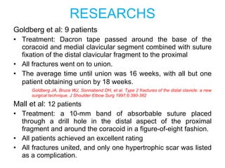
  • 67. RESEARCHS Goldberg et al: 9 patients Treatment: Dacron tape passed around the base of the coracoid and medial clavicular segment combined with suture fixation of the distal clavicular fragment to the proximal All fractures went on to union. The average time until union was 16 weeks, with all but one patient obtaining union by 18 weeks. Goldberg JA, Bruce WJ, Sonnabend DH, et al. Type 2 fractures of the distal clavicle: a new surgical technique. J Shoulder Elbow Surg 1997;6:380-382 Mall et al: 12 patients Treatment: a 10-mm band of absorbable suture placed through a drill hole in the distal aspect of the proximal fragment and around the coracoid in a figure-of-eight fashion. All patients achieved an excellent rating All fractures united, and only one hypertrophic scar was listed as a complication.
  • 68. 3. Coracoclavicular Stabilization With or Without Excision of the Lateral Clavicular Segment Indications: Comminuted lateral clavicular segment; Older patients in whom potential healing of a small lateral clavicular segment would be difficult; Underlying acromioclavicular arthropathy; Fractures lateral to the trapezoid origin; Fractures that involve the coracoclavicular ligament origin as an inferiorly displaced fragment.
  • 69. Coracoclavicular Screw - An appreciable rate of fixation failure due to screw cutout or loosening - Removed at three months
  • 71. TREATMENT These fractures are usually managed nonoperatively unless fracture displacement produces superior mediastinal compromise. In these circumstances, an emergent attempt at closed reduction should be made with open reduction performed next if this is unsuccessful. Internal fixation The use modified Balser plate and use of Mersilene or other strong braided interosseous suture
  • 72. *Complications of nonoperative treatment - Nonunion (1-5%) - Decreased shoulder strength and endurance   *Complications of operative treatment (10%- 30%) Hardware complications : 30% of patient request plate removal. Adhesive capsulitis Infection (~4.8%) Hardware irritation requiring removal (~8%) Mechanical failure (~1.4 %)